
Abstract
Molecular imaging is a form of nanotechnology that enables the noninvasive examination of biological processes in vivo. Radiopharmaceutical agents are used to target biochemical markers, permitting their detection and evaluation. Early visualization of molecular variations indicative of pathophysiological processes can aid in patient diagnoses and management decisions. Molecular imaging is performed by introducing into the body molecular probes, which are often contrast agents that have been nanoengineered to target and tether to molecules, thus enabling their radiologic identification. Through a nanoengineering process, ultrasound contrast agents can be targeted to specific molecules, extending ultrasound’s capabilities from the tissue to molecular level. Molecular ultrasound, or targeted contrast-enhanced ultrasound (TCEUS), has recently emerged as a popular molecular imaging technique due to its ability to provide real-time anatomic and functional information without ionizing radiation. However, molecular ultrasound represents a novel form of molecular imaging and consequently remains largely preclinical. This review explores the commonalities of TCEUS across several molecular targets and points to the need for standardization of kinetic behavior analysis. The literature underscores evidence gaps and the need for additional research. The application of TCEUS is unlimited but needs further standardization to ensure that future research studies are comparable.
Introduction
As the clinical translation of contrast-enhanced ultrasound (CEUS) is applied to the liver and other structural anatomic locations, it is important to look at the next experimental application of contrast microbubbles. To this end, a review of the literature was needed not only to understand the history of CEUS but also to capture the growing body of literature surrounding the ability to capture imaging of cellular-level processes. Diagnostic pressure to visualize disease at its earliest stage of development has pushed researchers to descend from the structural anatomy to the level of cellular pathways of disease.
The first application of CEUS imaging was reported in 1968 by Gramiak and Shah, 1 in which a saline mixture was discovered to improve visualization of the aortic root. Today, the use of ultrasound contrast agents is common practice during echocardiograms for left ventricular opacification and left ventricular endocardial border definition. 2 In addition, the American Society of Echocardiography practice guidelines recommend its use in a variety of other clinical scenarios to enhance echocardiographic image quality.3,4 More recently in the United States, CEUS has been approved for its second indication, which is to further aid in the characterization of liver lesions.
However, ultrasound contrast agents are used extensively in Europe, Canada, and Asia as an enhanced method of detecting blood perfusion at the tissue level.5–9 This is being done in a wide variety of tissue types and pathologies, as the 2011 European Federation of Societies for Ultrasound in Medicine and Biology Guidelines recommend its use in 18 different applications, spanning more than 75 indications. 10 The most notable noncardiac applications for CEUS involve the evaluation of abdominal lesions, specifically those of the liver and kidney.2,11 It has even been hypothesized that further research in this area will allow ultrasound to play a competitive role in abdominal imaging, relative to computed tomography (CT) and magnetic resonance imaging (MRI). 12 Supporting this idea, Ding et al. 13 reported a sensitivity and specificity of 96% and 97%, respectively, in the detection of liver lesions. These findings were corroborated in a 2013 review of the literature, which reported the overall effectiveness of CEUS for the detection of liver lesions to maintain a sensitivity of 86% to 100% and a specificity of 80% to 100%. 14
Its widespread use in these countries is supported by multiple years of in vitro and in vivo research, which have established the properties of ultrasound contrast agents.15,16 Therefore, current research in this area focuses on developing innovative applications for CEUS imaging. 2 One area in which such innovative applications are being investigated is imaging of the musculoskeletal system. This line of inquiry is based on ultrasound contrast’s ability to enhance visualization of the macro- and microvasculature, which affords the potential of detecting hyperemia present in the early stages of many musculoskeletal pathologies.17–19 Studies investigating musculoskeletal CEUS applications have demonstrated significant differences in microvasculature blood flow detection within a variety of joints in the digits, wrists, and knees of individuals with rheumatoid arthritis compared with normal controls.20–22 This research has facilitated its clinical use in the evaluation of a variety of pathologies, beyond rheumatoid arthritis, such as bone erosions, synovitis, tendinopathies, enthesopathies, carpal tunnel syndrome, and osteoarthritis.23–25
Another research focus in the area of ultrasound contrast explores its ability to provide biochemical and physiological information at the molecular level. Through a nanoengineering process, ultrasound contrast agents are able to be targeted to specific biological markers in vivo.26,27 This is known as targeted contrast-enhanced ultrasound (TCEUS), or molecular ultrasound imaging, and its applications are currently being investigated in a variety of tissue types and pathologies.
Targeted Contrast-Enhanced Ultrasound Imaging
The first use of molecular ultrasound imaging, or TCEUS, was reported by Unger et al. 28 for the detection of thrombus formation. Since then, numerous studies have investigated TCEUS applications for the targeting and detection of a variety of biological markers that are thought to be present in the early stages of various disease processes.29,30 Ultrasound contrast agents are restricted to the vascular compartment; therefore, they are ideal for investigating disease processes taking place on the endothelial surface.9,31,32 Subsequently, disease processes such as angiogenesis, inflammation, and thrombus formation are the primary focus of TCEUS research.33–35
One such application being investigated is molecular imaging of the angiogenic process, in an attempt to establish an early diagnostic tool for pathological processes such as tumor formation. Multiple preclinical animal studies have reported successful findings in this area by targeting various integrins, as these are present in the initial stages of angiogenesis.36–44 Other preclinical animal studies investigating angiogenesis have also reported successful findings while targeting different vascular endothelial growth factor receptors for the application of early tumor detection.40,42–53 The most commonly targeted angiogenic markers are alpha-v beta-3 integrin (αvβ3) and vascular endothelial growth factor receptor 2 (VEGFR-2), and they could have major implications in the future of cancer diagnosis and treatment. 54 Although most research has been directed at targeting angiogenic markers, the targeting of inflammatory markers is another application for which molecular ultrasound imaging is being investigated. This stems from the hypothesis that, due to the vast number of diseases in which inflammation presents, the future clinical advantages of this application could outweigh those of angiogenesis. 55
Proof of principle
The inflammatory process is mediated by the initial capturing and adhesion of rolling leukocytes by cell adhesion molecules (CAMs) on the endothelial surface.29,56–60 Such CAMs include P-selectin, intercellular adhesion molecule 1 (ICAM-1), and vascular cell adhesion molecule 1 (VCAM-1). Each of these CAMs has been found to correlate with various stages of inflammation severity and are therefore felt to be early inflammatory biomarkers.9,34,61–65 To this end, the determination of their presence could aid in an early diagnosis of inflammation. 66 For molecular ultrasound imaging to play a role in the early detection of inflammation, its ability to detect inflammatory biochemical markers must be investigated. This has led to numerous preclinical proof of principle studies that have explored microbubbles targeted to various inflammatory biochemical markers, in an assortment of tissues (Table 1).
In Vitro and In Vivo Molecular Ultrasound Imaging Proof of Principle Studies Investigating Inflammation While Targeting P-Selectin, VCAM-1, and ICAM-1.
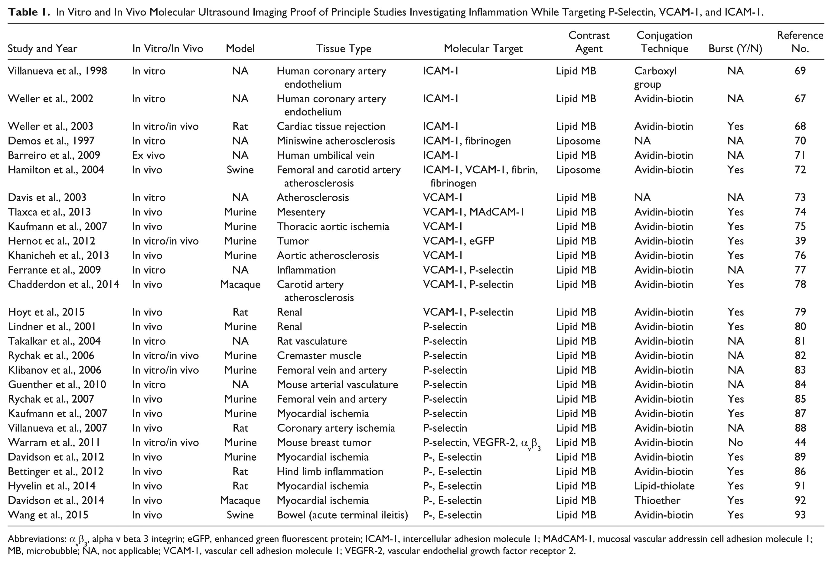
Abbreviations: αvβ3, alpha v beta 3 integrin; eGFP, enhanced green fluorescent protein; ICAM-1, intercellular adhesion molecule 1; MAdCAM-1, mucosal vascular addressin cell adhesion molecule 1; MB, microbubble; NA, not applicable; VCAM-1, vascular cell adhesion molecule 1; VEGFR-2, vascular endothelial growth factor receptor 2.
Weller et al.67,68 investigated TCEUS targeted to ICAM-1 in cardiac tissue, for the detection of early stage cardiac transplant rejection, and reported a statistically significant increase in microbubble binding both in vitro and in vivo. In a similar study, signal intensities obtained in TCEUS imaging targeted to ICAM-1 were found to be significantly elevated in an inflamed coronary artery.
69
Successful detection of ICAM-1 targeted microbubbles has also been reported while investigating markers of atherosclerosis.
70
VCAM-1 targeted TCEUS imaging has also been investigated to determine its role in the early detection of inflammation. Promising results have been reported while investigating inflammation of the bowel, kidneys, vasculature, and soft tissue, as well as in tumor detection.39,71–79 Last, particular success has been attained while targeting P-selectin with TCEUS during preclinical in vivo studies. In a study examining renal tissue inflammation, microvascular adhesion of ultrasound contrast microbubbles to P-selectin was found within an inflamed kidney, resulting in an enhanced sonographic signal.
80
Another application has been established by the successful targeting of P-selectin proteins in inflamed muscle tissue and vasculature.81–86 Furthermore, Kaufmann et al.
87
demonstrated 10 times more binding of microbubbles targeted to P-selectin than controls while examining myocardial ischemia and reperfusion. Similar findings were reported by Villanueva et al.,
88
also examining P-selectin targeted microbubbles in myocardial ischemia and reperfusion. This has been further supported by other comparable studies.89
–
These studies have provided evidence of the ability of targeted ultrasound contrast to bind to P-selectin, ICAM-1, and VCAM-1 for the early evaluation of inflammation. Successful visualization of these inflammatory biochemical markers has been accomplished in a variety of pathophysiological processes and tissues. However, the absence of the exploration of TCEUS imaging for the application of evaluating neural tissue inflammation following a traumatic spinal cord injury was notable, thus providing rationale for its investigation.
Longitudinal
In addition to serving as a diagnostic tool, ultrasound imaging has advantages that create an opportunity for targeted microbubbles to be used as a method of disease monitoring.35,66,93,94 Due to its acoustic properties, molecular ultrasound poses no threat of adverse effects caused by overexposure to ionizing radiation, making its use as a surveillance tool ideal.9,27,94,95 Repeated molecular ultrasound imaging studies provide information regarding disease progression, which can be used to gauge disease advancement and monitor therapy effectiveness.66,94 This will provide clinicians with the unique opportunity of modifying patient management at an early stage, likely before clinical symptoms have manifested.31,35,46
Proof of principle studies have provided evidence that molecular ultrasound imaging is capable of detecting biochemical markers in a variety of tissues, for the advancement of its use as a diagnostic tool. Disease monitoring via molecular ultrasound imaging would require a high level of sensitivity for the detection of biochemical markers at all levels of expression. Therefore, it is unfortunate that most proof of principle studies have been cross-sectional in design and provide evidence of molecular detection effectiveness at only a single stage of disease and severity. Furthermore, this is often performed during the acute phase of disease, in which biochemical markers are expressed at high levels. This provides no evidence of molecular ultrasound imaging’s capabilities for detecting late stages of disease and makes this information unsuitable for its translational use in disease surveillance. To appropriately provide evidence for this potential application, it is necessary that preclinical longitudinal studies be performed to evaluate TCEUS’s ability to detect targeted molecules at varying levels of expression. Longitudinal designed studies represent the next step in molecular ultrasound research, as information gained will advance the knowledge of its effectiveness and aid in proper bench-to-bedside translation.43,45,96 It is unfortunate that the number of published longitudinal studies is minimal compared to that of proof of principle studies (Table 2).
In Vitro and In Vivo Studies Investigating the Utility of Molecular Ultrasound Imaging for Detecting Molecular Markers of Disease at Varying Levels of Expression.
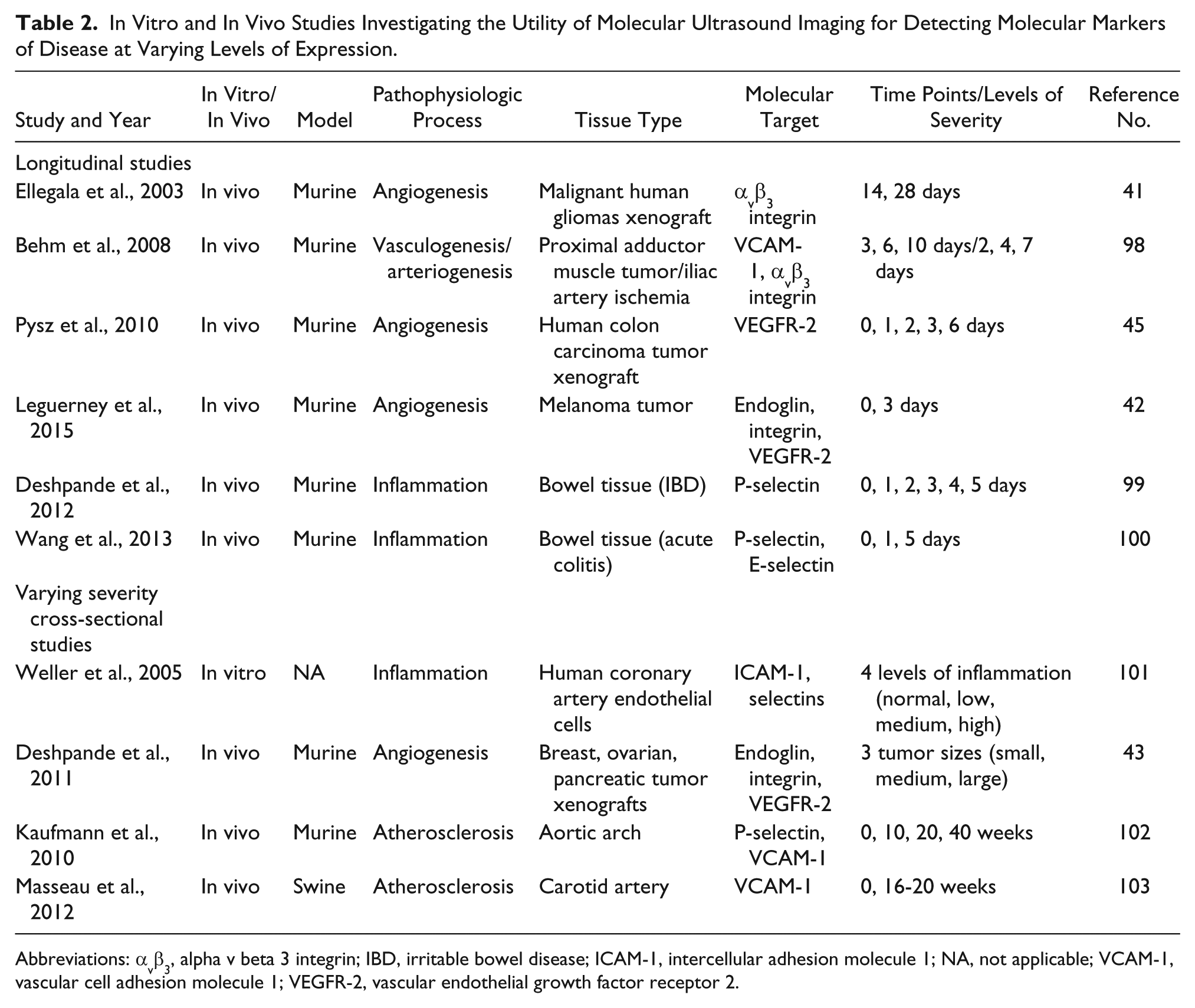
Abbreviations: αvβ3, alpha v beta 3 integrin; IBD, irritable bowel disease; ICAM-1, intercellular adhesion molecule 1; NA, not applicable; VCAM-1, vascular cell adhesion molecule 1; VEGFR-2, vascular endothelial growth factor receptor 2.
The majority of these studies were performed in vivo and investigated the longitudinal monitoring of tumor growth by examining angiogenesis. This has been accomplished most frequently by targeting microbubbles to various integrins and/or VEGFR-2.41,42,45,97 A few longitudinal TCEUS studies have been performed using antibodies targeted against P- and E-selectin for the evaluation of inflammation; however, these investigations are limited to tissues of the bowel.98,99,100
In addition to these longitudinal studies, other cross-sectional studies do exist that incorporate subjects with different levels of disease. Although not longitudinal, these studies were conducted with the same goal of providing evidence of molecular ultrasound’s use in disease surveillance. Early research using this model was performed by Weller et al. 101 in 2005, who demonstrated in vitro that the adhesion strength of targeted microbubbles to ICAM-1 had a strong linear relationship with inflammation severity (level of ICAM-1 expression). In vivo studies have investigated carcinoma by using tumors of different sizes to represent severity. 43 This was accomplished by targeting multiple angiogenic markers in various tumor types. 43 Other in vivo studies have explored molecular ultrasound’s use in the evaluation of atherosclerosis, often targeting CAMs such as P-selectin and VCAM-1.102,103
These studies represent innovation in molecular ultrasound imaging research. They were conducted with the objective of extending knowledge beyond proof of principle information, by providing evidence regarding molecular ultrasound’s ability to detect biochemical markers at varying levels of expression. By doing so, they have taken the next step toward the clinical translation of TCEUS imaging.43,45,96
The variety of tissue types in which targeted ultrasound contrast agents have been successfully detected during inflammation is indeed impressive; further research is needed to provide evidence on the appropriate dosing of contrast agents. Contrast dosing information must be obtained for ultrasound contrast imaging to be appropriately, effectively, and safely translated to humans.92,96 The volume of contrast agent administered is directly related to obtained signal intensities and, in turn, the sensitivity of the TCEUS.104,105 This further entails the need for established dosing parameters, so as to ensure accurate interpretation of the scan.104,105 Dosing parameters also need to be considered for pediatric applications, so that toxicity risks are minimized, while still achieving a clinically appropriate scan.
Analytics of Kinetic Behavior
In addition to investigations of TCEUS applications, techniques of image analysis and interpretation should also be examined. One popular method of ascertaining information from contrast imaging is through the analysis of the contrast agent’s temporal behavior. 106 Temporal behavior refers to the activity of the contrast agent over time. This idea was first introduced in the 1990s and is primarily used and investigated in MRI. 106 Enhancement kinetics are represented semiquantitatively in the form of time intensity curves (TICs). 107 Time intensity curves are generated by placing a region of interest over the area being examined. The signal intensity of the image, which has been enhanced via the contrast agent, is calculated and plotted over time to provide a visual depiction of the contrast agent’s temporal behavior. 107 Known by a variety of terms, TIC shape analysis can then be conducted, in which the curve representing signal intensity is classified based on its shape. 108
As mentioned previously, the use of TICs to obtain information regarding the perfusion of an area has largely been in the field of MRI. 106 Time intensity curves are primarily used in situations in which malignancy of a tumor is suspected, and knowledge of its perfusion characteristics is necessary. 107 Researchers who investigate TIC analysis applications attempt to define distinctive TIC patterns and assign them to physiologic or pathologic findings.106,109 Classification of TICs focuses more on the shape of the curve and relies less on their quantitative absolute values. 107 This idea has been referred to as TIC shape analysis and will be referenced as such for the remainder of this article. 107 Yankeelov and Gore 110 have coined this idea “curve-ology,” whereas Kuhl et al. 107 referred to this method of TIC analysis as “enhancement kinetics.” Kuhl et al. 111 continued to describe three classifications of TIC shape analysis, based on the shape of the initial signal intensity wash-in phase and the wash-out phase components of the TIC. A type 1 curve shape depicts a continuous enhancement of signal intensity that increases over time. 111 A type 2 curve is characterized by a wash-in phase that reaches a peak and then maintains a plateau during the wash-out phase after approximately 2 to 3 minutes of imaging. 111 And finally, Kuhl et al. 111 defined a type 3 curve as a wash-out curve, where a decrease in signal intensity is seen approximately 2 to 3 minutes after peak signal intensity is achieved in the wash-out phase. These TIC shape analysis classifications were determined through evidence obtained by the same researchers, which showed that 57% of malignant breast lesions produced a type 2 curve, whereas 83% of benign lesions produced either a type 1 or type 3 curve. 111
In a more recent study, van Rijswijk et al. 109 investigated the use of TIC shape analysis in the evaluation of soft tissue sarcomas. In this study, researchers used contrast-enhanced MRI to obtain TICs representing soft tissue sarcoma perfusion. These TICs were then subjectively placed into five different categories based on their shape. 109 A type 1 curve was categorized by the absence of a slope in the wash-in phase and therefore the absence of enhancement. 109 A type 2 curve depicts a gradual and continuous increase in signal intensity for approximately 5 minutes, with no steep slope in the wash-in phase. 109 Type 3 was defined as a curve containing a rapid slope in the wash-in phase, followed by a plateau in the wash-out phase. 109 Similar to type 3, type 4 was characterized by a rapid slope in the wash-in phase; however, a decrease in signal intensity is seen in the wash-out phase. 109 Finally, van Rijswijk et al. 109 defined a type 5 TIC as a curve with a rapid signal intensity slope in the wash-in phase, followed by a sustained signal intensity increase in the wash-out phase. Throughout this study, curve types 3 through 5 were seen, as the investigators concluded that the only curve feature that was consistently associated with malignancy was early enhancement. 109
Recently, the utility of TIC shape analysis has been investigated in CEUS imaging. As briefly described previously, ultrasound contrast agents comprise microbubbles consisting of a gaseous core, stabilized by an outer shell made of lipid, protein, or polymer.9,15,104,112–115 Once introduced into circulation, ultrasound contrast microbubbles interact with the incident ultrasonic beam, which causes them to oscillate and produce an enhanced sound wave that is returned to the transducer. This is due primarily to the large difference in acoustic impedance between the gas inside the contrast microbubble and the surrounding tissue.9,15,17,66,94,116–119 The returned enhanced sound wave produces an enhanced signal intensity, which can then be displayed in the form of a TIC, just as in MRI. Using this analysis technique in CEUS, multiple clinical conditions have been evaluated, including the monitoring of liver lesions. 120 In a review conducted by Cosgrove and Lassau, 19 TICs were obtained from CEUS imaging studies that had been performed longitudinally on a metastatic breast tumor while the patient underwent antivascular therapy. It is interesting that TIC shape analysis demonstrated a decrease in size and a difference in shape of the curve as treatment continued. 19 In this case, CEUS TIC shape analysis was found to be a more sensitive measure than CT. 19 Furthermore, Lassau et al. 121 reported very similar findings, in which changes in CEUS TIC shape analysis representing liver perfusion were observed longitudinally in an individual receiving treatment for hepatocellular carcinoma. Last, it is important to note that, just as demonstrated in the MRI literature, a wide spectrum of nomenclature has been used to describe CEUS TIC shape analysis. Terms such as perfusion kinetics, contrast uptake curves, and time variance imaging were all reported in studies that examined CEUS TICs.121–123
Conclusion
Molecular imaging has the potential to detect protein activity at the cellular level. This pathophysiologic information can be leveraged to better recognize the formation of disease processes while still in the acute stages. Targeted contrast-enhanced ultrasound makes use of nanoengineering to create microbubbles that can be targeted and tethered to molecules at the site of diagnostic inquiry. Once tethered, the microbubbles are insonated, which provides a resonance frequency that increases the signal-to-noise ratio through the amplification of reflections toward the transducer. Raw signal data have the potential to predict the behavior of cellular processes below structural anatomy.
Analysis of the temporal behavior of the TCEUS signal allows for both quantitative and qualitative measures. This technology is still in its infancy and therefore is in need of standardization to ensure that the microbubbles’ kinetic behavior can be reproducibly analyzed.
Since a lack of standardization exists for analyzing microbubble kinetic behavior, MRI TICs were investigated to determine if these techniques translate. An actual comparison of the analytic techniques would be required to make an informed determination on which are the most sensitive for describing TCEUS kinetics.
Footnotes
Declaration of Conflicting Interests
The authors declared no potential conflicts of interest with respect to the research, authorship, and/or publication of this article.
Funding
The authors received no financial support for the research, authorship, and/or publication of this article.