
Abstract
A 48-year-old male who had undergone left inguinal orchidectomy for testicular tumor 10 days previously presented with left inguinal and scrotal swelling. The scrotal sonogram showed what appeared to be two testes, with a mass within the left testicular area. Real-time scanning revealed that the left “testis” was thick fluid/hematoma with a more organized hematoma within.
Introduction
Scrotal ultrasound remains the established first-line investigation in many presentations of scrotal pathology, in follow-up cases, and as a screening tool in clinical practice.1,2 Scrotal ultrasound is harmless to the patient, relatively rapid and inexpensive, and produces high resolution images that reveal a spectrum of diagnoses from normal to pathology as well as ambiguity.3,4 The differentiation between intra- and extratesticular lesions is essential and has a direct impact on patient management. A scrotal sonogram easily demonstrates the location of a scrotal mass, enabling timely surgical intervention if needed. Follow-up scans are rarely required routinely post scrotal surgery, but in certain cases ultrasound can be useful to assess for surgical complications or subsequent suspected pathology. The case presented is an unusual appearance obtained during a postorchidectomy follow-up that provided ambiguous images of an atypical postoperative hematoma mimicking the removed testis. This case is an example of the advantage of real-time sonography, where static images may have given rise to significant false interpretation.
Case Report
The patient was examined using a Logiq 9 ultrasound scanner (GE Medical Systems, Slough, UK) employing 12/9 MHz linear transducer. The patient was a 48-year-old male with a history of left inguinal orchidectomy for intratesticular tumor (pathologically confirmed combined nonseminoma/seminoma) 10 days previously. The patient presented with painless left scrotal and left inguinal swelling (Figure 1).
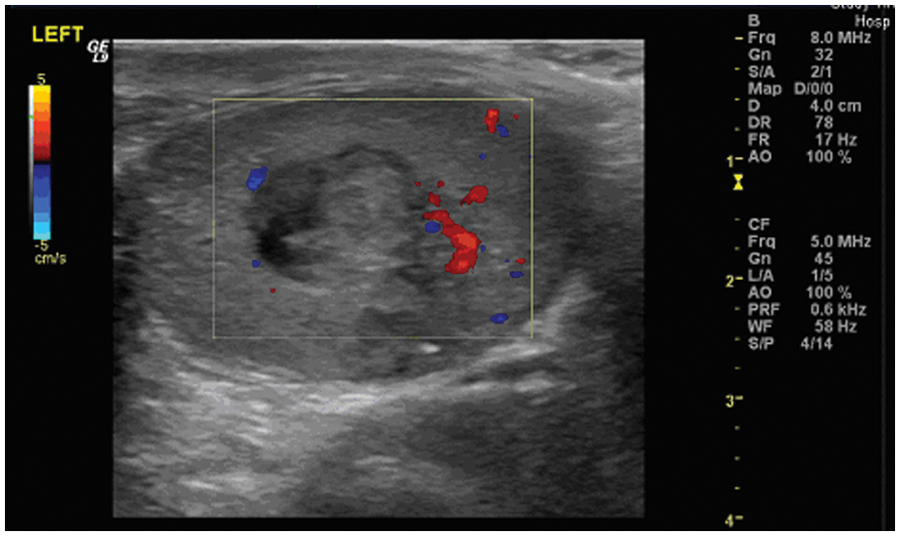
Avascular mass in the left testicular region mimicking intratesticular pathology
A superficial hematoma was demonstrated within the left inguinal region (Figure 2). The initial transverse scrotal sonogram demonstrated the unexpected appearance of what appeared to be right and left testes within the scrotum (Figure 3). The “testes” appeared equal and uniform in echogenicity, with an apparent hyperechoic mass within the left testis (Figure 4). The patient and surgical notes confirmed that orchidectomy had definitely taken place. Eventually, upon real-time scanning after several minutes, the “left testis” was seen to be slightly turbid, swirling, thick fluid filling the left hemiscrotum, with a more complex well-defined avascular mass within. No spectral Doppler signal or color Doppler filling were seen during the course of the examination in this region. Ultrasound features in real time suggested thick blood with an organizing hematoma. The right testis appeared to be normal, with a small peripheral extratesticular linear calcification.
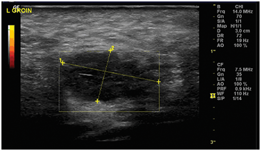
Image of a superficial left inguinal hematoma
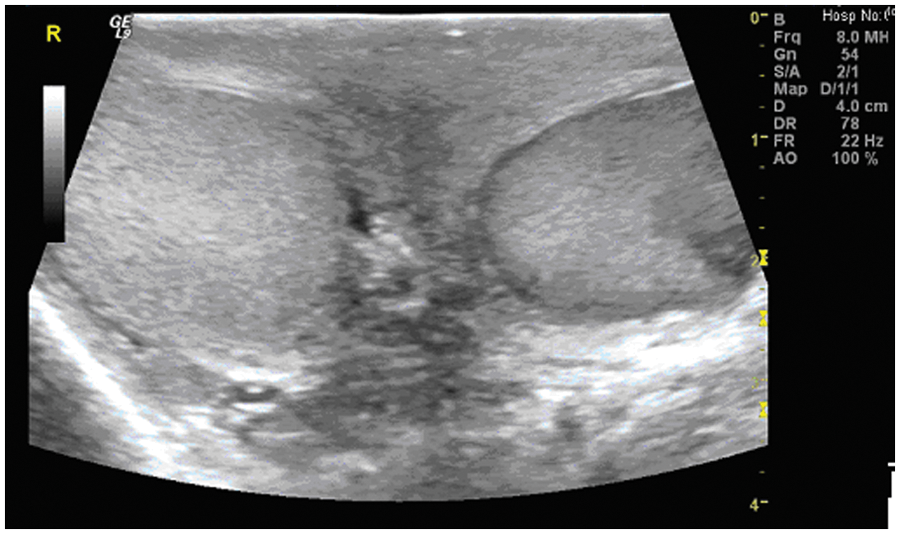
Still frame image with the appearance suggesting two testes, with the mass seen on the left side
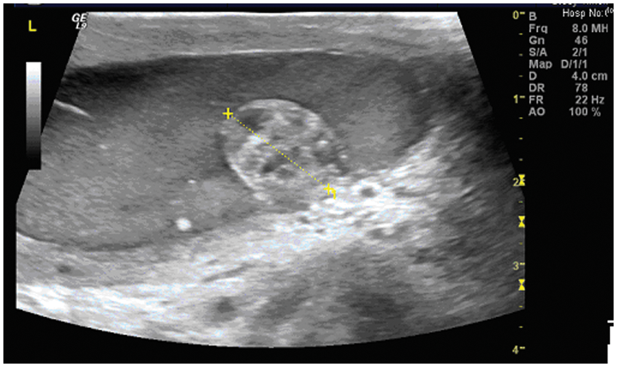
Hyperechoic 1.5 cm mass in the left testicular region mimicking intratesticular pathology
Discussion
Scrotal pathology ranges from the benign and innocuous, most commonly extratesticular, to aggressive malignant lesions, most commonly intratesticular. 5 The incidence of testicular tumors is relatively rare and the overall prognosis is usually excellent.6,7 Radical orchidectomy is one aspect of the definitive treatment of testicular cancer, with potential postoperative complications including infection and scrotal hematoma. Scrotal sonography is a key element in the diagnosis of potential scrotal pathology, defining the location, ultrasound characteristics of any lesion, and possible diagnosis of a range of conditions; postoperative sonography is also of value when clinically indicated.
There are many potential caveats in the field of medical ultrasound, with innumerable normal variants, pathology mimicking lesions, and a variety of unusual images. False positive scrotal sonograms have been previously reported,8,9 and the sonographic differentiation between intratesticular tumor and hematoma can be difficult. 10 There are a range of sonographic appearances post orchidectomy, 11 with scrotal hematoma a recognized complication. A hematoma may have variable echo pattern, ranging from cystic to heterogeneous to solid. The images presented in this case, however, were atypical and somewhat unusual. Because of this, the advantage of real-time ultrasound scanning was seen for accurate interpretation and reporting. The gray-scale images in isolation would have made it impossible to reach the correct conclusion; if presented with these images, a reasonable report would include the possibility of tumor within the left testis, with a question of failed orchidectomy. The patient in this case ultimately was successfully treated conservatively.
Footnotes
Acknowledgements
The authors would like to acknowledge the encouragement and assistance of Dr. A. Stockdale and Professor C. Hutchinson in preparing this article.
Declaration of Conflicting Interests
The authors declared no potential conflicts of interest with respect to the research, authorship, and/or publication of this article.
Funding
The authors received no financial support for the research, authorship, and/or publication of this article.