
Abstract
Enzyme-linked immunosorbent assay (ELISA) automation for routine operation in a small research environment would be very attractive. A portable fully automated low-cost immunoassay system was designed, developed, and evaluated with several protein analytes. It features disposable capillary columns as the reaction sites and uses real-time calibration for improved accuracy. It reduces the overall assay time to less than 75 min with the ability of easy adaptation of new testing targets. The running cost is extremely low due to the nature of automation, as well as reduced material requirements. Details about system configuration, components selection, disposable fabrication, system assembly, and operation are reported. The performance of the system was initially established with a rabbit immunoglobulin G (IgG) assay, and an example of assay adaptation with an interleukin 6 (IL6) assay is shown. This system is ideal for research use, but could work for broader testing applications with further optimization.
Introduction
Since the invention of enzyme-linked immunosorbent assay (ELISA) more than half a century ago, it has been used as a research and diagnostic tool in medicine and pathology, as well as control standards in industry and clinical testing. The greatest advantages of ELISA are great sensitivity and specificity with the proper antibodies customized for specific applications.
Although ELISA technologies have improved a lot since its debut, the most commonly used platform is still 96-well (less common are 384-well and 1536-well) plates for most researchers and lab operators. Standard multistep manual operations remain the major assay procedures for an ELISA test. Many companies, such as Bio-Tek, developed automation instruments (plate loader, plate washer, etc.) to pair with a plate reader to automate some of these assay procedures. However, this pseudoautomation process only mitigates the load in a big lab environment, such as test centers, since it requires multiple expensive and bulky benchtop or floor-stand units. Furthermore, traditional ELISAs are time-consuming (normally 4–6 h for a sandwich ELISA) and always require calibration for best accuracy. In a small research or test lab environment, it is very inefficient and costly to run a few sample tests with ELISA on a daily basis. Thus, an easy-to-use, cost-effective, and automated rapid immunoassay system could reduce these labor-intensive manual operations.
Recent developments of microfluidic technologies (especially the concept of lab-on-a-chip [LOC]) have opened a door for further miniaturization and automation of traditional immunoassays.1–4 The microfluidic system is ideal for ELISA automation because of many advantages: easy automation, minimized sample requirement, better reaction kinetics due to the large ratio of surface area to volume, rapid assay procedure, and reduced size for portable/handheld applications.1,3–7 Currently in the market, there are several portable immunoassay systems available for point-of-care applications (Samsung Labgeo, Philips Minicare, Micropoint Mlabs, Perlong Medial FIA8200, etc.), but they are either costly to maintain or use exotic techniques that lack versatility. These factors are extremely important for small research environments. Here, we are reporting a fully automated capillary-based immunoassay system featuring customizable assays, low maintenance cost, rapid assay detection, and reliable test results. Because of its similarity to traditional microplate-based assay, it can easily adapt new assay methods for various applications, including the measurements of microorganisms, macromolecules, and small molecules.
Capillary tubing is a great candidate as a microfluidic reactor. Compared with LOC designs, it costs little because mass production technology of long capillary tubing is very mature (check companies like PolyMicro and Paradigm Optics). The quality control of dimensions and surface properties for these capillaries could easily surpass that of most LOCs. Furthermore, it is versatile in materials and sizes that could fit for different assay requirements. Thus, it is attractive to have a capillary-based automated immunoassay system for research applications. Su et al. patented a capillary-based immunoassay system design. 8 They have successfully demonstrated the adaptation of multiple pathogen assays with the system.9–11 This prototype device features a configuration for sequential reagent loading and preprogrammed assay protocol to run multiple tests with reduced assay time and minimized cross-contamination. However, it does not fit for real application, mainly because of its cumbersome operational protocols. The mounting of capillary columns on the instrument is difficult for the system even for experienced users. The signal reliability and sensitivity are not good enough for the detection of many protein biomarkers, due to the pulsation movement introduced by the peristaltic pump and the low detection limit requirement. Here, we are reporting a new system design to address these issues and demonstrate its applications for protein biomarker analysis. The future applications of the system could be expanded to the detection of food-borne pathogens for food safety, homeland defense, the military, environmental monitoring, chemical processing, and medical or clinical diagnostics.
Materials and Methods
Materials
Ninety-six-well Maxisorp Nunc plates are purchased from VWR (Radnor, PA). Acrylic and polycarbonate (PC) capillary columns (different internal [ID] and external diameters) are from Paradigm Optics (Vancouver, WA). The interleukin 6 (IL6) ELISA kit (3460-1H-20) is from Mabtech (Cincinnati, OH). Rabbit immunoglobulin G (IgG) ELISA reagents (211-005-109, 711-035-152, and 011-000-003) are from Jackson Immuno (West Grove, PA). Starting block with T20 (37543) and UltraTMB (34028) are from Thermo Scientific (Waltham, MA). Coating buffer (421701) is from BioLegend (San Diego, CA), and washing solution 10× TBST (786-161) is from Gbiosciences (St. Louis, MO). CHEMUSB4-VIS-NIR VIS Spectrophotometer with built-in cuvette holder and light source is from Ocean Optics (Dunedin, FL). The 585.1 Style Ultra Micro Flow Cell with 1.5 mm window is from Spectrecology (Dunedin, FL). The system valves (MLP777-601, MLP777-605, and MLP778-605) are from IDEX (Lake Forrest, IL). The peristaltic pump (SP100VO) is from APT Instruments (Omaha, NE). The syringe pumps (PSD4) used are from Hamilton (Reno, NV). All fluidic connectors are purchased from IDEX or made in-house. The control board, disposable components, and enclosure are made in-house. All other prototyping materials are from McMaster (Elmhurst, IL).
System Configurations
The overall idea of this system is to use multiple capillary columns as the bioreactor. Valves and pumps work together to deliver different reagents from a reagent cartridge through each capillary, and finally through the detector, in a programmed way, which is achieved with a dedicated control board and graphical user interface (GUI). The entire assay procedures are actually similar to those of traditional plate-based ELISA without introducing any exotic steps. The main challenge is how these components are configured to work in the same way for different capillaries without introducing cross-contaminations. As shown in Figure 1 , two designs have been tested. The pull-only design ( Fig. 1 , left) is similar to the system reported before 8 because of its simplicity. In this design, all reagents (R) are pulled through the capillary columns (C) with a single pull pump (P1) (normally a peristaltic pump since it costs less and no refilling is required). Four valves (S, V1, V2, and B) are used to select reagents, capillary columns, and bypassing the detector, respectively, during the assay process. This design worked very well for several applications. However, the reliability tends to be low due to the nature of peristaltic pumping and the pulling mode itself, which affects the detection process even more when there is bubble generated under vacuum in the flow path. This is more critical for the detection step since all the signal is measured with a small substrate fluidic plug (<20 µL) passing through a flow cell detector. The pulse movement of the liquid plug greatly limits the capability to bring down the limit of detection (LOD). Instead, a new pull–push design ( Fig. 1 , right) was developed to increase system reliability and improve sensitivity.
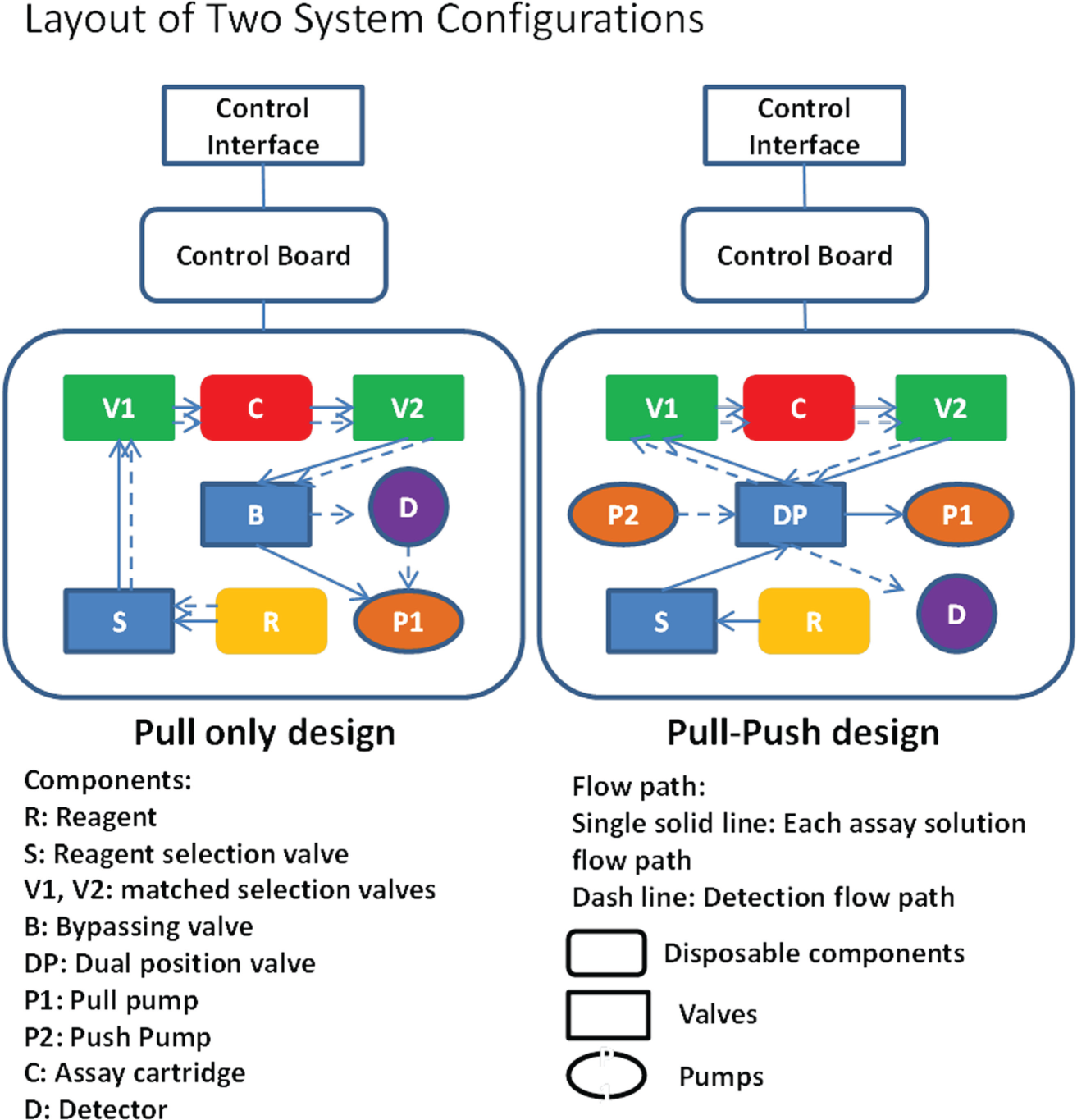
Layouts of two configurations for capillary-based automation immunoassay system (pull only and pull–push). All blocks show the components included as a completed system and explained in the figure. The solid-line arrows show the flow path of reagent delivery, and the dash-line arrows show the flow path during detection.
In the pull–push design, a second pump (P2) is introduced to push reagents (R) through the capillaries (C) and detector (D), instead of pulling through after switching of a dual-position valve (DP). Other reagents are delivered in a similar way with the primary pump (P1) in pull mode. Both pumps are high-quality syringe pumps from Hamilton instead of peristaltic pumps. This article mainly focuses on the pull–push design because of its improved performance, and a more detailed description of this design is shown later in Figure 4 .
As for the detector, since most traditional ELISAs use optical detection (mainly absorbance-based measurement), the immunoassay system maintains the same detection methods for easier assay adaptation. A microfluidic flow through a cell-type absorbance module from Ocean Optics (CHEMUSB4-VIS-NIR VIS Spectrophotometer) is integrated.
Assay Formats
One main target for the system is easy adaptation of current immunoassays used with traditional 96-well plate platforms. Thus, the idea of keeping the overall assay procedure similar to the traditional method has great benefit to reduce the platform transition cost. Another target for the system is to minimize the assay time by introducing microfluidic bioreaction sites, which could perform an accelerated assay protocol without sacrificing the performance. It has been noticed that immunoassays with great performance achieved on 96-well plates do not always translate to great performance in a flow-through capillary system; however, those assays that work well in an accelerated microfluidic platform would definitely work well on the 96-well platform with classical protocols (data not shown). Thus, it is still a valid approach to use a 96-well plate platform for antibody prescreening and assay optimization before adaptation on the capillary-based immunoassay system. The assay conditions obtained from a 96-well plate platform could act as the starting point for further optimization in the flow system.
In theory, most immunoassay methods could be adapted to the flow-through system, but we mainly focused on sandwich ELISA, as shown in Figure 2 . These stepwise assays are best suited for flow-through without the need of mixing. Although using enzyme-conjugated secondary antibody ( Fig. 2 , left) is best to minimize the assay time, it is sometimes limited by both sensitivity requirements and its availability. A more common approach is to use a tertiary antibody ( Fig. 2 , middle). 12 However, many times this additional step will not improve the sensitivity, which becomes more and more important in current biomarker research. The biotin-(strept)avidin system is a widely used method to enhance ELISA performance due to its high specificity and strong binding. 12 Although it is still limited by the availability of biotinylated secondary antibodies, it could provide the best assay performance in many cases, and a commercial biotin conjugation kit is available (such as the biotin labeling kit from Dojindo, Gaithersburg, MD). Both methods with tertiary antibody or (strept)avidin have extra steps, which increases the overall assay time. However, these reagents could often be premixed with previous reagents to reduce steps and not affect performance. Thus, it is the balance of time and sensitivity, depending on the assay requirements. As for the substrate, either a horseradish peroxidase (HRP)– or alkaline phosphatase (AP)–based system could be used. We mainly discuss HRP-based assay development here due to the fact that our initial samples are serum/plasma and there is residual AP activity from these samples (data not shown) that could affect the biomarker detection.
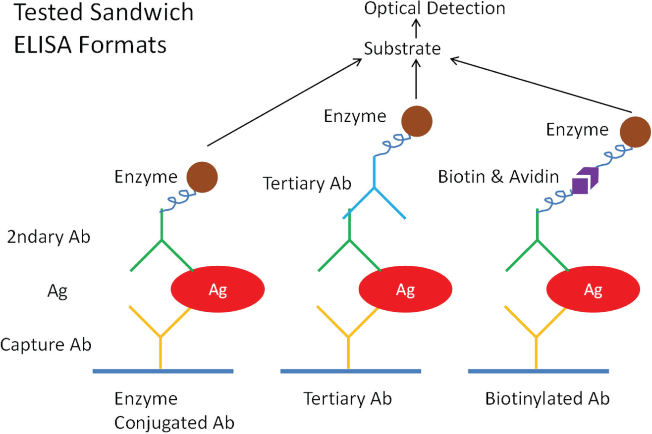
Three sandwich ELISA formats tested with a capillary-based automation system: enzymatic secondary antibody (left), enzymatic tertiary antibody (middle), and biotinylated secondary antibody (right). Faster assay could be achieved with the enzymatic secondary antibody because of less assay steps, while the other two formats could provide better assay sensitivity.
As for the assay procedure, it is a direct conversion from a static 96-well plate platform to a flow-through system with similar static incubation steps. Each capillary column acts as an individual assay site that has been precoated with capture antibody and fully blocked with blocking solution and stored dry in the refrigerator before use. When assay starts, all the columns will be primed with blocking buffer and all reagents are primed through a common channel. Samples/standards/controls are first loaded to each individual capillary one by one and incubated for a specific time. Washing steps are programmed to wash each capillary and keep the same incubation time frame. All other reagents in the order of secondary antibody, HRP-conjugated tertiary antibody or streptavidin-HRP, washing, and substrate solutions are loaded in the same way to all capillaries in a flow–stop for incubation–flow pattern. After the final incubation with the substrate, the developed solution is pushed through a flow cell for absorbance measurement at 625 nm wavelength. All steps are similar to those for the static 96-well plate platform, except a much shorter incubation time is required due to the nature of the large ratio of surface area to volume of the reaction site. An acidic solution may be used as a stop solution to denature residual enzyme activities in the common flow path before final measurement and improve the assay performance.
Capillary Column and Assay Cartridge
The ratio of surface area to volume is inversely proportional to the diameter of the capillary tube. The incubation time required to achieve the same level of adsorption is inversely proportional to the square of the area-to-volume ratio. 13 The larger the ratio of surface area to volume, the shorter the assay time that is required to obtain a similar assay performance. Thus, capillaries with smaller IDs are expected to be better to reduce the assay time. However, the size of capillaries cannot be too small because small ID tubing often causes problems in the flow due to high back pressure and an increased chance of clogging. The size of the tubing has to be balanced for performance and overall system reliability. Dimensions varying from 125 to 1000 µm tubings were tested, and 500 µm was determined to be the best size for the current system.
The second factor to consider about the capillary columns is the length. Ideally, shorter is better to reduce sample and the reagent volume requirement. However, it is also limited by the volume requirement in the subsequent detection step. The downstream absorbance measurement is performed in a flow cell, which has an internal volume of 18 µL. Thus, 10 cm of 500 µm ID capillary columns is used (roughly 19.6 µL volume).
The third factor is the material of capillaries. Since the capillary columns are the reaction sites of ELISA, the choice could greatly affect the assay performance, especially the first primary antibody immobilization step. Two antibody coating methods are commonly used for coating the surface of these capillary columns: physisorption and chemical binding. PC, polystyrene (PS), cyclic olefin copolymer (COC), and various polytetrafluoroethylene (PTFE) capillaries have demonstrated good results with physisorption coating, while fused silica and acrylic demonstrated success with chemical binding. Both antibody coating methods are relatively easy to perform. With a tubing connector and a syringe filled with reagents, a long capillary tubing could be coated at once with great uniformity. Among the materials tested, PC, acrylic, and fused silica capillaries work the best for our purpose.
The major challenge in developing a capillary-based automated system is to make the engagement of capillary columns to the system in a user-friendly manner. In the previously reported design, 8 users have to mount six columns with 12 connectors on the instrument before each test. It is not only labor-intensive, but also error-prone due to potential damages to the ends of capillaries during assembly. It is difficult even for trained lab technicians. To address this issue, a capillary housing is designed to quickly engage and disengage all capillary columns in one simple step. The convenience is achieved with redesigned magnetic connector technology. 14
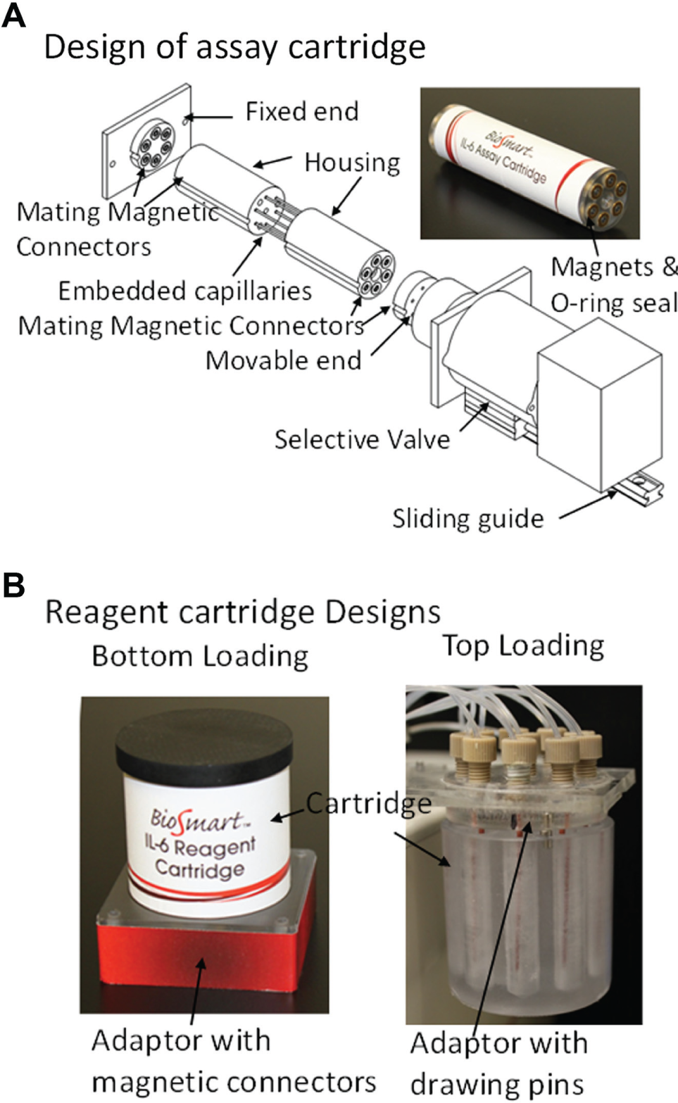
In current cartridge design shown in Figure 3A , up to six precoated capillary columns are embedded in the magnetic connector–enabled housing unit. The capillaries could be mounted to the system at the same time with a built-in self-alignment feature. Furthermore, since different capillaries (dimensions, materials, etc.) can be used with the same housing unit, it is easy for users to switch assays or try different assay methods. This is very cost-efficient for a research lab environment. As an example, the current setup uses 10 cm long, 500 µm ID capillaries (~20 µL internal volume). All these capillaries could be prepared in the same batch and cut from the same long tubing to minimize column-to-column variations. Either PC or acrylic could be used depending on the assay methods. The performance of assays could be easily evaluated by using different specs of capillary columns.
(
Magnetic connectors were used at both ends of the cartridge to simplify the procedure of loading and unloading. These magnetic enhanced connectors use simple magnetic actions and a properly designed O-ring seal to facilitate the fluidic connections. These gold-plated magnetic connectors can ensure easy fluidic connection to withstand up to 100 psi for an extended lifetime. When multiple magnetic connectors are used, the cartridge also features self-alignment. Since both ends need to be engaged when loading the cartridge, one side of the mating piece is designed to be movable ( Fig. 3A ). It is connected directly on the selection valve and mounted on a slider. The cartridge could be engaged and disengaged easily.
Reagent Cartridge Designs
Another disposable component of the system is the reagent cartridge, which is filled with all the reagents before test. For a system that is capable of running five tests simultaneously, it will take up to 10 reagents, including 5 standards/samples/controls, washing buffer, 2 reaction reagents, substrate solution, and stop solution. It is extremely difficult for the user to manage all 10 reagents for every test. Thus, a more user-friendly reagent loading mechanism has been implemented. As shown in Figure 3B , methods of bottom loading and top loading have been successfully implemented.
The bottom loading design ( Fig. 3B , left) is also enabled with magnetic connector technology. In this design, all the reagent reservoirs are paired with magnetic connectors and match a mating magnetic adapter at the bottom. Once engaged, the reservoirs are connected to individual channels and could be pulled away from the bottom with a combination of pump and valve. Users just need to slide the cartridge in the adaptor slot. There is one main concern about this design, though. It tends to leak after cartridge removal since only air pressure could be used to hold these solutions in the used cartridge after removal, which is error-prone. A more straightforward method is to use multiple loading pins to draw reagents out of the reservoirs from the top ( Fig. 3B , right). A series of tubings are combined together in an adapter that is mated with the cartridge. Three magnet pairs are used for simple cartridge engagement and disengagement. This design eliminates the potential leaking problem and is more user-friendly. Both designs were successfully tested in the system.
Integration of Flow Components
The flow path of the system is a combination of pumps and valves, and they are highly configurable. In the current setup, one 10-way selector valve is used to specifically load solutions, including washing buffer, blocking buffer, stop solution, secondary antibody solution, substrate solution, and up to five sample/standard/control solutions. Both the reagent cartridge and assay cartridge are designed for disposable use and could be injection molded to lower the cost further. The magnetic connector technology provides a simple and reliable cartridge exchange mechanism. In the current system, the reagent cartridge has 10 wells to accommodate all the solutions ( Fig. 3B ). The assay cartridge features five antibody-coated microcolumns (each column is 10 cm long with 500 µm ID) that can run five samples simultaneously or run controls and standards together with samples.
As shown in Figure 1 , left, the pull-only configuration is simple to develop and successfully used for food pathogen analysis. 10 However, this configuration was unable to meet the sensitivity and reliability requirement when testing many protein biomarkers (data not shown). A major problem for this configuration comes from the drawing mode with a peristaltic pump. The pulsation pumping during detection greatly affects the signal reliability. Instead, an improved system with pull–push design for reagent loading and detection could retain the simplicity while achieving better reliability ( Fig. 1 , right). The detailed scheme is shown in Figure 4 . Two flow paths have been combined in the system: one for reagent loading and the other for detection. The core component is a six-way two-position valve. Once switched, the assay cartridge would be switched from one flow path to another without disturbing the solutions in all the components. A 10-way selector is used for reagent selection. The reagents will be routed through the capillaries in the assay cartridge with a pair of selection valves according to assay protocols. All the waste solutions are drawn out with the same drawing syringe pump to a waste container. Since the drawing pumping is more susceptible to bubble formation or impurities, an in-line flow sensor was included for real-time flow monitoring. Meanwhile, a pressure sensor could also be added to monitor the pressure changes in the flow path. After the final step of substrate loading, the injector is actuated to switch to the detection flow path. The colored substrate solutions in the capillary columns are pushed through the detector one by one with a pushing syringe pump. The flow with a pushing pump has proved to be more reliable and decreases the chance of bubble formation, thus increasing the overall system performance.
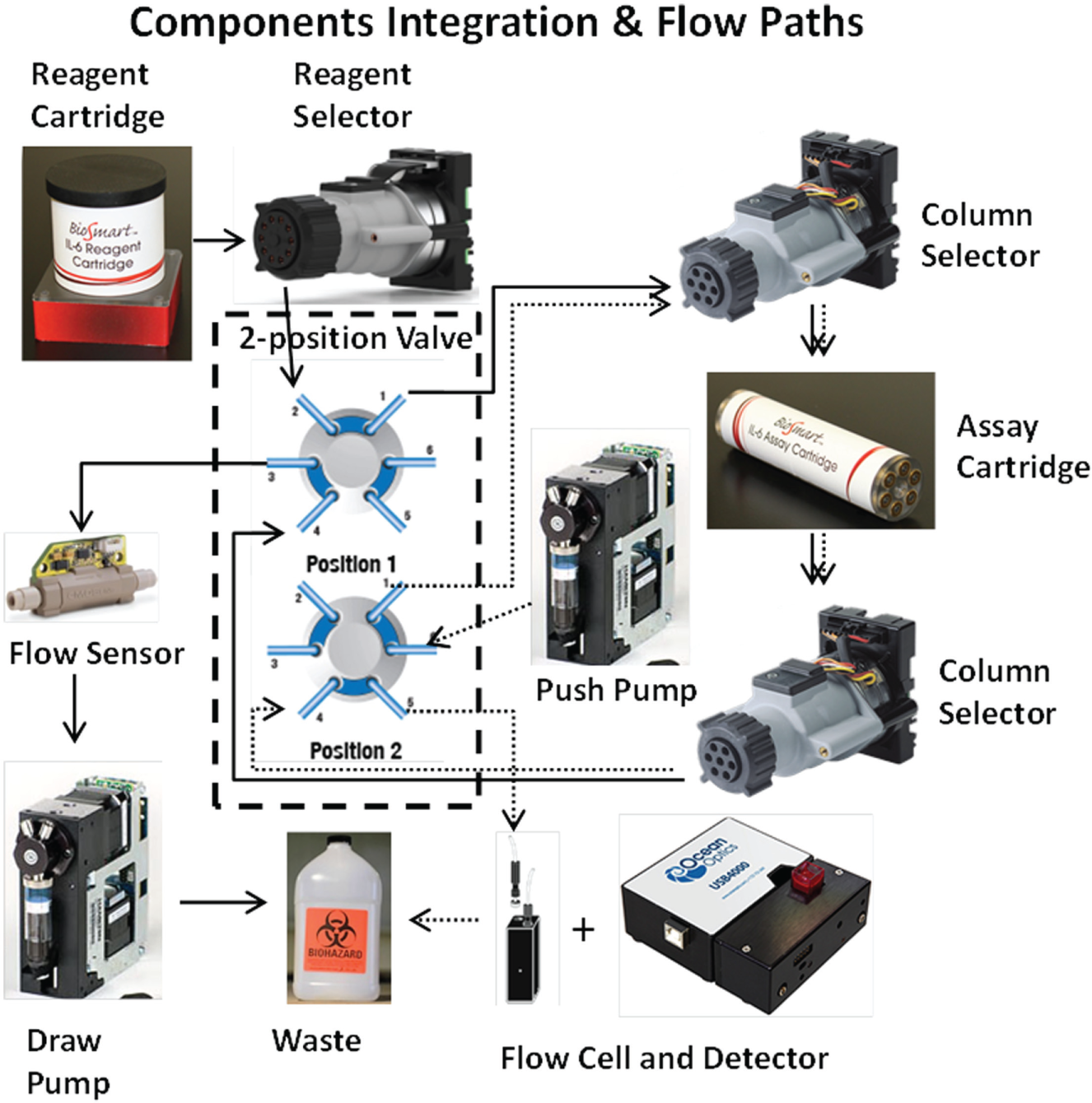
Integration of cartridges, pumps, and valves in the pull–push mode. Two flow paths are used to separate reagent delivery and the detection procedure with a two-position valve. The flow from the reagent cartridge to the waste is shown with solid-line arrows, and the detection flow path is shown with dotted-line arrows. A flow sensor is used to monitor the flow conditions, and the final detection is in a flow cell with a spectrometer.
Assay Test
For evaluation of the integrated immunoassay system, a demonstration rabbit IgG assay was first tested. The assay was initially developed with a 96-well plate platform for antibody screening and initial protocol development. The assay format uses HRP-conjugated secondary antibody, as shown in Figure 2 , left. After that, the protocols for the flow-through system were developed. In detail, acrylic capillary columns were used. Mouse anti-rabbit monoclonal capture antibody (10 µg/mL) was coated on the wall of a long capillary (>5 m) overnight with the chemical binding method. It was later blocked with blocking buffer for at least 2 h before being completely dried. The dried capillary was finally cut into 10 cm long columns and stored in a desiccator in the refrigerator or assembled with the assay cartridge. All other reagents were preloaded to the reagent cartridge, including 1 µg/mL HRP-conjugated donkey anti-rabbit secondary antibody, blocking buffer, washing buffer, substrate, stop solution, and spiked various concentrations of rabbit IgG in fetal bovine serum (FBS). The assay process started with 20 min of antigen incubation, followed by 10 min of secondary antibody incubation and 12 min of substrate development before the detection. The overall time is about 60 min after adding all steps of washing. The optical detection was performed in real time. Different sample loading orders were assessed to check the reliability of the system.
Later, a similar approach was used to demonstrate easy assay adaptation for new analytes to the system with interleukin 6 (IL6) as an example, as well as to explore the feasibility of practical applications with this system. Kits for the IL6 assay were purchased from Mabtech. The commercial IL6 ELISA assay uses a biotin-streptavidin system to enhance the sensitivity, as shown in Figure 2 , right. In detail, 4 µg/mL mouse anti-IL6 monoclonal capture antibody was coated on the wall of a long acrylic capillary (>5 m) overnight. It was later filled with blocking buffer and blocked for at least 2 h before dried. The dried capillary was cut into 10 cm long columns and stored in the desiccator in the refrigerator or assembled with the assay cartridge. All other reagents were preloaded to the reagent cartridge, including 1 µg/mL biotinylated mouse anti-IL6 secondary antibody, 0.5 µg/mL streptavidin-HRP, washing buffer, substrate, stop solution, and spiked IL6 solutions at various concentrations in human serum. The assay procedure started with 20 min of antigen incubation, followed by 15 min of secondary antibody incubation, 10 min of biotin-steptavidin incubation, and 12 min of substrate development before the detection. The overall time is about 75 min after adding all steps of washing. The signal is automatically analyzed to perform baseline correction and peak integration. Measured concentration is reported based on the internal calibrators. A master calibration curve was established for improved performance based on spiked tests.
Results and Discussion
Prototype of Fully Integrated Immunoassay System
The current prototype of the immunoassay system is shown in Figure 5 , which is a fully automated microfluidic-based assay system. It was initially developed for traumatic brain injury biomarker analysis, but could be easily used for other similar bioanalytes. The core components of the system include a universal control board, control software, selected syringe pumps, and rotary valves. All other modules, including reagent cartridge, detector, and assay cartridge, can be replaced, depending on the assay requirements. New modules, such as temperature controller and mixers, can be easily added if necessary. The flexibility of the modular design makes the adaptation of new assays much simpler and more efficient. The overall size of the device is about 17 × 7 × 7 in., which can fit on a small benchtop space and be moved around fairly easily.
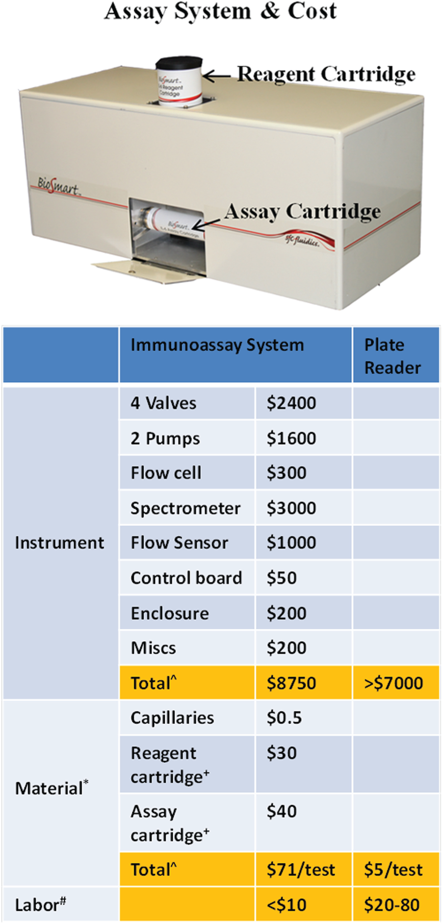
Fully integrated immunoassay system with assembled reagent and assay cartridges. The bottom table shows the estimated cost comparison with a low-end plate reader. ^, total cost with off-the-shelf components and materials. The projected cost for the instrument is less than $5000 and for material less than $2/test with molded pieces. *, estimated with two samples per test and reagent cost excluded, since it varies with different tests. +, cost of machined piece. #, estimated technician hourly rate of $20/h.
The overall setup and running cost for this immunoassay system is lower than that for running a traditional 96-well plate assay. As shown in the table of Figure 5 , the initial instrument cost is not low due to the many off-the-shelf components used. The projected final cost should be well below $5000, which is comparable to that of a low-end plate reader. The initial material costs are also high due to machining, but will come down to less than $2 with molding. The main cost savings are from the labor cost (save up to 80%). The full automation of manual ELISA steps greatly reduces the overall operation cost for day-to-day applications.
The core technique in the immunoassay system is capillary-based immunoassay, which has many advantages: (1) nature for flow-through, and thus easy for solution exchange; (2) easy system automation; (3) larger ratio of surface area to volume to save time; (4) extreme low cost, disposable; (5) relatively high quality of capillary tubing; and (6) multiple material options for different analytes (plastic and fused silica, etc.).
The overall target of this system is to achieve fast and accurate assay measurements with flexibility to change assays. The key concept implemented to improve the assay performance with automation herein is the combination of microfluidics with assays. Microfeatures enable a large ratio of surface area to volume, so that for diffusion-limited assays (including most ELISAs since the kinetics of the antibody/antigen reaction is much faster than diffusion), the theoretical required assay time and assay volume are greatly reduced (the actual number varies based on different designs). The automation feature is achieved from the inherent fluidic mode with the interface to precise fluidic control. A magnetic connector–enabled disposable cartridge design simplifies the loading and unloading process and reduces user errors.
The operation of the system is very straightforward. An example of the complete assay procedure is described below:
Start the system and the control software.
Load the reagent cartridge and insert the assay cartridge.
Load the sample to the defined sample reservoir.
Select the proper assay protocol from the GUI.
Start the program, and the result is reported in 60–75 min.
The user-involved time could be less than 5 min, which is a great improvement over that of traditional 96-well plate platforms.
Flow Profile
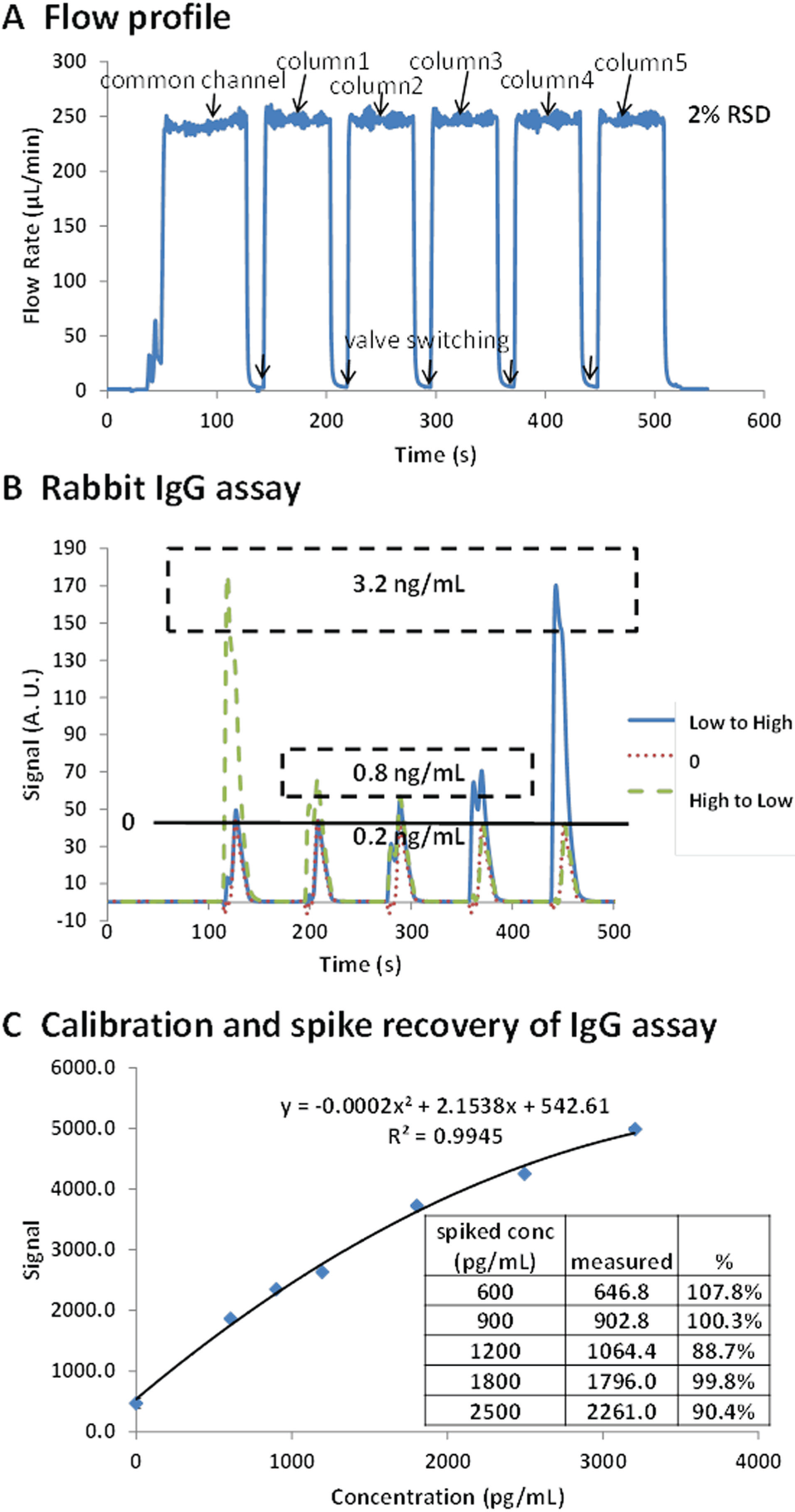
One common concern of pumping in drawing mode is the variation of the flow. The vacuum created to drive the solution is greatly affected by the pumping conditions, including, but not limited to, pump properties, valves, connectors, and solution properties. Bubbles tend to form under vacuum conditions and can introduce variations to both the assay reaction and detection process. For the best result, all the solutions have to be degassed under a vacuum degasser before use. The system also needs to be primed well at the beginning of each test to avoid any bubble trapping, which is built into the protocol for complete automation. Tiny bubbles occasionally form in the system during the test, but would not affect the average flow and solution delivery because of their size and numbers. A typical section of flow profile of switching between five capillary columns is shown in Figure 6A . To deliver a reagent through the capillaries, it is first primed through a common channel to minimize the possible initial variations. Later, it is pulled/pushed through each capillary column in a preprogrammed order. The flow variation is about 2% with a Hamilton syringe pump in drawing mode, improved from the APT peristaltic pump (~5%). The general flow rate used in all tests was set at ~250 µL/min. Faster flow introduces more shear force to the surface and consumes excessive reagents for each solution exchange step, while slower flow takes longer to deliver each reagent. Thus, a 250 µL/min flow rate was selected based on the balance between assay time and performance.
(
Rabbit IgG Immunoassay Test
The first fully automated immunoassay was demonstrated with rabbit IgG assay, and one set of test results is shown in Figure 6B . Samples are prepared in FBS to simulate real situations. Three test results are plotted together for comparison. One cartridge for concentration 0 (FBS only) acts as the negative control and establishes the baseline for concentration 0. The column-to-column variations are rather small, with all five peak signals between 40 and 43. The assay results with IgG concentrations from low to high (order of 0, 0, 0.2, 0.8, and 3.2 ng/mL) and from high to low (order of 3.2, 0.8, 0.2, 0 and 0 ng/mL) are overlaid together. Not only are the signal levels similar for the same concentrations at two different tests, but also all the peak positions are matched well. The detection limit is about 0.2 ng/mL. This is comparable to commercial assay kits (e.g., Abcam AB187400). This comparison study also confirms minimal cross-contamination in the fluidic system, even when several flow paths are shared between reagents. Good cleaning steps and a well-defined program are the keys to minimizing cross-contamination.
Later, a series of rabbit IgG assays were performed to check the accuracy of the system with spiked samples. In these tests, a duplicate of sample concentration was measured against internal standard concentrations of 0, 0.3, and 3.2 ng/mL. The assumption of a linear relationship of the signal and concentration is actually not perfect, as shown in Figure 6C . The signals tend to increase less at higher sample concentrations. Thus, a simple 3-point linear calibration might not be enough to get great accuracy, especially when sample concentrations are high or low. An effective approach to solve this issue is to develop a master calibration curve, as the polynomial fit curve shown in Figure 6C . In each test, test standards and sample results would be matched to the master curve first for an offset adjustment, followed by the calculation of samples with adjusted calibrator signals. This would improve the accuracy while still keeping the advantages of simple real-time calibration. The recovery table inserted in Figure 6C shows that the inaccuracy of different concentration measurements is less than 13%, which is on a par with many manual 96-well plate ELISAs.
This system is designed for small-scale routine ELISA analysis due to the limitation of sample numbers. However, it is possible to scale up with more capillary columns for more sample analyses with other multiway pumps (e.g., up to seven sample tests with 10-way selection valves). Considering its simplicity with automation and much shorter assay time (~60 min), this immunoassay system could be a great fit for lab automation.
Adaptation of New Assays
Another advantage for this immunoassay system is its flexibility to adapt new assays. Because the capillary-based system is essentially very similar in reagents and assay procedures compared with 96-well plate-based assays, the conversion is straightforward. In theory, any assays that can be captured on site for quantifications could be a good fit for the new immunoassay system. For a new targeted assay, a 96-well plate will be first used to verify if the performance meets the requirement. Later, try to find the best material and antibody coating methods for capillary columns and check the assay performance with the immunoassay system. Tweak the assay conditions to optimize the performance until meeting the requirements. After internal verification, it would be ready for routine applications. It is worth noting that it is possible to premix several reagents together in one step to reduce the assay time.
Many assays targeting potential traumatic brain injury biomarkers have been successfully transferred to the immunoassay system, including IL6, s100β, glial fibrillary acidic protein (GFAP), UCH-L1, and brain-derived neurotrophic factor (BDNF). The overall transition time could be as few as 2 weeks. As an example, the transfer of the IL6 assay began with commercially available antibody kits (Mabtech). After verification with a 96-well plate, an initial protocol was developed based on the plate results to test the immunoassay system. It has additional steps compared with the rabbit IgG assay, and the overall assay time is 75 min. The current antibody coating method on acrylic capillaries with covalent bonding worked very well, which significantly reduced the transition difficulties. The assay conditions were further tweaked (antibody concentrations, time of each step, washing steps, etc.) after initial demonstration of the concept. A similar set of master calibration curves could be obtained from multiple spiking tests to improve the performance ( Fig. 7 ). The spike recovery is generally within 20%, and the detection limit is 50 pg/mL in human serum samples. After further verification by comparing with 96-well plate assays, it is ready for daily use. The whole process was less than 2 weeks.
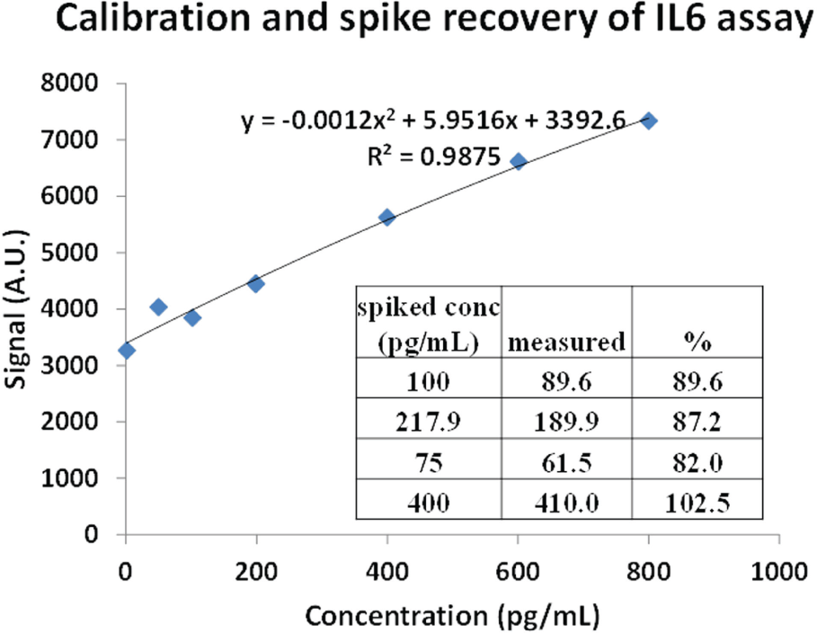
Spike recovery test of IL6 in spiked human serum with an immunoassay system and the generated master calibration curve. The recovery rate is shown in the inserted table.
Since different applications have different assay requirements, the feasibility of using the immunoassay system should be determined after careful initial assessment. Common issues that might occur during assay transfer include, but are not limited to, high background signal due to nonspecific adsorption, low signal level due to poor antibody coating, and poor signal gradient due to slow reaction kinetics. Thus, adaptation of these assays might require a skilled lab technician with knowledge of assay optimization.
Conclusion
A fully automated low-cost immunoassay instrument has been developed to simplify routine ELISA tests in research applications. It features easy adaptation of new assays because of its similarity to the traditional 96-well plate platform. The key component of the system is a capillary-based reaction cartridge, which has various options to adapt different assays. The assay time is greatly reduced to 60–75 min, with the only manual operations being loading disposables and samples, and starting the program. The loading and unloading of disposables is user-friendly with enabled magnetic-assisted connections. The system performance is on a par with that of many traditional ELISA platforms. The running cost is extremely low compared with other LOC technologies and traditional plate reader–based systems. It is a great tool for small-scale routine ELISA operations to relieve labor-intensive manual operations and minimize human errors. It fits the research environment very well and could work for broader testing applications (food, environmental, and clinical) with further optimization.
Footnotes
Declaration of Conflicting Interests
The authors declared no potential conflicts of interest with respect to the research, authorship, and/or publication of this article.
Funding
The authors disclosed receipt of the following financial support for the research, authorship, and/or publication of this article: Work described in this paper is financially supported by CDMRP grant W81XWH-09-0523. The content is solely the responsibility of the authors and does not necessarily represent the official views of CDMRP.
References
Supplementary Material
Please find the following supplemental material available below.
For Open Access articles published under a Creative Commons License, all supplemental material carries the same license as the article it is associated with.
For non-Open Access articles published, all supplemental material carries a non-exclusive license, and permission requests for re-use of supplemental material or any part of supplemental material shall be sent directly to the copyright owner as specified in the copyright notice associated with the article.