
Abstract
Conventional approaches to bacterial identification and drug susceptibility testing typically rely on culture-based approaches that take 2 to 7 days to return results. The long turnaround times contribute to the spread of infectious disease, negative patient outcomes, and the misuse of antibiotics that can contribute to antibiotic resistance. To provide new solutions enabling faster bacterial analysis, a variety of approaches are under development that leverage single-cell analysis, microfluidic concentration and detection strategies, and ultrasensitive readout mechanisms. This review discusses recent advances in this area and the potential of new technologies to enable more effective management of infectious disease.
Introduction
The accurate identification of pathogens underlying infectious disease is an essential capability for clinical medicine. Moreover, turnaround time is critical as treatments of blood, urine, and flesh infections all benefit from rapid diagnosis.1–5 The diagnosis of bacterial blood infections that can lead to sepsis is particularly time-sensitive. For this indication, every hour that an infection remains untreated with effective antibiotics decreases the survival rate of the patient by 7.6%. 6 Infectious disease diagnostic tests must be able to pinpoint causative agents so that antibiotics can be selected effectively, but it is also critical that drug-resistance phenotypes are detected to ensure that effective antimicrobial therapy is administered.7–9
Most infectious disease testing is performed using conventional microbiological techniques that rely on the culture of clinical samples and subsequent antimicrobial susceptibility testing.10,11 Bacteria must be cultured to increase their numbers to levels detectable by optical density–based methods, recultured as pure isolates, and then analyzed for antibiotic resistance. Broth dilution or antimicrobial gradient methods allow the calculation of minimum inhibitory concentrations of therapeutics and the identification of resistant phenotypes. While these are manual methods, automated testing systems are also employed in clinical testing labs that allow highly standardized testing. Nonetheless, the overall turnaround times of these tests are long, often 72 h or longer, which necessitates the treatment of serious infections without test results or the delay of treatment for less serious infections, leading to infectious disease spread or the development of complications. 12
One solution that has been pursued to shorten the delays caused by culture-based infectious disease testing is the development of molecular-level assays that employ enzymatic amplification of pathogen nucleic acids. Rather than waiting for bacteria to multiply to sufficient levels for analysis, genomic DNA is amplified using techniques like polymerase chain reaction (PCR) and detected using fluorescence readout. 13 This approach can reduce the time required for analysis from days to hours. A variety of commercially available molecular methods are now used clinically for infectious disease diagnosis and are making an impact on the effectiveness of infectious disease management.14–19 While this approach presents a means to speed infectious disease diagnosis, molecular analysis faces limitations when broad resistance profiles must be surveyed. For example, broad-spectrum β-lactamase activity can be observed in bacterial strains carrying a variety of resistance genes. 20 PCR-based methods have limited multiplexing and therefore are difficult to apply to large families of sequences. Moreover, previously uncharacterized resistance factors are not amenable to molecular-level testing. Therefore, it is of interest to develop next-generation technologies that speed the direct phenotypic testing of bacteria and monitor the effects of antimicrobial therapies.
This review summarizes recent progress toward the development of new technologies for infectious disease testing and antibiotic-resistance profiling. Phenotypic tests that can more rapidly report on drug susceptibility are becoming more sophisticated, especially when coupled with microfluidic approaches for concentrating and analyzing bacteria. New analytical techniques are also catalyzing progress in this area, allowing highly sensitive measurements that return rapid results.
Single-Cell Methods for Bacterial Analysis
Existing microbiology-based methods for bacterial identification and antibiotic resistance testing rely on monitoring cultures containing high numbers of bacterial cells and require the growth of the culture to progress until the numbers of cells can be read out spectroscopically. Recent advances in single-cell monitoring21–38 may provide new solutions that can speed bacterial analysis by monitoring changes in growth on a cell-by-cell level. Single-cell microbioreactors 30 and microscale incubators 27 provide a means to culture bacteria in very small volumes, and new types of single-cell analysis methods24,25,34 allow rapid readout of bacterial viability. This type of direct analysis eliminates the need for extensive culture as very small numbers of bacteria can be analyzed.
A variety of methods have been tested to enable the readout of bacterial viability at the single-cell level. Monitoring surface-bound magnetic beads,39,40 the direct magnetic properties of cells, 25 bacteria confined to fluidic channels, 34 and morphological changes that occur upon antibiotic exposure23,24,39,40 all represent promising methods for bacterial profiling. For all of these approaches, algorithm development is critical given the heterogeneity of bacterial cells in clinical specimens.
An early advance in the development of bacterial analysis devices with single-cell resolution relied on the use of dielectrophoretic focusing of bacteria within an array of interdigitated electrodes. 34 Optical tracking of the captured cells then allowed their division to be monitored. Proof-of-concept experiments indicated that this method could successfully test Escherichia coli for polymyxin susceptibility. Multiplexing and parallel testing of multiple drugs could be achieved using this method if appropriate automated instrumentation was developed.
One of the most advanced single-cell systems is a high-throughput approach that tracks the morphology of bacterial cells as they are exposed to different antibacterial drugs ( Fig. 1 ). 24 The cells are encapsulated within a microscale slab made from agarose, and nutrients and antimicrobials are supplied to the trapped cells. Over a 3-h time window, cell division is monitored to determine the effects of the antibiotics. A variety of behaviors can be observed with this device in addition to cell division, including swelling and filamentary formation, and these phenotypes can be included in the determination of whether bacteria are resistant or susceptible to a given drug.
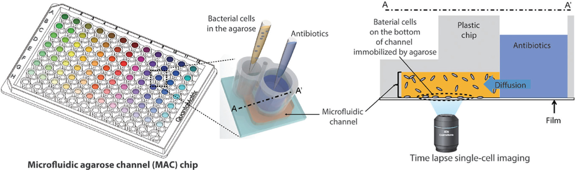
A high-throughput agarose channel chip for analysis of antibiotic susceptibility. Bacterial cells embedded in agarose are deposited within the chip, and an adjacent well contains an antibiotic to be tested. Bacterial morphology is monitored using time-lapse bright-field microscopy to gauge the response of the cells to different antimicrobials. Reprinted from Choi et al. 24 with permission.
Using this technique, 189 clinical isolates were analyzed, including cultures of E. coli, Pseudomonas aeruginosa, Klebsiella pneumoniae, Staphylococcus aureus, and Enterococcus spp. The set of bacteria tested included specimens exhibiting extended spectrum β-lactamase activity, imipenem resistance, methicillin resistance, and vancomycin resistance, and the isolates were exposed to a variety of antimicrobial agents. The morphological analysis was conducted in parallel with the gold-standard broth microdilution assay (BMD), and discrepancy rates less than 10% were observed. The high levels of performance observed in this study indicate that this approach can compete with existing methods, especially given that it delivers results in under 3 h as opposed to the 66 h required using conventional methods.
Microfluidic Approaches for Rapid Bacterial Identification
A major application area for microfluidic technology development is the miniaturization of biological assays. The ability to process and analyze biological and clinical samples is central to the automation of bacterial identification and antibiotic resistance profiling, and a variety of advances have been made using microfluidic technologies.41–50 Several droplet-based systems have been reported that allow small numbers of bacteria to be analyzed for antimicrobial resistance, and inertial and gradient microfluidic systems have achieved turnaround times of just a few hours and the ability to process whole-blood samples. Adaptations of microfluidic systems incorporating paper-based devices 51 and smartphone-mediated analysis 52 may also facilitate bacterial profiling in limited resource settings.
Studies of simple microfluidic devices where gradients of antimicrobial drugs are generated showed that comparable results can be achieved with miniaturized approaches relative to bulk culture analyses. 47 Unlabeled bacteria can be visualized and their growth kinetics measured in situ, but little improvement in turnaround time was achieved with early systems. However, a more complex linear gradient device allowed culture in 3D and the generation of high-resolution growth curves in 2.5 to 4 h. 44 This device was tested with five antibiotics against three different strains of gram-positive and gram-negative bacteria, and good agreement was observed using this device compared with gold-standard methods.
Inertial microfluidics, an approach that relies on focusing cells using differential lateral migration of cells across streamlines, has also been applied to profiling bacteria and is particularly effective when whole blood is the target sample to be analyzed ( Fig. 2 ). 43 Bacteria are separated from blood cells with reasonable efficiencies (>65%), even when present at low abundance. To speed analysis, this approach was coupled with quantitative RNA detection using the commercially available Nanostring (Nanostring, Seattle, WA, USA) technology. The sample processing and bacterial analysis workflow could be completed in 8 h. While this turnaround time is longer than for some of the other systems described above, it represents a complete sample-to-answer protocol that enables the analysis of whole blood.
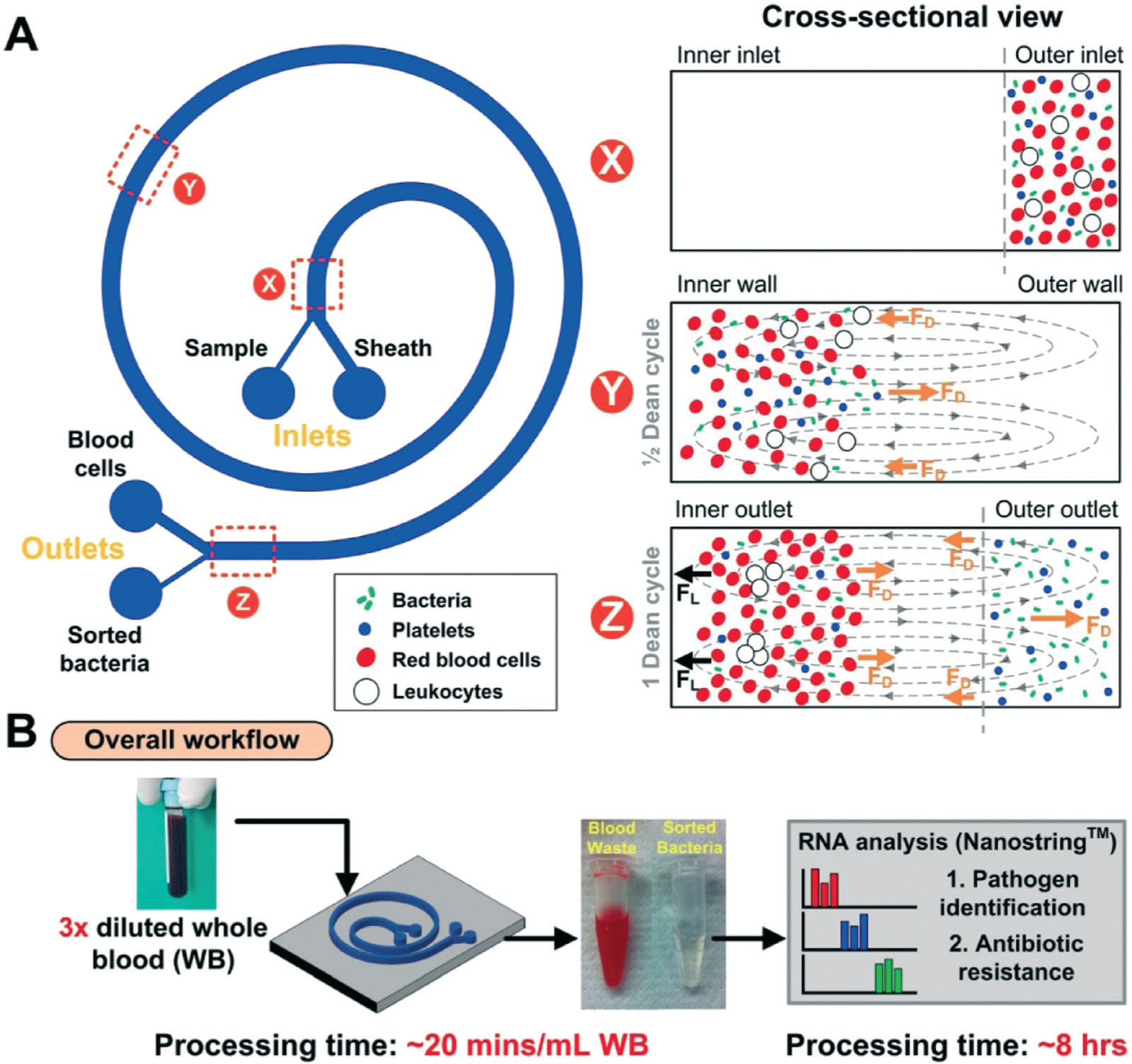
Inertial microfluidics for bacterial isolation and analysis. (
The encapsulation of bacterial cells in micro- and nanodroplets coupled with microfluidic manipulation presents another means to track the phenotypic properties of infectious pathogens. One of the first systems to use this type of approach confined bacterial cells in nanoliter plugs, and using an approach termed stochastic confinement, the detection and profiling of specific bacteria could be accelerated depending on the volume of the plug used ( Fig. 3 ). 53 A more automated droplet-based system allowed high-throughput analysis of multiple bacterial types and antibiotics with a turnaround time of several hours. 54 A recent breakthrough in the use of droplet microfluidics used a DNAzyme as a sensor to identify droplets containing bacterial cells, even in the presence of a high level of whole blood ( Fig. 4 ). 46 This system has not yet been adapted for antibiotic resistance profiling, but it should be amenable to this type of analysis.
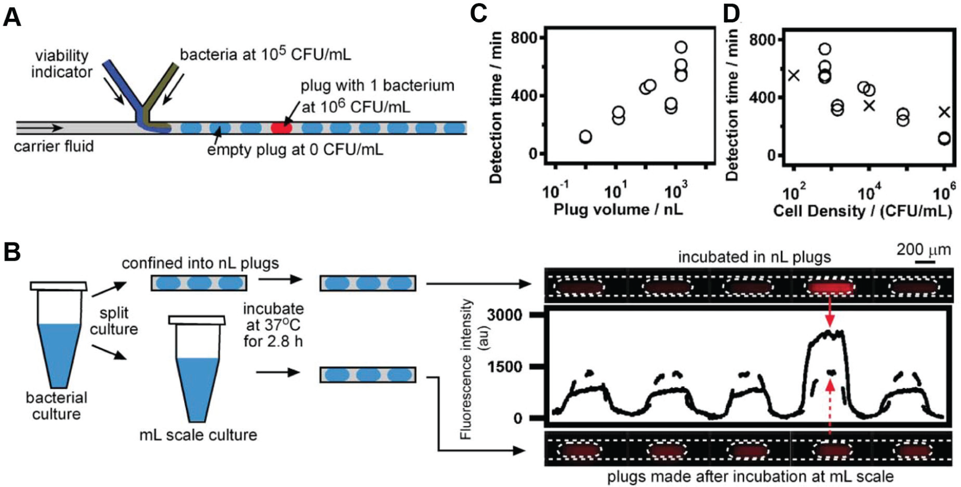
Stochastic confinement of bacteria in nanoliter droplets. (
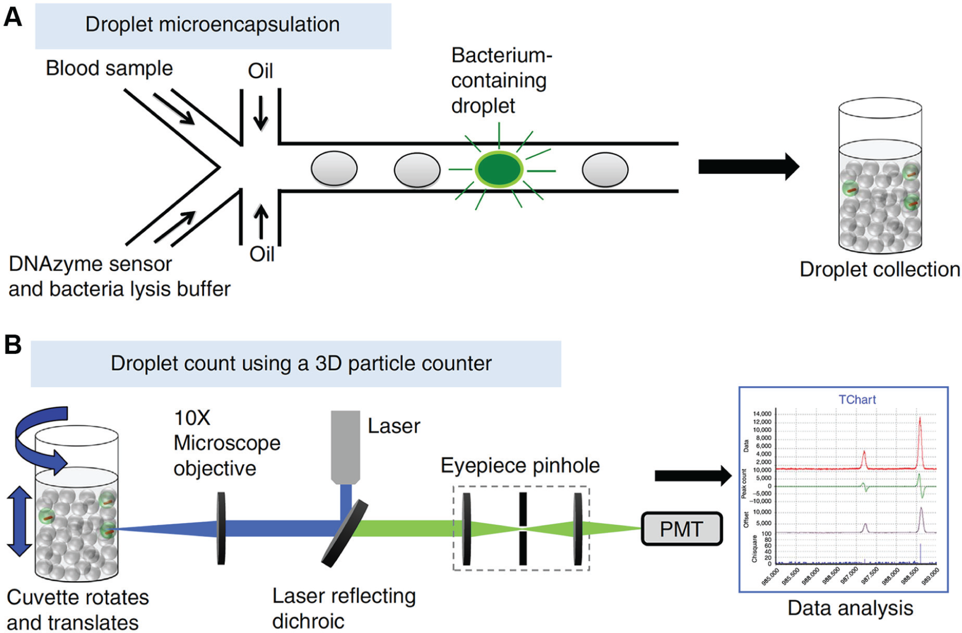
A digital droplet system for detection of bacteria in blood. (
Ultrasensitive Detection Approaches
Another area that is central to the generation of advanced bacterial profiling systems is the development of highly sensitive bacterial detection methods. Conventional detection systems rely on optical density measurements, which are not very sensitive. By replacing this detection modality with other analysis methods, faster detection could be achieved by leveraging enhanced sensitivity to read out phenotypic information more rapidly. A variety of electrophoretic, 55 electrochemical,56–76 mechanical,77,78 and mass spectrometry–based79,80 systems have been developed for this application that may speed the identification of antibiotic-resistant bacteria.
Mass spectrometry, and specifically matrix-assisted laser desorption time-of-flight mass spectrometry (MALDI-TOF MS), is a powerful method for bacterial identification ( Fig. 5 ). It can be used to identify specific bacteria types and resistance enzymes and has successfully been used to analyze a variety of bacterial strains.81–83 MALDI-TOF can also be used to analyze bacterial isolates for the ability to hydrolyze antibiotics to detect antibiotic resistance.84,85 This allows phenotypic profiling of bacteria, but the complex instrumentation and time requirements may not offer a significant advantage relative to culture-based approaches.
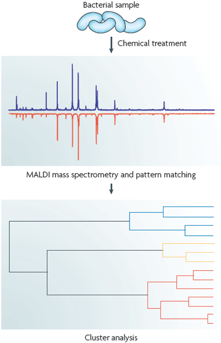
Matrix-assisted laser desorption (MALDI) mass spectrometry for bacterial identification. Mass spectrometry can be used to identify bacteria and their resistance patterns via the profiling of proteins or nucleic acids. Spectral fingerprinting combined with cluster analysis can enable highly specific bacterial characterization. Reprinted with permission from Sauer and Kliem. 81
Mechanical transducers present another promising means of reading out the responses of single bacterial cells.77,78 An atomic force microscopy-based approach demonstrated that a cantilever could be used to detect bacteria at low abundance ( Fig. 6 ), 77 characterize metabolic activity, and read out the response to antibiotics on the order of a few minutes. Mechanical readout of changes to the bacterial membrane appeared to report on the presence and response of bacteria to antimicrobials. The potential speed of this readout approach is very impressive, but the need for vibrational isolation may make this readout mechanism difficult to incorporate in clinical instrumentation.
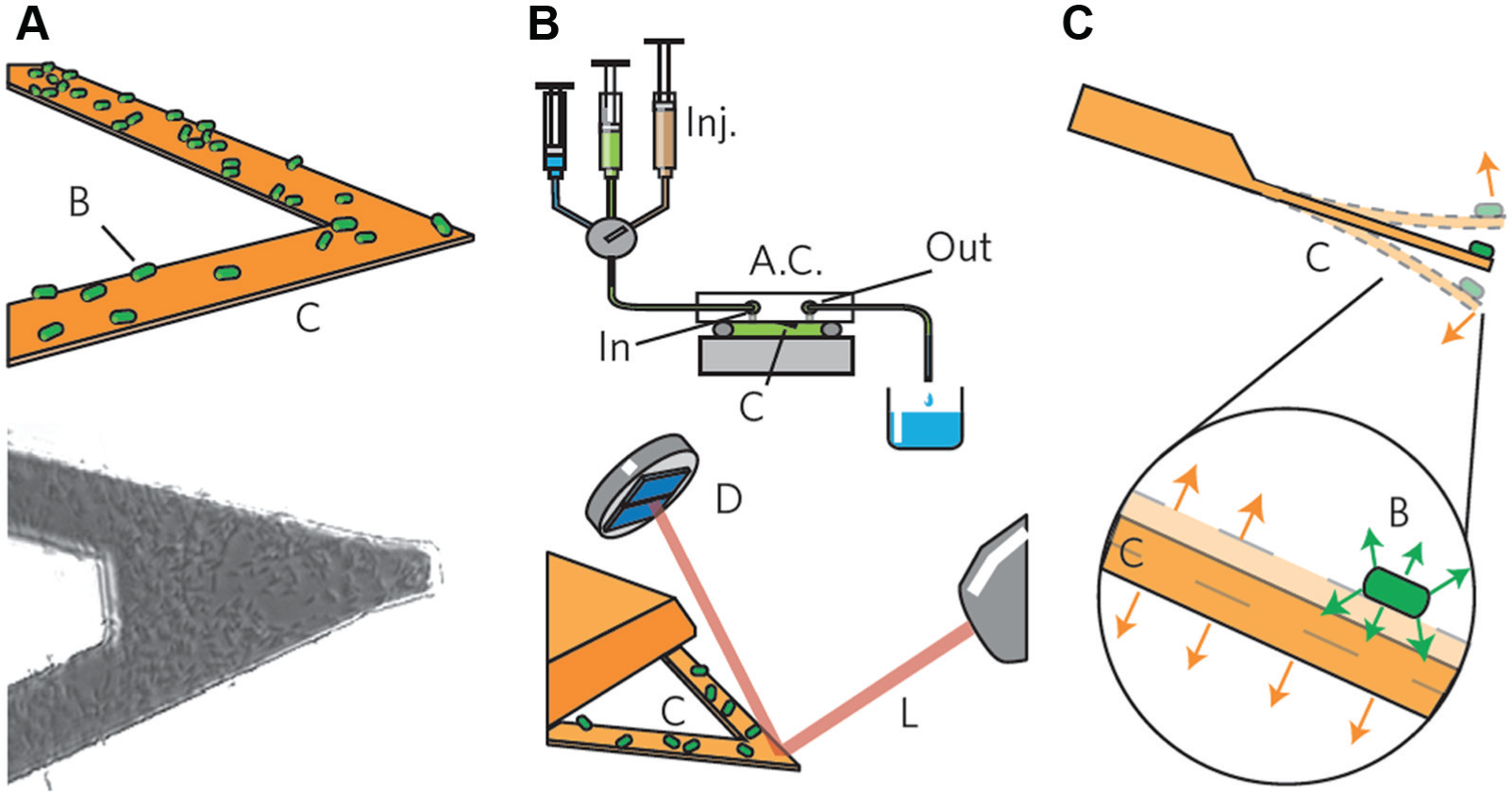
A nanomechanical sensor for bacteria. (
Electrochemical sensors have been used in a variety of systems for the identification of bacteria and identification of resistance phenotypes. An electrochemical system focused on the analysis of ribosomal RNAs has been shown to be effective for the analysis of urinary pathogens,36,41,52,67,68,72,75,76,86 and the amenability of electrochemical sensors to incorporation in highly multiplexed arrays makes this approach a good fit with the need for looking a large panels of markers to survey resistance genes comprehensively. Another electrochemical system for bacterial analysis relying on large surface area nanostructured microelectrodes58,64,71 has been deployed to detect very low levels of bacteria by targeting messenger RNA (mRNA) sequences and other biomarkers56,60,61,65,66,73,74 and can also be multiplexed to analyze large analyte panels. 65
Electrochemical readout can also be used to speed phenotypic testing of bacteria within miniature testing cells. A variety of electrochemical reporters for cell viability are known,59,69,70 presenting an alternative to optical reporters that may present advantages for conducting high-sensitivity viability analysis and drug susceptibility measurements. One device reported recently used the dye resazurin to monitor cells trapped in nanoliter wells within a microfabricated device ( Fig. 7 ). 57 At concentrations as low as 1 cfu/µL, the response of bacteria to a given antibiotic could be read out within 1 h. Proof-of-concept for this approach was shown using E. coli and K. pneumoniae exposed to ampicillin and ciprofloxacin, and similar approximate minimum inhibitory concentration values were obtained when the electrochemical data were compared with that collected using conventional culture-based testing.
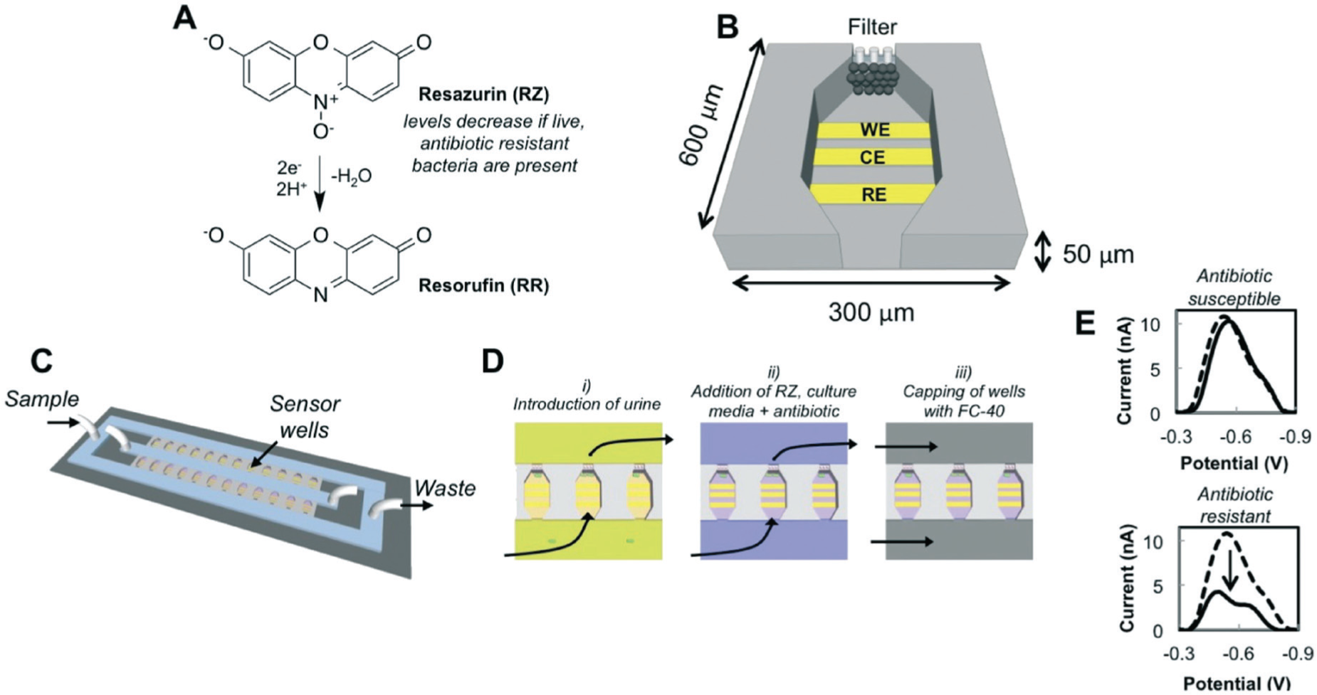
A device for electrochemical bacterial phenotyping. (
In sum, a variety of new approaches are being deployed to combat antibiotic resistance by providing faster methods for bacterial identification and profiling. Single-cell methods have been developed that compare favorably to gold-standard microbiological tests, and new microfluidic approaches are also producing promising advances that will speed test turnaround times. The development of new analytical methods that can detect bacteria faster using mechanical and electrochemical transducers will bring enhanced sensitivity to this problem. While all of these techniques face challenges related to streamlining sample preparation and system integration, it is anticipated that these translational barriers will be addressed as technologies mature. With many new technologies advancing toward the clinic, the slow turnaround of conventional bacterial testing approaches should become a thing of the past.
Footnotes
Declaration of Conflicting Interests
The author declared no potential conflicts of interest with respect to the research, authorship, and/or publication of this article.
Funding
The author wishes to acknowledge the support provided by the Natural Sciences and Engineering Research Council of Canada, and the Canadian Institutes of Health Research for the studies described in this article that were conducted in her laboratories.