
Abstract
Aim:
This study aims to assess the influence of resin removal treatment regimes on the surface topography and compressive strength of de-bonded ceramic surfaces.
Material and methods:
Sixty-five lithium disilicate ceramic (LDC) discs were prepared, cleaned, and polished with carbide paper. All samples were etched using 9.6% hydrofluoric acid (HFA). Fifteen samples were taken as positive controls; the remaining 50 samples were subjected to the process of silanization. Resin build-up using dual-cure cement was performed incrementally and light cured. Based on different methods of resin cleaning from de-bonded LDC, the samples were divided into five groups, n=10 each: group 1 (no treatment), group 2 (slow-speed diamond bur), group 3 (1 min heat treatment), Group 4 (6 min heat treatment), and group 5 (sandblasting with Al2O3). Following resin removal, LDC samples were tested under compressive failure load in a universal testing machine. Five disc specimens from each group were sputter coated with gold for scanning electron microscopy (SEM). Analysis of variance (ANOVA) and Tukey’s post hoc test was used for descriptive statistics. Level of significance was established at p<0.05.
Results:
The highest compressive strength with significant difference among all experimental groups was found in group 5 (321.54 ± 13.25 MPa) (p<0.05). The lowest compressive strength values, presenting significant difference compared with all other groups, were displayed in group 1 (158.57 ± 5.22 MPa) (p<0.05). Compressive strength among group 2 (231.54 ± 15.55 MPa), group 3 (237.81 ± 10.81 MPa), and group 4 (255.53 ± 8.95 MPa) specimens was statistically comparable (p>0.05). On SEM, heat-treated specimens confirmed coarser granules, with mild porosities and roughening, whereas sandblasted specimens exhibited consistent evenness with moderate porosity and loss of glazed surface.
Conclusion:
De-bonded LDC surface, treated with heat treatment and sandblasting procedures, exhibited removal of residual resin and significantly high compressive strength compared with non-cleansed ceramic surface.
Keywords
Introduction
To meet the aesthetic requirement of dental restoration, lithium disilicate ceramic (LDC) has gained popularity compared with traditional metal porcelain.1,2 High acceptance of all ceramic crowns and partial coverage restorations over leucite-reinforced glass ceramics and metal porcelain are due to superior biocompatibility and translucency, and better thermal properties and enhanced flexural strength.3,4 Apart from the unparalleled physical properties of LDC, for predictable treatment outcome the bonding of all ceramic materials to dentin remains the mainstay.5,6
To improve bonding of LDC to dentin, pre-treatment using hydrofluoric acid (HFA), silane (Si), and sandblasting with Al2O3 is performed. 7 The process improves the surface energy of LDC, and increases surface area and surface roughness. 8 Another method to improve retention of all ceramic restoration is preparation of the tooth structure. However, the process is still under debate as it results in irreversible damage to teeth and dentin exposure with low bond integrity.9,10 Despite all care and effort, the de-bond ratio for all ceramic restorations remains as high as 11%.11,12
In 90% of de-bonded cases cement is adhered to the ceramic. This adhered cement contaminates the ceramic surface, interferes with bonding, prevents infiltration of adhesive, and compromises etching quality.1,11 Therefore, to recreate the desired surface topography of ceramic it is of utmost importance to remove the cement before the re-bonding procedure.13,14 The conventional method to remove resin before re-cementing is treatment with HFA.15,16 Other methods include removal of resin with burs and heat treatment of de-bonded ceramic before re-cementing, creating a clean, porous, and retentive surface.15,17
Work by Santos et al., 18 Koodaryan et al., 1 and Román-Rodríguez et al. 15 has already assessed the effect of different resin-cleaning procedures from ceramic surface before re-cementing with regard to bond integrity and surface roughness. However, no studies have been done to assess the compressive strength of LDC cleaning with different protocols before re-bonding. Moreover, limited studies are available assessing surface topography of LDC after retreatment of de-bonded restorations.19,15 It is hypothesized that de-bonded LDC treated with different regimes (heat treatment, air abrasion, and diamond bur) will show significant difference in the surface topography and compressive strength of the material. Therefore, the aim of this study was to assess the influence of resin removal treatment regimes on the surface topography and compressive strength of de-bonded ceramic surfaces.
Material and methods
Sixty-five LDC (IPS e max Ivoclar, Liechtenstein) discs having diameter of 8 mm and thickness of 3 mm were prepared using the lost wax technique and hot pressing. The surface of the specimen was cleaned with distilled water; this was followed by 120 s bath in 99.9% ethyl alcohol solution (B-Grade Anhydrous Ethyl Alcohol) and air drying for 60 s. The bonding surfaces of specimens were glazed according to manufacturer recommendation and polished with a semi-automatic polishing machine (MetaServ 250, Buehler, Lake Bluff, IL) with silicon carbide paper (300 grit and 600 grit). Following polishing, the samples were cleaned ultrasonically in distilled water (Nestle Pure, Germany) for 300 s.
Bonding surfaces were etched using 9.6% HFA (IPS ceramic etching gel Ivoclar vivadent) 60 s, washed (30 s), and air-dried. Fifteen samples were taken as positive control and no further treatment was done. The remaining 50 samples were subjected to the process of silanization (Monobond Plus ceramic primer Ivoclar, vivadent) for 60 s and air-dried. Resin build-up using dual cure cement (Calibra, Dentsply, Caulk) was performed in increments on each ceramic disk already placed in rubber mould (3 × 3 mm) and light cured (Bluephase G2, Ivoclar, Vivadent) for 45 s using light at intensity of 450 mW/cm2. After removal of the rubber mould the samples were again light cured for 10 s.
Specimens were placed in thermocycler (Nova Inc., Konya, Turkey) with a dwell time of 45 s at 5–55°C for 10,000 cycles to simulate oral conditions. All samples were placed in a universal testing machine (Elista TSTM 02500, Elista Corp). Downward force parallel to the ceramic resin interface at a transversal velocity of 1 mm/min was applied until fracture. Based on different methods of resin removal from LDC the samples were divided into five groups with 10 samples each.
Compressive strength
Following resin removal by different protocols, LDC samples were tested under compressive failure load (ASTM C39/C39M–20) in a universal testing machine (Elista TSTM 02500, Elista Corp) with a cross-head speed of 1.0 mm/min. Using a round-ended metal probe, force was applied at the center of the samples for equal distribution. Compressive strength of the specimens was calculated as

Where, f: is force in newton (megapascal MPa); d: diameter of sample in millimeter (mm)
Surface topography
For scanning electron microscopy (SEM) (SEM, JSM-6335F; Jeol, Tokyo, Japan) five ceramic specimens from each group were sputter coated with gold (6 nm thickness) at 40 mA for 250 s. Five samples from the positive control group were assessed for surface topography after HFA treatment without resin binding. SEM images were captured at 10 kV at ×1000 magnification under wide field mode for visual reading
Statistical analysis
Data for compressive strength testing were tabulated using statistical program for social science (SPSS version 21, Inc., Chicago, US). Analysis of variance (ANOVA) and Tukey’s post hoc test at a significance level of (p<0.05) was used for descriptive statistics.
Results
The data of compressive strength exhibited normal distribution according to Kolmogorov–Smirnov test. For compressive strength values, ANOVA showed significant difference among all investigated groups (p<0.05). The highest compressive strength values, displaying significant difference with all experimental groups, were found in group 5 (sandblasted for 60 s with Al2O3) (321.54 ± 13.25 MPa) (p<0.05). The lowest compressive strength values, presenting significant difference compared with all other groups, were found in group 1 (negative control, with no treatment) (158.57 ± 5.22 MPa) (p<0.05) (Table 1).
Compressive strength values Means and SD for experimental groups using ANOVA and Tukey multiple comparisons test.
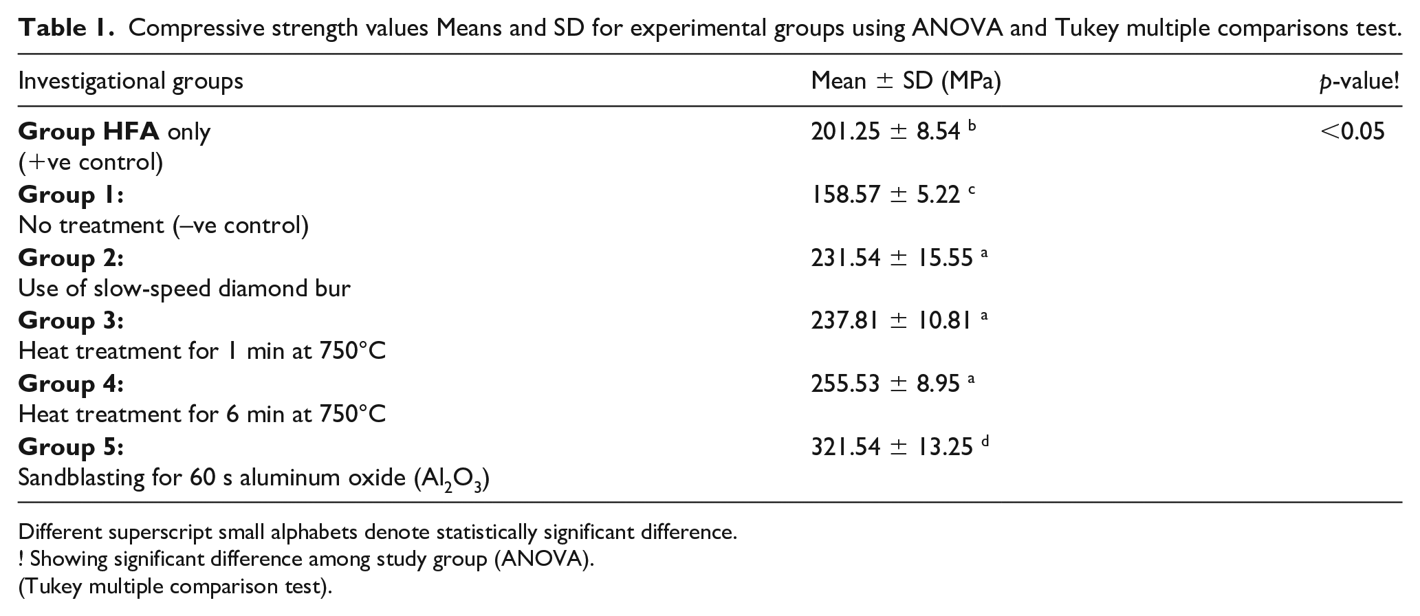
Different superscript small alphabets denote statistically significant difference.
! Showing significant difference among study group (ANOVA).
(Tukey multiple comparison test).
Among the experimental groups, group 2 (diamond bur) (231.54 ± 15.55 MPa), group 3 (heat treatment for 1 min) (237.81 ± 10.81 MPa), and group 4 (heat treatment for 6 min) (255.53 ± 8.95 MPa) were found to be statistically comparable (p>0.05). Group treated with HFA only (positive control) (201.25 ± 8.54 MPa) exhibited significant difference compared with all experimental groups (p<0.05) (Figure 1)

Compressive strength values Means and SD among different groups.
Surface topography
Examination by SEM revealed that positive control specimens showed a distinct pattern of LDC etched surface (Figure 2(a)). Furthermore, SEM image with no treatment (group 1) presented a resin-covered ceramic surface (Figure 2(b)). Moreover, photomicrograph images of LDC treated with high temperature to remove resin in group 3 and group 4 (Figure 2C and 2D) displayed coarser granules, with mild porosities, and uneven/rough surface with microcracks. In addition, specimens in group 5 (Al2O3 sandblasting) exhibited consistent evenness with moderate porosity and loss of glazed surface (Figure 2(e)).

SEM images of samples in different study groups at 10 kV ×1000 magnification.
Discussion
The present study was based on the hypothesis that de-bonded LDC treated with different regimes will show significant difference in the surface topography and compressive strength of the material. Interestingly, the hypothesis was accepted as different cleaning regimes exhibited significant change in compressive strength compared with control. In addition, ceramic resin removal treatments exhibited changes in ceramic topography. Compressive strength of a material is critical as it reflects the mechanical response of ceramics to intraoral forces produced in function and parafunction. Moreover, the method is simple, cost effective and provides comparative quantitative data with multiple comparison groups. 20
In the present study, cleaning of LDC with diamond bur (231.54 ± 15.55MPa), and heat treatments for 1 min (237.81 ± 10.81 MPa) and 6 min (heat treatment) (255.53 ± 8.95 MPa) displayed comparable compressive strength values. Slow-speed burs are efficient at chair side for the removal of resin before recementation. 21 However, if high-speed burs are used, they have the potential to harm the lithium surface. 1 Our finding is in harmony with work by Zachrisson and Arthun 22 stating that low-speed bur removes resin with less heat and does not compromise the restoration fit. There are two problems with this type of resin-cleaning protocol; first, the procedure is operator dependent and it has high liability of error. 23 This is evident from the wide standard deviation (SD) of compressive strengths among bur-treated specimens in the existing study. Second, difficulty of differentiating between adhesive resin and ceramic because of similar color tone may compromise the LDC structure, making it weaker and prone to fracture.19,24
LDC cleaned by heat treatments at 750°C showed minimal to no residual resin on the surface. However, SEM imaging confirmed coarser granules, with mild porosities and roughening (Figure 2(c, d)). Moreover, in other areas a melting effect of glass ceramic and cracks was observed, caused by high temperature resulting in loss of depth of surface porosities, leading to weakening of the material. 25 Heat treatment significantly removed the resin and produced a surface with better compressive strength compared with control. A plausible explanation for this outcome is a low melting point causing burning out of resin at high temperature.26,27 This finding is in line with work done by Román-Rodríguez et al., 15 Pineda-Vásquez et al., 19 and Della and Barghi. 28 The drawback with this type of treatment is that it is time-consuming and requires technical support from the laboratory for cleaning and retreatment procedures. 19
The lowest compressive strength values were observed in group 1 with no treatment (158.57 ± 5.22 MPa), followed by samples treated by HFA (201.25 ± 8.54 MPa). Low compressive values in the HFA group can be attributed to the inadequate resin removal by HFA before cementing. This observation can be correlated with the SEM images showing inadequate removal of resin from ceramic surface pores (Figure 2(a)) limiting ceramic surface area. In the author’s opinion, the only possible advantage to this type of cleaning regime is chairside availability. These results suggest that use of HFA only to dissolve resin before re-cementing should not be permissible, as it compromises the compressive strength of the material. Cleaning the surface with heat or bur treatment followed by HFA and silanization is recommended. These recommendations are supported by Pineda-Vásquez et al. 19 and Román-Rodríguez et al. 15
The highest compressive strength was exhibited by samples from group 5, which were sandblasted with Al2O3. Aluminum tri-oxide particles at high speed generate an impact, resulting in removal of resin on the ceramic surface. However, a study by Addison et al. 29 stated that the sandblasting procedure degrades the strength of the material. Fortunately, this was not the case in our study. This divergent outcome can be attributed to diameter of the Al2O3 particles sand blasted, angle of impact, impact velocity of particles, and characteristics of the blasted material.30,31 SEM images for specimens in this category displayed mixed removal of resin, showing a needle-like appearance with moderate porosity, increased surface area, and high-energy surface (Figure 2(e)).
The results of this study are limited due to the in vitro testing settings, type of material, and cleaning protocol used. A clinically relevant aspect of bond strength after ceramic cleaning and re-bonding was not assessed in the present study. In addition, contemporary techniques including use of micro-ablation with laser prototypes now commonly used in dental procedures may also be explored for resin removal. Therefore, further studies assessing the effect of conventional and contemporary (laser application) ceramic cleaning regimes on the rebond strength of LDC to resin cements are recommended. Based on the current findings, it can be recommended that heat treatment (6 min at 750°C) followed by sandblasting of the ceramic for re-bonding of the de-bonded ceramic veneers or other restorations will produce a stronger ceramic with high surface energy for reliable clinical adhesive bond.
Conclusion
De-bonded LDC surface treated with heat treatment and a sandblasting procedure exhibited removal of residual resin and high compressive strength compared with a non-cleansed ceramic surface. De-bonded ceramics treated with HFA only displayed a weakened ceramic structure, and therefore this is not recommended.
Footnotes
Acknowledgements
The author would like to thank the Deanship of Scientific Research at Prince Sattam Bin Abdulaziz University for their support in this project
Declaration of conflicting interests
The author declared no potential conflicts of interest with respect to the research, authorship, and/or publication of this article.
Funding
The authors disclosed receipt of the following financial support for the research, authorship, and/or publication of this article: The author is grateful to the Deanship of Scientific research at Prince Sattam Bin Abdulaziz University for funding this research.