
Abstract
Acoustic droplet ejection (ADE) enables crystallization experiments at the low-nanoliter scale, resulting in rapid vapor diffusion equilibration dynamics and efficient reagent usage in the empirical discovery of structure-enabling protein crystallization conditions. We extend our validation of this technology applied to the diverse physicochemical property space of aqueous crystallization reagents where dynamic fluid analysis coupled to ADE aids in accurate and precise dispensations. Addition of crystallization seed stocks, chemical additives, or small-molecule ligands effectively modulates crystallization, and we here provide examples in optimization of crystal morphology and diffraction quality by the acoustic delivery of ultra-small volumes of these cofactors. Additional applications are discussed, including set up of in situ proteolysis and alternate geometries of crystallization that leverage the small scale afforded by acoustic delivery. Finally, we describe parameters of a system of automation in which the acoustic liquid handler is integrated with a robotic arm, plate centrifuge, peeler, sealer, and stacks, which allows unattended high-throughput crystallization experimentation.
Keywords
Introduction
Transfer of liquids via acoustic energy provides accuracy and precision at single-digit nanoliter scale regimes in which contact and tip-based methodologies face physical impediments.1,2 Acoustic droplet ejection (ADE) is achieved by providing focused acoustic energy at a liquid-air interface to overcome surface tension and eject a droplet upward that is typically captured on an overhanging inverted target plate. By modulating the ultrasonic frequency, time, and amplitude, a given transducer can be calibrated for a particular unit-volume delivery, ranging from subpicoliter to several-microliter scale (see Ellson et al. in this issue). As implemented in the Labcyte Echo550 instrument, the transducer is calibrated for a 2.5 nL volume droplet ejection. The ultrasonic pulses occur on millisecond time scales, allowing rapid sequential pulses to achieve larger volumes. The method is a true non-contact dispensation, as no physical device (e.g., tips or tubing) touches the solution to effect delivery, yielding advantages in zero per transfer lost volume and no cross-contamination between samples.
For the biological sciences, the technology was initially optimized for solvents common to high-throughput applications but has expanded into diverse areas and assay formats driven by robust performance, speed, and miniaturization.3,4 Protein crystallization, for example, presents a particularly challenging milieu for acoustic drop ejection, as the experimentation requires transfer of chemical constituents in aqueous solution with very diverse physical properties. Pioneering work established the efficacy of acoustic delivery in this relatively empirical process and delineated advantages gained from manually adjusting the acoustic energy amplitude and duration on a well-by-well basis to accommodate the different solutions. 5 However, the manual nature of the energy tuning in this first iteration was an impediment to streamlined automation. Leveraging the ability of lower energy acoustic pulses to interrogate the depth and physical properties of a liquid, methods were developed to use such data for automatic adjustment of the required energy parameters to achieve appropriate delivery. This method, termed dynamic fluid analysis in the Labcyte implementation, enables facile operation across a broad range of different physicochemical reagents without user intervention or prior knowledge of plate contents and thus represents a critical development for ADE application to protein crystallography and successful transfer of its many varied test crystallization solutions.
Here we characterize the performance of acoustic drop ejection with dynamic fluid analysis on an Echo system with several hundred common crystallization reagents, benchmarked against high-concentration stocks of what were previously characterized as more challenging solutions. 5 Applying this technology to several real-world crystallization processes allows comparison of crystallization between “standard” larger and extremely small volumes, including addition of seeds, additives, or small-molecule ligands that leverage the nanoliter-scale acoustic delivery. We further describe parameters of an integrated protein crystallization system that includes the Echo ADE supported by a robotic arm, plate stackers, plate peeler, plate sealer, and centrifuge. Acoustic technology has potential impact in other stages of protein crystallography, documented in prior and ongoing creative examples of microcrystal suspension transfer to aid seeding of protein crystallization, 6 mounting crystals for data collection, 7 growing crystals in situ for direct data collection to enable efficient structure-based screening of ligands, 8 and delivering nanocrystals for x-ray free electron laser analyses. 9
Materials and Methods
Hardware and Consumables
A Cybi-Well liquid handler (CyBio AG, Jena, Germany) was used to prefill the reservoirs in crystallization destination plates (96-well round-bottom CrystalQuick plate, cat. No. 609820; Greiner Bio-One, Monroe, NC) and source plates (384-well polypropylene microplate, cat. No. P-05525; Labcyte, Sunnyvale, CA). The volumes were 10 µL and 25 µL, respectively.
All acoustic liquid transfers were performed on an Echo 550 (Labcyte).
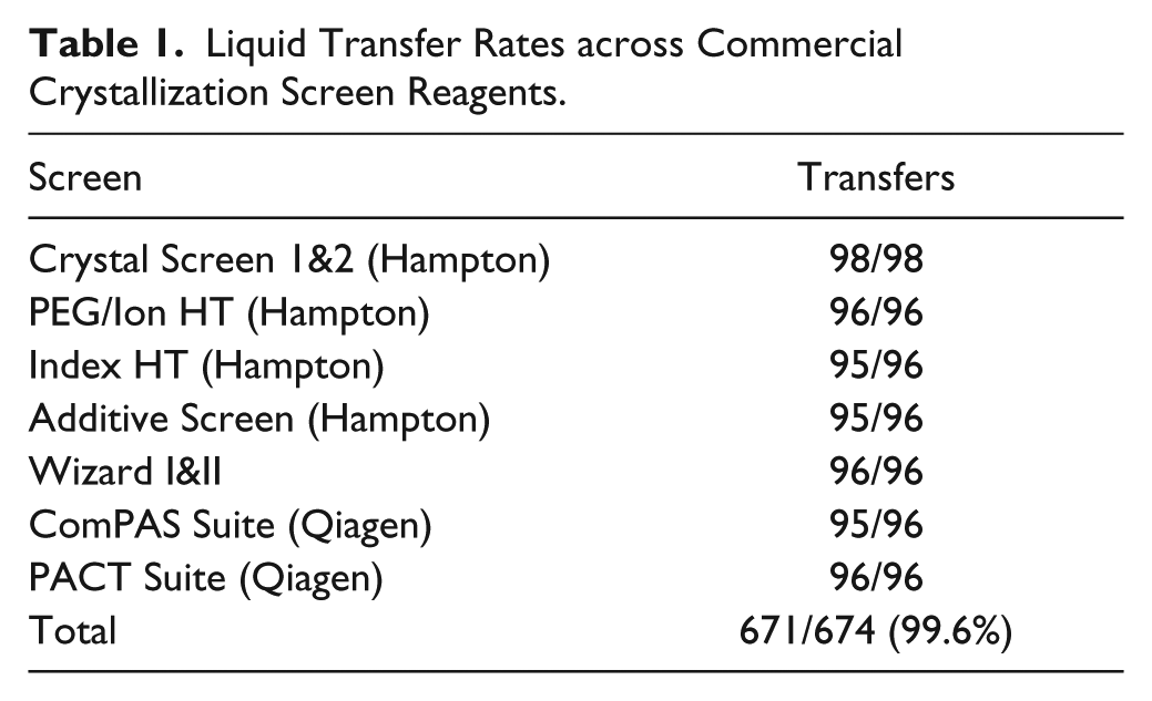
Coefficients of variation (CV) values for liquid transfer were defined as the ratio of the standard deviation to the mean for replicates of 64 measurements. Fluorescein was added to concentrated stocks of crystallization reagents and volumes transferred were quantitated by monitoring fluorescein emission at 521 nm on a Biotek reader. Qualitative measures of successful liquid transfer across seven commercial crystallization screens ( Table 1 ) were monitored by use of a colorimetric moisture-sensitive paper (TeeJet Technologies, P/N 20301-1N).
Liquid Transfer Rates across Commercial Crystallization Screen Reagents.
High-throughput crystallization experiments were performed using the Access workstation (Labcyte), which integrates the Echo with a centrifuge (model 05187-001; Agilent Technologies, Santa Clara, CA), a sealer (KAPS-500; K Biosystems, Basildon, UK), and a peeler (model XP-A; Brooks Life Science Systems, Poway, CA). MicroClime Environmental lids (Labcyte) were used during runs to prevent sample evaporation from peeled trays before acoustic liquid transfers. Crystallization trays were stored and imaged using a Crystal Farm imager (Brooks Life Science Systems, Poway, CA). Crystallization kits were purchased from Hampton Research (Aliso Viejo, CA), Qiagen (Germantown, MD), and MiTeGen (Ithaca, NY).
Application Software
Protocols dictating the transfer volume, location, and sequence of the crystallization reagents and protein samples were generated by Echo Array Maker software. Tempo Automation Control software imports Echo protocols and coordinates Echo liquid-handling actions, robotic plate movements, and tasks performed by integrated devices into an optimized schedule.
Protein Reagents and Crystallization
All proteins were purified in house except for human IL13, which was purchased from R&D systems (cat. No. 213-IL/CF; Minneapolis, MN). Angiopoietin 2 protein was used to screen against the JCSG+ screen (Qiagen) with Echo and Mosquito (TTP Labtech, UK) liquid handlers. Crystalliza-tion hits were identified under the following conditions: ATPase family AAA domain-containing protein 2 (ATAD2) apo crystals were obtained using crystallization reagent 0.1 M Bis-Tris pH 6.5, 0.2 M ammonium acetate, and 45% methyl-pentane diol (MPD); ATAD2 was also crystallized with 0.1 M HEPES, pH 7.0, 15% polyethylene glycol (PEG) 20000. ATAD2/histone peptide complex was crystallized with 0.1 M sodium citrate tribasic dihydrate, pH 5.5, 20% PEG 4000, and 18% isopropanol. IL13/Lebrikizumab Fab was crystallized with 20 mM HEPES, pH 7.2, 0.1 M NaCl, and 2% glycerol. Vascular endothelial growth factor (VEGF)/peptide was crystallized with equal volumes of protein and 25% PEG 1500. Fab Hy146 was crystallized with 10 nL protein and 10 nL of the crystallization reagent containing 0.1 M Tris pH 8.5, 0.2 M mono ammonium dihydrogen phosphate and 50% MPD. Optimization experiments used 0.15 M NDSB-195 as an additive and reagent No. 35 (0.02 M HEPES, pH 6.8, 0.16% azelaic acid, 0.16% m-benzenedisulfonic acid, 0.16% mellitic acid, 0.16% pyromellitic acid, and 0.16% trans-cinnamic acid) from the Silver Bullet kit (Hampton Research). The final concentration of the Silver Bullet mixture in the drop is 25% (v/v).
To screen for crystallization hits, the drops were set with 30 nL of crystallography reagent and 30 nL of protein samples unless otherwise noted. To optimize crystal growth, seeds (2.5 nL) and additive/detergent (5 nL) were added to drops consisting of 50 nL of protein and 42.5 nL of crystallography reagents. When using Silver Bullets for optimization, the drops contained 50 nL of protein, 25 nL of crystallography reagent, 22.5 nL of Silver Bullet reagents, and 2.5 nL seeds. In co-crystallization experiments, 2.5 nL of compound solution in DMSO was transferred directly into crystallization drops with 50 nL of protein and 50 nL of crystallography reagents.
Results
The Labcyte Echo performs liquid transfer between a source plate, commonly 384-well format, and a destination plate that is inverted above it ( Fig. 1 ). A transducer positioned beneath the source plate transmits acoustic energy via water as a coupling fluid to the source plate. The focal distance for each well’s volume is determined in a rapid survey pass on incoming source plates by measuring acoustic reflections from the various phase interfaces. This acoustic interrogation was extended to annotate fluid properties and thereby determine suitable energy settings for best drop ejection performance. The versatile CP calibration developed at Labcyte uses two low-powered acoustic energy pings to perturb the liquid meniscus and gauge the response in several rapid iterations. This expanded profile of the liquid property enables more precise energy calibration and thus promised robust automated dispensation without manual intervention or plate-specific inputs, allowing multiple fluid types in a single plate. We used several approaches to gauge the improved performance. Earlier work had established several classes of challenging reagents for ADE. High concentrations of MPD were problematic in absorbing acoustic energy; isopropanol’s particular surface tension properties and high concentrations of higher-molecular-weight PEG could lead to variability in delivery. Performance of the new calibration set was tested on these challenging concentrated stocks. Table 2 shows the CV values for the volumetric accuracy of delivery of these solutions, which were all better than 5% v/v. Table 1 charts a qualitative measure of liquid transfer to moisture-sensitive paper for seven common crystallization screens. Whereas working with a fixed energy reported 93% success rates of delivery (out of 480 conditions, largely due to failures to transfer solutions containing significant amounts of MPD), 5 here with dynamic fluid analysis overall transfers were noted at 99.6% success (671 of 674 conditions).
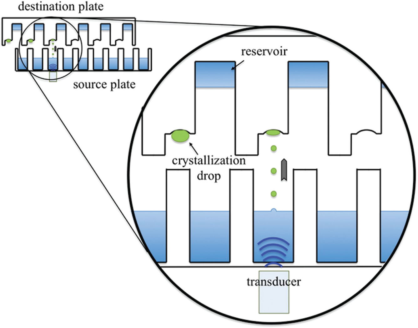
Schematic of acoustic droplet ejection (ADE) between a source plate and inverted destination plate. Lateral movements of both plates allow flexible addressing. The transducer positioned beneath the source well provides focused acoustic energy in millisecond-scale time pulses. The meniscus of the source well is deformed and a droplet ejected in an upward trajectory toward the destination surface. For protein crystallization plates, we prefill a reservoir with ~10 µL of crystallization reagent. ADE is used to deliver small volumes of the crystallization and protein reagents to a crystallization droplet (colored green to differentiate) on a shelf adjacent to the reservoir. The plate is then sealed to allow contained vapor diffusion between the reservoir and the droplet to drive crystallization drop concentration.
Coefficients of Variation (CV) for Delivery of High-Concentration Stock Reagents.
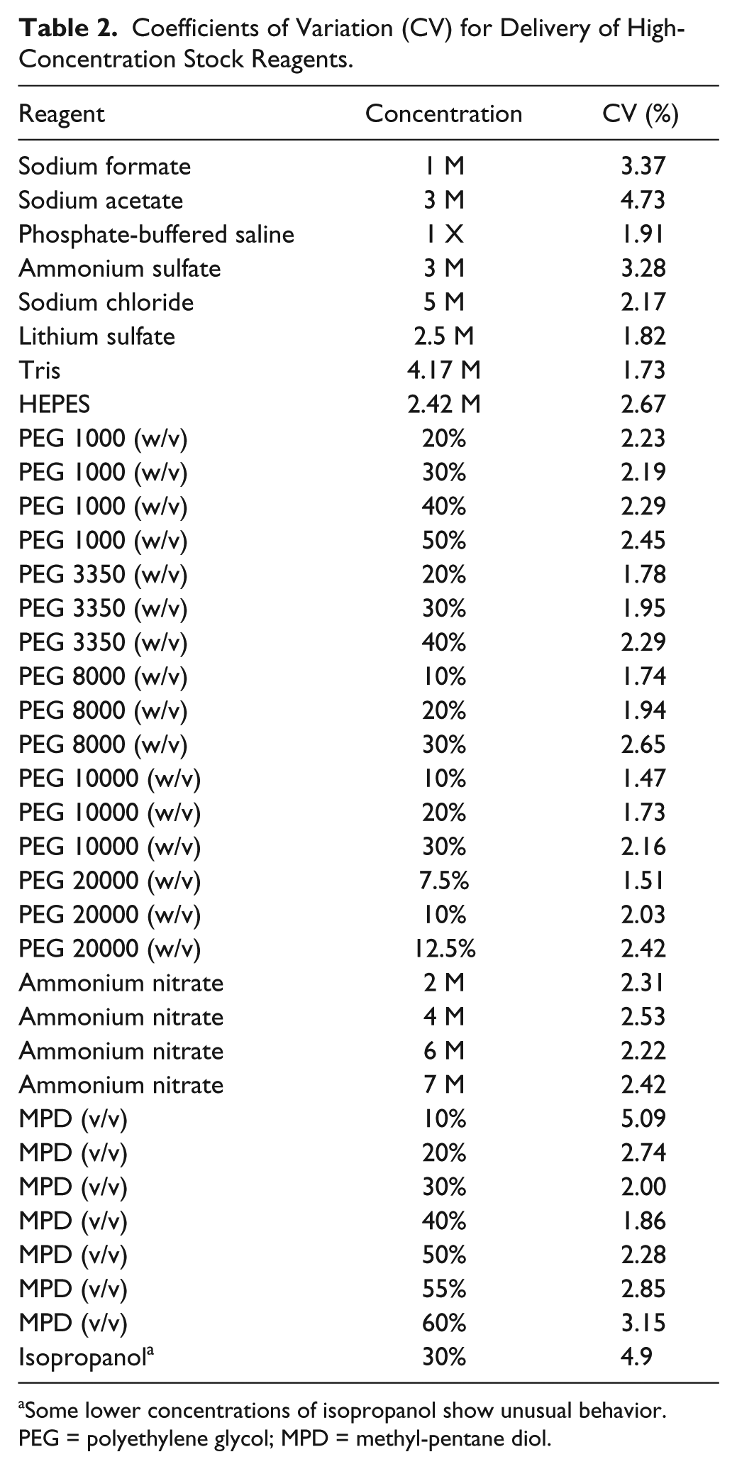
Some lower concentrations of isopropanol show unusual behavior. PEG = polyethylene glycol; MPD = methyl-pentane diol.
Guided by this improved performance, we validated that crystallization systems we used in our research were well reproduced at these smaller scales characteristic of ADE. We used the Echo to replicate the common sitting drop vapor diffusion crystallization format, in which a small drop of concentrated protein is mixed with various crystallization solutions and positioned on a crystallization shelf over a larger reservoir of the crystallization solution that drives concentration of components in the crystallization droplet ( Fig. 1 ). Several Labcyte Echo software control protocols can be used to map drop delivery location (e.g., Cherry Pick, Plate Reformat, Array Maker). Some crystallization plates have an uncommon layout relative to standard high-throughput plates. We used the Labcyte Array Maker software, which allows arbitrary droplet positioning and enables targeting of multiple locations within individual wells of the 96-well crystallization plate format. This flexibility permits testing of multiple protein targets, volumes, or reagent combinations in an efficient plate-economical manner. Our standard high-throughput approach employs a Mosquito (TTP Labtech) to set up trials with drop volumes of 100 nL protein plus 100 nL crystallization solution with reservoirs of ~100 uL. For the Echo system, we prefill crystallization trays with 10 µL of the reservoir solution (a limited volume because this tray is inverted during droplet setup and larger volumes of less viscous solutions would drip out; Fig. 1 ). The 384-well source plate contains three 96-condition screens, and unused wells are used for protein stocks, seed solutions, or additives (alternatively, four screens are included and the protein is provided in a second source plate). The acoustic drop ejection then makes two passes, producing spot-on-spot delivery of the crystallization solution and the protein, routinely using 30 nL volumes of each. Although very small-volume crystallization trials may affect the balance between crystal nucleation and growth, 10 we readily reproduced crystallization of protein samples, observing similar morphologies and growth on both the Mosquito- and Echo-derived trials ( Fig. 2 ).
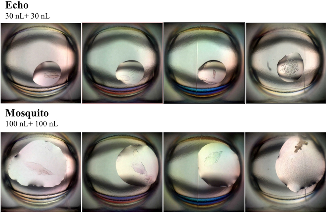
Comparison of crystallization behavior of angiopoietin 2 in crystallization drops of 30 nL protein plus 30 nL crystallization solution set up using acoustic droplet ejection (ADE) versus 100 nL components set up using a Mosquito system. Crystal morphologies are similar in each case, and the crystals were observed at earlier time points in the smaller ADE drops.
The smooth gradation in accessible volumes while preserving the delivery method reduces potential difficulties in translating successful crystallization from ultra-small-volume screening drops to larger production drops, which has been an issue when methodologies or size scales are altered more dramatically.10,11 Figure 3 shows consistent crystal growth for ATAD2 being explored at various size crystallization drops ranging from 10 nL to 800 nL protein volumes. In some systems, we observe a relatively similar size crystal across different volumes, and in other cases, such as this one, the crystal size scales proportionally to the drop size. However, even here, crystals are readily visible at the smallest tested volume (10 nL). Facile programming of different volumes allows routine exploration of this variable, both in absolute size and in the adjustment of the ratio of protein:crystallization solution within the drops.
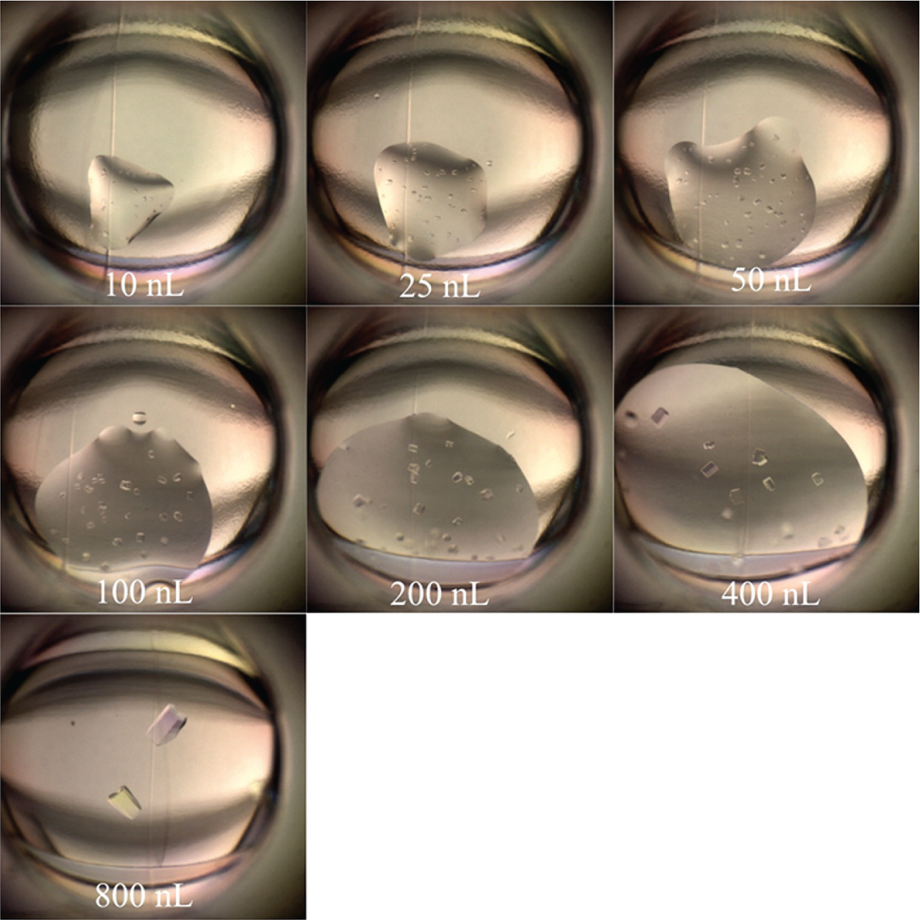
Crystallization of ATAD2 at protein volumes from 10 nL to 800 nL using acoustic droplet ejection. The protein was crystallized with 0.1 M Bis-Tris pH 7, 2.5 M ammonium sulfate at 19 °C. Crystallization is apparent in all drops, demonstrating reproducibility across this volume range. Crystal size scales with drop size for this protein, highlighting effective low-volume screening and translation to larger volume production drops.
In our practical use of the system, it is convenient to store the source plates containing the crystallization screens for some duration and reuse. To determine the longevity of the crystallization screen in the polypropylene plate, we examined the pattern of crystallization hits observed for our angiopoietin 2 test system against a single screen from a stored plate at various time points. Figure 4 demonstrates minimal impact on crystallization behavior in this system after 4 wk of source plate storage. At the final time points, it was interesting to note two additional conditions that showed crystal hits. Although a positive outcome for crystallization, this does suggest that these wells are experiencing some measure of change. The Echo system survey pass was used to chart well volumes at each time point. Most wells showed minimal loss, and only a few wells near the corners of plates showed noticeable sensitivity to evaporation in this metric. Further measures are recommended to control humidity and evaporation when storing source plates beyond 4 wk.
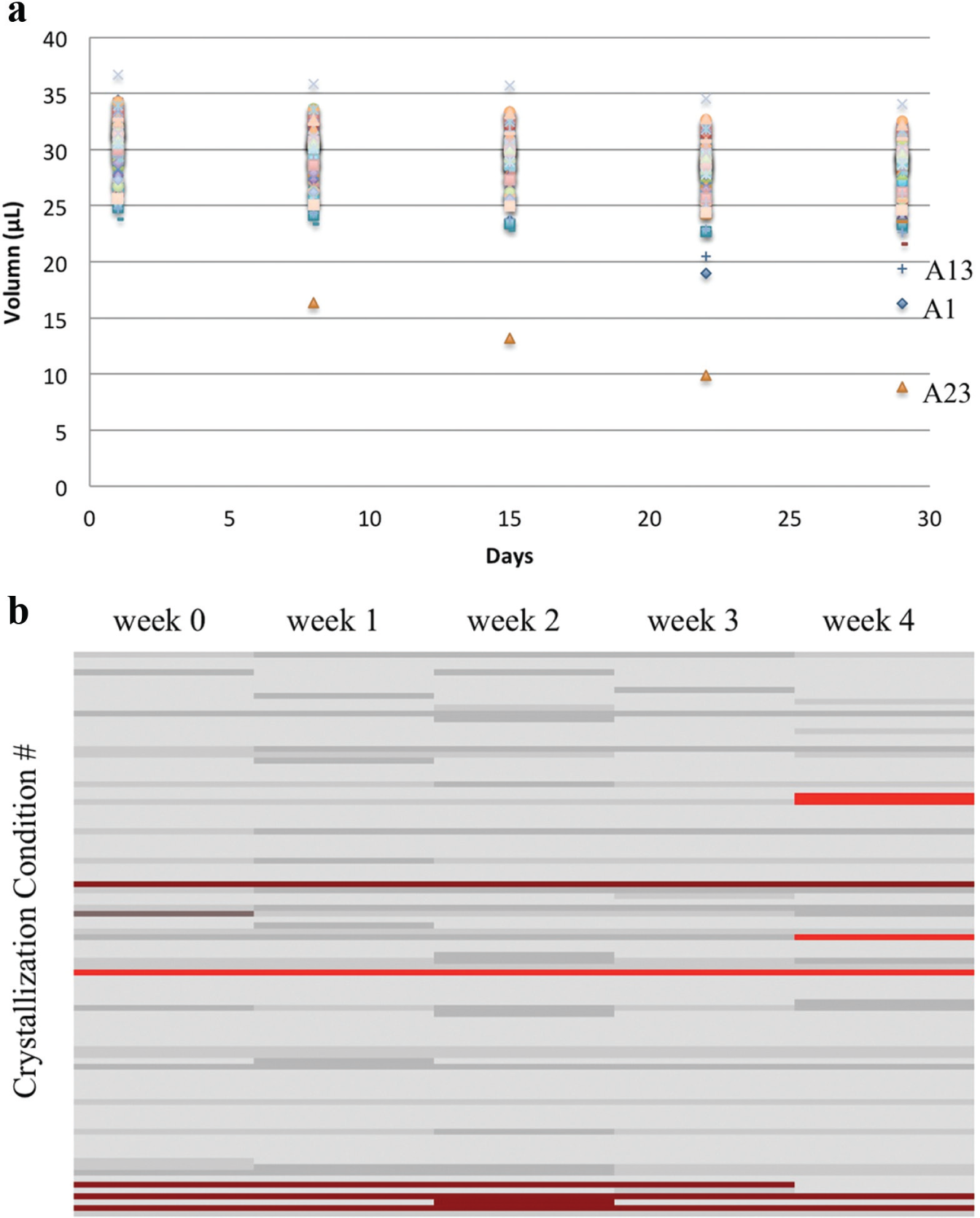
Characterization of source plate storage. (
Ultra-small-volume screening by ADE has been fruitful in identifying crystallization conditions on several novel target campaigns in our research. Figure 5 highlights examples, including ATAD2 ( Fig. 5a–c ), which crystallized from solutions containing 18% isopropanol, 45% MPD, or 15% PEG 20000, helping validate the performance of the dynamic fluid analysis in delivery of these more challenging components. We also show a crystal of IL13 bound to Lebrikizumab Fab ( Fig. 5d ), where this single crystal from a small-volume Echo-based trial led to the structure solution. 12 A VEGF complex ( Fig. 5e ) and a Fab crystal ( Fig. 5f ) are additional examples of varied crystal morphologies, and the latter condition also includes 50% MPD.
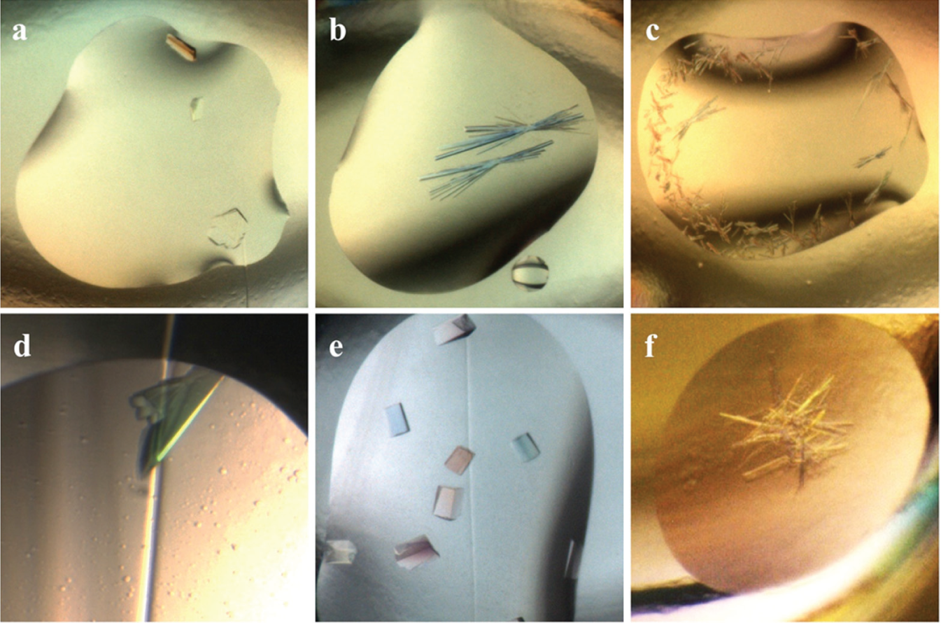
Crystallization hits identified using acoustic droplet ejection including solutions of previously challenging reagents and demonstrating diffraction-quality crystal production in nanoliter-scale volumes. (
Further work in optimization of such real-world examples highlights the utility of being able to deliver single-nanoliter-scale volumes of crystallization seeds, 6 additive compounds, or small-molecule ligands to facilitate crystallization. Figure 6 shows two proteins that demonstrated dramatic improvement from micro-crystalline initial hits toward large, well-formed, distinct crystals upon addition of additives ( Fig. 6a ), “Silver Bullet” additive cocktails ( Fig. 6b ), and microcrystalline seed suspensions. Typically, limitations on minimal dilutions of the additives require their exploration at relatively large drop sizes, but as the Echo can deliver these reagents in 2.5 nL increments, the investigation used only 50 nL protein per drop.
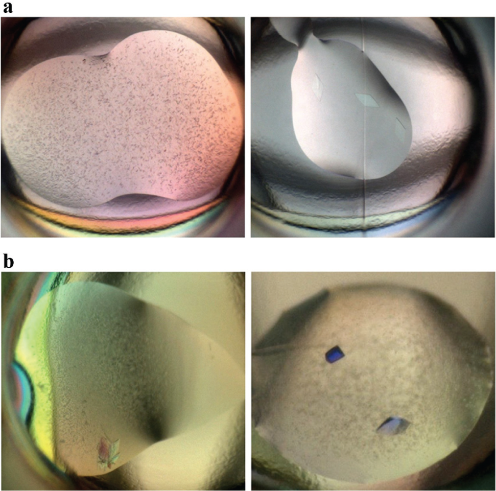
Crystal optimization by direct addition of additives and seeds. (
The Echo system can be readily integrated with plate-handling automation to enable higher-throughput multiplate operation, an attractive feature for the empirical challenges of protein crystallization. We designed a system that integrates the Echo, a plate peeler, a plate sealer, and a plate centrifuge around an Access robotic arm and several plate stacks (
Fig. 7
;
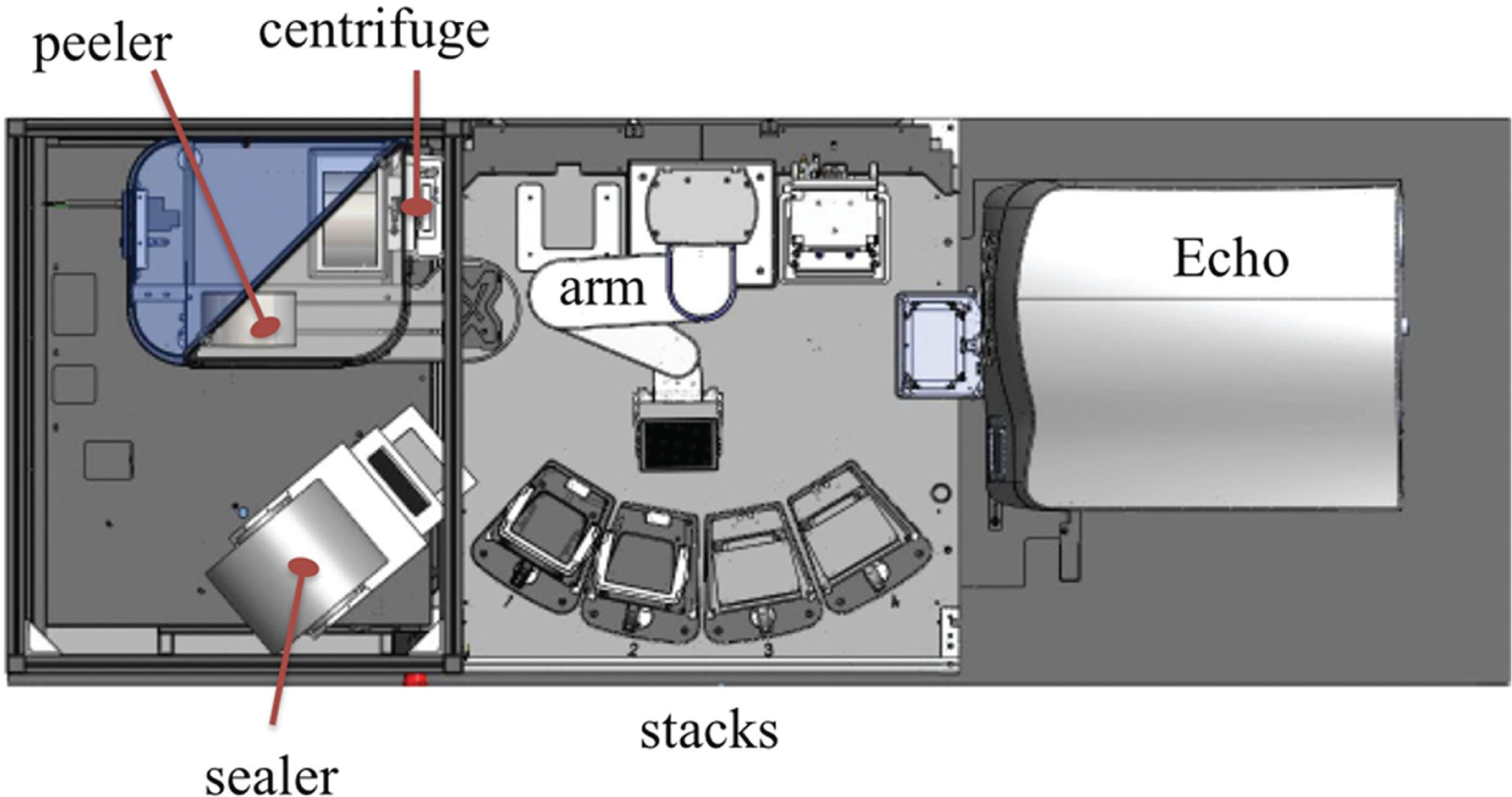
Schematic of integrated automation with Access system arm hub at center of Echo ADE, stacks, sealer, peeler, and centrifuge units.
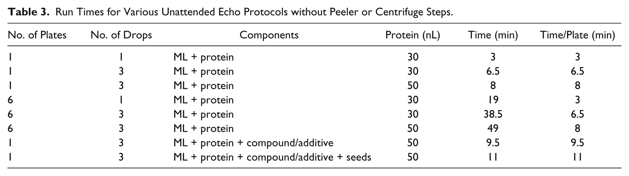
Run Times for Various Unattended Echo Protocols without Peeler or Centrifuge Steps.
Discussion
The discovery of robust protein crystallization conditions has traditionally been a resource-intensive method and was thus poised to benefit greatly from the extreme miniaturization afforded by ADE. The ability of ADE to set sub-50-nanoliter-scale crystallization experiments was rapidly established, 5 showing good reproducibility, protein-reagent economy, and rapid droplet equilibration for crystal growth and, importantly, demonstrating that in this size regime crystallization nucleation and growth behaved similarly as has been documented for larger drops. These characteristics facilitate productive translation between scales and systems. ADE performance at fixed settings was functional for protein crystallization, but the potential advantages of automated liquid calibrations to handle more diverse aqueous solutions with precision and accuracy were compelling, not only for crystallography but for many aqueous-based systems. As such, the dynamic fluid analysis feature is a critical development in the robust automation of ADE for protein crystallization. We have demonstrated here its improved delivery of key reagents, quantified for high-concentration stock solutions, as well as in practical crystallization successes. Our tested crystallization systems include those that require delivery of known challenging components such as MPD, isopropanol, and PEG 20000. The fact that these systems were reproducible and translated readily to other techniques confirms the improvements in performance realized by the additional fluid analysis.
The method has further been effective in optimizing crystallization through delivery of single-digit nanoliter volumes of additives and seeds. 6 We have recently extended this to less common practices, such as addition of proteases for in situ limited proteolysis in aid of crystallization. Similarly, these small drops can be used to explore binding of small-molecule ligands in structure-based design campaigns. Because ADE was initially optimized for handling small-molecule compounds in DMSO, the system is adept at delivering precise quantities of these reagents into the crystallization droplets, whether for co-crystallization or soaking experiments. Provided a source plate of DMSO-solubilized compounds, an extra pass on the Echo serves to add 96 compounds to 96 different crystallization drops in ~40 s. These trials can be followed more traditionally with suitable crystals harvested and mounted for data collection, allowing rapid prosecution of biochemical or biophysical screening hit lists for structures.
In a related note, a recent publication reported an elaboration of this idea to enable crystallography-based screening directly using ADE to position multiple crystallization drops in situ on x-ray source micromesh mounts. 8 Although this required some customized hardware, it bypasses manual steps of crystal mounting and streamlines crystal exchange time during synchrotron data collection. The efficacy of these approaches is dependent on a relatively robust crystallization system.
The extremely small-volume crystallization drops also raise concerns of evaporation effects. In practice, with the lids and sealer in the system, we describe that the 10 nL plus 10 nL drops yield successful crystallization, although we start to note a small percentage of nonoverlapping droplets at this size (data not shown). With proper humidity control, it would be interesting to investigate smaller setups, because on the standard Echo transducer, another fourfold decrease to 2.5 nL protein volumes is accessible. Plate modifications with smaller entrance apertures punched in films have been examined. 13 With customized crystallization chambers, one could use acoustic energy to achieve sub-nanoliter-scale crystallization trials. With the smaller droplets, the ability to image for crystallization success is also an important consideration. These drops do offer a shallower depth of field, and we found that images of the 10 nL drop trials produced by our Crystal Farm imager were readily interpretable without special adjustments. Again, investigations into much smaller droplets will likely require a more sensitive approach such as ultraviolet or second-order nonlinear imaging of chiral crystals imaging techniques. 14
One advantage of our current implementation is the use of standard crystallization plates, but their larger-volume design premise leaves some limitations. First, we currently prefill the reservoir with 10 uL of solution on a separate instrument, whereas smaller reservoirs that can be efficiently set on the Echo system itself would further streamline the process. We have succeeded in crystallizing proteins when we instead used the Echo ADE to set a 1 to 2 uL reservoir in a small depression adjacent to a 20 nL crystallization droplet. This proof of concept would allow much higher density experimentation and would further benefit from a plate design that reduced the air volume present in the standard crystallization plates currently used to avoid complete dehydration of the crystallization droplet.
The delivery of high-concentration stock solutions was also intended to allow mix-on-the-fly assembly of crystallization drops from separate component chemical solutions. Although feasible in terms of liquid delivery, as documented in Table 2 , the technique suffers technically from the limited volume of the target drop (e.g., 50 nL), such that only a relatively coarse set of dilutions is possible for a given reagent, even with the 2.5 nL single-delivery volume. Larger-volume customized drops can be more readily concocted, although time and mixing considerations exist. Nonetheless, this may allow more systematic analyses of protein phase behavior against given reagents to guide successful crystallization, as demonstrated previously in a microfluidic setting. 15
We have also considered delivery of the extremely viscous lipidic cubic phase used as a crystallization matrix for many membrane proteins. 16 However, it was difficult to develop a method for transfer of components without exposing the sensitive protein sample to methanol or other possibly denaturing reagents. The lipidic sponge phase is a less viscous alternative, although its success for membrane protein crystallization is less documented. 17
One intriguing, unique feature of the acoustic energy methods is the ability to move liquids from reservoir to a suspended drop within a sealed plate. In this fashion, very small additions of reservoir solution to the crystallization experiment could be achieved at chosen time points beyond the initial setup, allowing dynamic modulation of the crystallization environment and some level of “steering” a drop and its protein contents along a solubility phase diagram. This would require the accurate placement of the suspended drop relative to the ejection target, but one can envision an enclosed apparatus in which there is a combination of microfluidic channels or acoustic steering 18 and other emerging techniques that would be able to bring the protein and crystallization reagents together without evaporation risk.
In summary, our work describes implementation of state-of-the-art ADE technology for protein crystallization in an automated workflow with routine use of ~30 nL protein per crystallization drop. We identify several areas where a new iteration of plate design could further leverage the advantages of ADE to capitalize on efficient use of valuable protein reagents while enabling rapid identification of promising crystallization hits. Novel applications of the compelling acoustic technologies are actively being explored in several stages of protein crystallization and crystal preparation.
Footnotes
Acknowledgements
We thank Joe Barco and other scientists at Labcyte for discussion and the Genentech Structural Biology Expression group for constructs and expression work and Qianting Zhai for provision of Fab HY146.
Declaration of Conflicting Interests
The authors declared the following potential conflicts of interest with respect to the research, authorship, and/or publication of this article: P.W., C.N., M.U., R.M., and S.F.H are employees of Genentech, Inc. B.E. and D.H. are employees of Labcyte, Inc.
Funding
The authors received no financial support for the research, authorship, and/or publication of this article.
References
Supplementary Material
Please find the following supplemental material available below.
For Open Access articles published under a Creative Commons License, all supplemental material carries the same license as the article it is associated with.
For non-Open Access articles published, all supplemental material carries a non-exclusive license, and permission requests for re-use of supplemental material or any part of supplemental material shall be sent directly to the copyright owner as specified in the copyright notice associated with the article.