
Abstract
Because of intensive developments in recent years, the microfluidic system has become a powerful tool for biological analysis. Entire analytic protocols including sample pretreatment, sample/reagent manipulation, separation, reaction, and detection can be integrated into a single chip platform. A lot of demonstrations on the diagnostic applications related to genes, proteins, and cells have been reported because of their advantages associated with miniaturization, automation, sensitivity, and specificity. The aim of this article is to review recent developments in microfluidic systems for diagnostic applications. Based on the categories of various fluid-manipulating mechanisms and biological detection approaches, in-depth discussion of the microfluidic-based diagnostic systems is provided. Moreover, a brief discussion on materials and manufacturing techniques will be included. The current excellent integration of microfluidic systems and diagnostic applications suggests a solid foundation for the development of practical point-of-care devices.
Introduction
In the statistical report from the Department of Health, Taiwan, 1 the top killer over the past 28 years was cancer. On average, a person dies from cancer every 13 min 10 s. Early treatment is an effective method to reduce the death rate. However, there is no significant symptom in the early stage of cancer. Therefore, regular screening is the best prevention method for cancer. Currently, disease screening requires operating in a well-equipped laboratory and handling by well-trained personnel. This results in hesitation from the general public because the screening service in the hospital is expensive and time-consuming. To popularize screening for disease prevention, the development of low-cost, miniaturized, automated, and sensitive diagnostic devices has been extensively explored in recent years.
The microfluidic system, also called lab-on-chip, bio-chip, or micro-total-analysis-system, has been rapidly developed from early single-channel devices 2 to current complex analysis systems. 3 –5 The microfluidic system is often interpreted as a miniaturized version of a conventional laboratory. Entire diagnostic protocol can be performed automatically, including sample pretreatment, sample/reagent manipulation, separation, reaction, and detection. It provides a total solution from the application of the sample to the display of analysis results. Because of microfluidic systems’ miniaturization and automation, there are a number of advantages to their use, such as less sample/reagent consumption, reduced risk of contamination, less cost per analysis, lower power consumption, enhanced sensitivity and specificity, and higher reliability. Moreover, portability becomes realizable for the point-of-care diagnostic devices.
Recently, a broad spectrum of scientists and technologies has been involved in the development of microfluidic systems and their applications. 6 –10 Microfluidic systems can be fabricated from silicon, glass, quartz, and various polymeric materials. The silicon microfabrication process is well established from the microelectronic and MEMS industry; however, silicon is not optically transparent and is electrically conductive. Silicon-based microfluidic systems have limitations for the applications based on optical and electrochemical detection. Glass, quartz, and polymeric materials are alternative materials for fabricating microfluidic systems. They are less expensive, optically transparent, and not electrically conductive. Therefore, various functional microfluidic devices (e.g., micropumps, 11 –13 micromixers, 14,15 microvalves, 16,17 microfilters, 18,19 microreactors, 20 –22 and microseparators 23 ) were then developed. These devices were integrated into a single chip and worked cooperatively to perform a specific assay. Various biological analytical applications, such as DNA analysis, 24 –26 immunoassay, 27 –30 and cell analysis, 31 –34 have been demonstrated using microfluidic systems. For example, a microfluidic-based DNA analysis system integrated with microchannels, heaters, temperature sensors, and fluorescence detectors was capable of capturing the DNA, mixing the solutions together, amplifying the DNA, and separating and detecting those products. 35 Fluids were manipulated by electro-osmotic pumping, and the DNA analysis results were measured by a fluorescence detector. A strand displacement amplification experiment was conducted, and the specific target DNA was successfully amplified and detected. Another example was conducted by a microfluidic system integrated with the micro-pneumatic pumping mechanism for immunoassay. The peristaltic effect driven by time-phased deflection of polydimethylsiloxane (PDMS) membranes along the microchannel can be used to deliver liquid. The deflection of PDMS membranes can be actuated by compressed air 36 or thermal heater. 37 Immunoassay was performed by sequentially pumping sample and regents to the detection chamber, and the results were detected by optical measurement of absorbance. 36 These examples showed the power of microfluidic technology and its capability of performing complex analytical problems.
The aim of this article is to review recent developments on microfluidic systems for diagnostic applications. Fluid-manipulating mechanisms, biological detection schemes, materials, and manufacturing techniques will be discussed. Because the performance of liquid pumping and the biosensing detector can directly affect the results of the assays, this article focuses its discussion on fluid-manipulating mechanisms and biological detection schemes. These developments in microfluidics provide promising tools for miniaturized, integrated, automated, sensitive, and specific biological analysis in practical point-of-care applications.
Fluid Manipulation
For the development of microfluidic-based diagnostic devices, fluid manipulation by external pumps (e.g., syringe pump and peristaltic pump) is not appropriate because fluidic connections between the external pumps and the microfluidic system are difficult for untrained personnel to handle. Therefore, many strategies for on-chip fluid manipulation have been reported in the literature. Sample/reagents are applied to the microfluidic system and manipulated by the on-chip pumping mechanism. This is not only to reduce the handling fault but also to standardize the analytical conditions in each analysis. In this section, some pumping strategies for microfluidic diagnostic applications will be reviewed. Most can be roughly categorized into two groups: mechanical and nonmechanical approaches. Based on these approaches, the mechanical approach can be subdivided into several categories: piezoelectric, pneumatic, centrifugal, electrostatic, and electromagnetic actuations. The nonmechanical approach mainly includes electro-osmotic, electowetting based, magnetohydrodynamic, electrochemical, and capillary manipulations.
Mechanical Approach
Piezoelectric micropumps
A micropump actuated by piezoelectric material was proposed in the late 1980s. 38 A piezoelectric disc was used to actuate a membrane on the pump chamber by applying an alternative electric field to drive fluid. In 2003, peristaltic pumping driven by three piezoelectric discs located on three reaction chambers was demonstrated to drive fluids for DNA amplification. 39 As shown in Figure 1 , this chip contained three reaction chambers, which were stable at 90 °C, 72 °C, and 55 °C for PCR, a bidirectional peristaltic pump, and an optical integrated detector. The maximum flow rate of 3.14 µL/s at an operating frequency of 10 Hz was obtained in this piezoelectric micropump, and the micropump could withstand a pressure differential of 50 kPa. The PCR was achieved by introducing the reactant droplet of 1 µL into the inlet, then moving back and forth between these three reaction chambers driven by the bidirectional pump. After 20 to 30 thermal cycles, the PCR products were pumped into the reservoir to be collected and analyzed by gel electrophoresis. Fluid manipulated by the piezoelectric micropump can achieve high stroke volume, high actuation force, and fast mechanical response. However, the integration of the piezoelectric driving equipment is a challenge for the point-of-care diagnostic device.
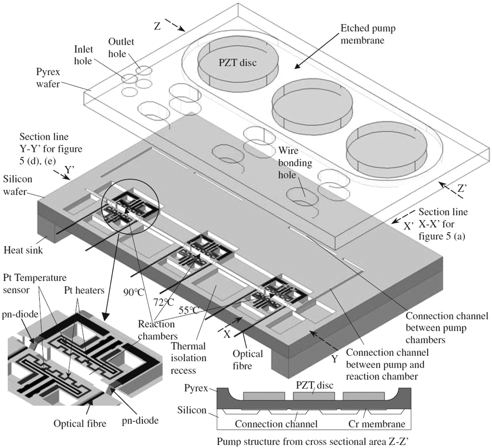
Schematic of the pump PCR chip. For simplification, the upper glass wafer and the lower silicon wafer are illustrated apart, although in the actual device, both wafers are connected by anodic bonding. The lower left insert figure shows an expanded view of the reaction chamber, and the lower right insert shows the cross section of the micropump. Copyright 2003. Reprinted from ref. 39 with permission from IOP Publishing Ltd.
Pneumatic micropumps
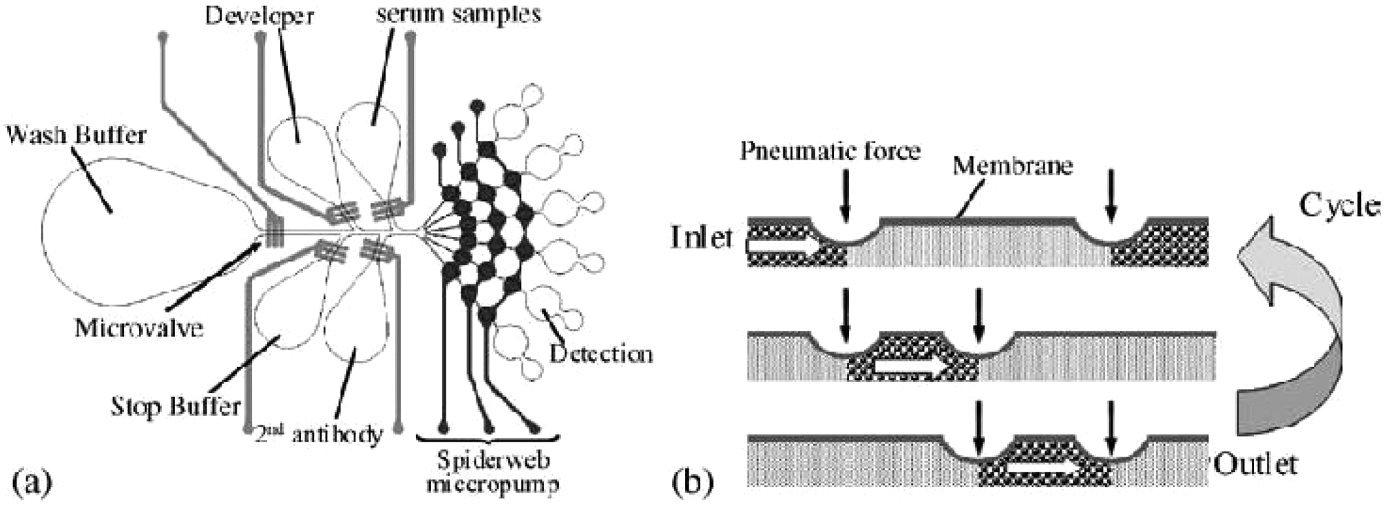
Based on the peristaltic effect, pneumatic micropumps have been developed by a series on/off actuation of three separate microvalves 40 or diaphragms 41 driven by compressed air. Three external electromagnetic valves were required to control the sequential actuation. Alternatively, one S-shaped air channel across the fluidic channel could accomplish the same peristaltic pumping. 42 Because the interconnecting air channel allowed for the sequential actuation of the fluidic channel, only one external electromagnetic valve was required to perform the pumping. The polydimethylsiloxane (PDMS) elastomer materials of low modulus are widely used in pneumatic micropumps. As shown in Figure 2 , the peristaltic effect driven by the time-phased deflection of PDMS membranes along the fluidic channel can be used to deliver liquid. A microfluidic system integrated with the pneumatic micropumps and microvalves can be used to determine hepatitis C virus (HCV) and syphilis from serum samples. 36 The detection process was automatic and began with bonding screening antigens (i.e., HCV and syphilis) to the detection areas. Then, the serum samples, washing buffer, horseradish peroxidase (HRP)–labeled secondary antibody, developing buffer, and stopping buffer in individual reservoirs were sequentially pumped to the detection areas by the “spiderweb” pneumatic micropumps. The results were detected by the measurement of absorbance. Moreover, an S-shaped pneumatic micropump has been used to drive the samples for the study of cell separation and nucleus collection using dielectrophoresis (DEP) forces. 42 The pumping rate can achieve 39.8 µL/min at a driving frequency of 28 Hz under a pressure of 25 psi. The viable and nonviable cells can be separated by the DEP forces and collected respectively in the specific reservoir. In addition, after cell lysis, the nucleus can also be collected using a similar scheme. However, the above pneumatic micropumps require an external air compressor and one or more high-speed valve connections. They are not fully integrated into the microfluidic system for fluid manipulation. To overcome this, a thermoactuated pneumatic micropump has been proposed and integrated with a tiny circuit module for operation. 43 On-chip thermal heaters were integrated into the system for pumping actuation. By heating air sealed in the chamber, the thermally induced volume change of air deformed the diaphragm, which can push fluid in toward the channel. This concept of actuation has no external air connection, a relatively simple device structure, and a compact system size.
(
Centrifugal force
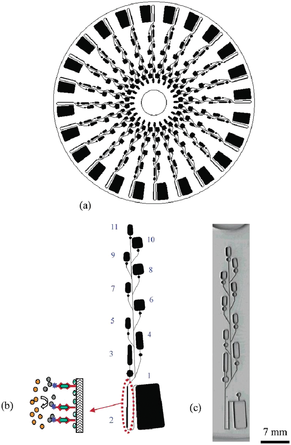
The centrifugal force has been demonstrated for the use of fluid manipulation in a compact disc (CD)–based microfluidic system. 44 –46 Fluids on the CD can be manipulated by the centrifugal force controlled by the rotational speed of the CD. The CD-based microfluidic system can perform several microfluidic functions (e.g., capillary valving, centrifugal pumping, and flow sequencing). As shown in Figure 3 , simultaneous and identical assays in parallel layouts can be fabricated on the CD. High-throughput screening of analytes can be realized. The CD-based microfluidic system is expected to have great commercial potential because of the mature developments of the CD technology (e.g., precision rotation control and optical reading). Enzyme-linked immunosorbent assay (ELISA) has been proposed to conduct on the CD-based microfluidic system for the demonstration of the clinical diagnostics. 46 Centrifugal and capillary forces were used to control the flow sequence of different solutions involved in the ELISA process. An analysis of rat IgG from hybridoma cell culture showed that the same detection range as the conventional method on the 96-well plate has been obtained with the advantages of less reagent consumption and shorter assay time. Another example was to determine α-fetoprotein (AFP), interleukin-6 (IL-6), and carcinoembryonic antigen (CEA). 47 A 200 nL sample was applied to the CD-based microfluidic system and passed through the microchannel packed with antibodies against AFP, IL-6, and CEA. Alexa 647–labeled detection antibody was used for the detection. The flow rate was controlled by altering the rotational speed, and the results were measured by a laser-induced fluorescent detector. The detection limits for AFP, IL-6, and CEA were, respectively, 0.15, 1.25, and 1.31 pmol/L. Up to 104 sandwich-type immunoassays were completed within 50 min. Excellent analytical efficiency within acceptable variations has been demonstrated when compared with the traditional assays performed by a 96-well ELISA plate.
(
Electrostatic micropumps
Electrostatic pumping is based on the membrane deflection driven by the Coulomb attraction force between oppositely charged plates. The deflected membrane is returned to its initial position when the plates are discharged. The electrostatic plates are charged and discharged by the application of appropriate voltage. Therefore, the pump chamber volume varies based on the alternate switching of applied voltage. Fluids can be forced to flow in the microchannel due to pressure difference induced by the membrane deflection in the pump chamber. Micropumps based on electrostatic actuation have been reported. 48,49 A dual diaphragm electrostatic micropump was developed, and it achieved a flow rate of 30 µL/min at a frequency of 30 Hz and operating voltage of 160 V. 49 Moreover, design and simulation of an electrostatic peristaltic micropump was reported for drug delivery applications. 50 The micropump was designed to satisfy most of the drug delivery requirements, but the actual fabrication and testing of the micropump was not reported. The advantages of electrostatic micropumps are low power consumption and fast response time. However, it has a complicated structure and requires high operating voltage. Few studies have been reported to integrate electrostatic micropumps into the microfluidic system for diagnostic applications.
Electromagnetic micropumps
Electromagnetic actuated pumping is typically based on the deflection of a flexible membrane driven by a permanent magnet and a set of drive coils. Either the magnet or coils may be attached to the membrane. When a current is applied through the coils, the resulting magnetic field generates an attractive or repulsive force between the magnet and coils for varying the pump chamber volume. Fluid can then be pumped to the microchannel. Electromagnetic actuation provides a large actuation force and requires low operating voltage. An electromagetic actuated polymethylmethacrylate (PMMA) valveless micropump was fabricated by standard micromachining techniques. 51 The micropump was a three-dimensional structure and consisted of two diffuser elements located at the inlet and outlet of the pump chamber. PDMS membrane with an integrated magnet made of NdFeB (neodymium, iron, and boron) magnetic powder was bonded to the pump chamber for actuation. A large stroke membrane deflection up to 200 µm was obtained using external actuation by a magnet. The micropump achieved a flow rate of 400 µL/min and back pressure of 1.2 kPa at resonant frequencies of 12 and 200 Hz, respectively. Although electromagnetic actuation provides a large actuation force, it does not benefit from scaling down in size because the electromagnetic force was reduced by the cube of the scaling factor. The complicated structure of the electromagnetic micropump may also hamper the integration of the microfluidic system for diagnostic applications.
Nonmechanical Approach
Electro-osmotic manipulation
Electro-osmosis is one of the electrokinetic phenomena that can manipulate electrolyte solution in a microchannel. When an ionic solution comes in contact with a solid surface (i.e., microchannel wall), an instantaneous electrical charge is acquired by the solid surface. An external electric field is then applied to two ends of the microchannel. A thin layer of cation-rich fluid adjacent to the solid surface starts moving toward the cathode. The bulk fluid is then manipulated by this boundary layer through the viscous interaction. A number of studies of electro-osmotic pumping have been reported. 52 –54 For example, an electro-osmotic micropump was fabricated by packing the 3.5 µm diameter nonporous silica particles into 500 to 700 µm diameter fused-silica capillaries using the silicate frit fabrication process. 52 The micropump achieved pressures in excess of 20 atm and flow rates of 3.6 µL/min for 2 kV applied potential. Also, a multistage electro-osmotic micropump was developed for higher output pressure. 53 The one- to three-stage electro-osmotic micropump was fabricated using 100 mm × 320 µm internal diameter columns packed with 2 µm porous silica particles, fused-silica capillaries, and stainless electrodes. Compared with the one-stage micropump, the output pressures of two- and three-stage micropumps were two to three times higher. The flow rates of two- and three-stage micropumps were identical with that of the one-stage micropump at the same driving voltage. Moreover, a high-pressure electro-osmotic micropump fabricated by a sol-gel process was reported. 54 A silica monolithic matrix with morphology of micro-scaled through pores was synthesized within the 100 µm inner diameter fused-silica capillary. The maximum flow rate and pressure for deionized water were 2.9 µL/min and 304 kPa, respectively, at 6 kV applied voltage. Electro-osmotic micropumps have the advantages of no moving parts involved, quiet operation, and bidirectional pumping controlled by switching the direction of the external electric field. The major limitation is the requirement of high voltage and electrically conductive solution.
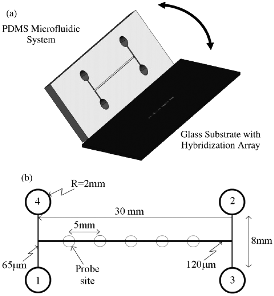
Based on the development of the electro-osmotic manipulation, microfluidic systems for various biological processes have been demonstrated recently. 24 , 55 –59 An electrokinetically controlled DNA hybridization microfluidic chip has been demonstrated and can perform all processes from sample dispensing to hybridization detection within 5 min. 24 The chip was composed of a PDMS upper substrate and a lower glass substrate that served as a substrate for the hybridization array, as shown in Figure 4 . The design consisted of an H-type channel structure containing immobilized single-stranded oligonucleotide probes. The electro-osmotic pumping can dispense the controlled samples of nanoliter volume directly to the hybridization array and remove nonspecific adsorption. Hybridization, washing, and scanning procedures can be conducted simultaneously. Detection levels as low as 50 pM were recorded using an epifluorescence microscope. Moreover, the microfluidic chip for real-time PCR has been reported using a Joule heating effect. 55 The chip was fabricated by PDMS and glass substrates. Joule heating was generated in an electrokinetically driven microfluidic chip by the current flow through the buffer solution. A DNA fragment of Escherichia coli O157:H7 stx1 was successfully amplified by a two-temperature TaqMan real-time PCR. Alternatively, enhancement of heterogeneous immunoassays using AC electro-osmosis was proposed to improve the rate of antibody binding to the sensor surface. 56 AC electro-osmosis can generate rotational fluid flow near the sensor surface (i.e., electrodes), where the AC electric field was applied. Immunoassay was performed and showed that the binding of secondary antibody on the sensor surface was significantly enhanced due to the possibility of local mixing. Enhancement of about 1.9 times at the center of the sensor and 6.7 times at the edges of the sensor was reported compared with a nonmixed counterpart. Also, electro-osmotic manipulation in the microfluidic chip has been demonstrated for the online monitoring of glucagon secretion from pancreatic islets of Langerhans. 57 Groups of islets were quantitatively monitored for changes in glucagon secretion as the glucose concentration was decreased from 15 to 1 mM. These examples show that a broad spectrum of applications can be achieved by the electro-osmotic manipulation in microfluidic systems.
(
Electrowetting manipulation
Manipulation of droplets on a microfluidic system can be performed by electrowetting forces generated when an electrical potential is applied to an electrode in an array. 60 –63 As shown in Figure 5 , by applying electrical potentials to sequential electrodes, a droplet can be dispensed from a reservoir, transported to any position on the array, and merged with other droplets to perform reactions. This approach suggested that the configuration is easier than the microchannel-based system. The flexibility of these electrowetting-based systems makes them suitable for a wide variety of applications. For instance, a prototype device using electrowetting manipulation has been developed to perform multiplexed enzyme analyses. 64 Droplets of alkaline phosphatase and fluorescein diphosphate were merged and mixed on the device, and then the fluorescent product was detected by the fluorescence plate reader. The detection limit was achieved at ~7.0 × 10⁻²⁰ M. Also, heterogeneous immunoassays have been demonstrated by efficient handling of magnetic microbeads using electrowetting manipulation. 65 A sample droplet and a reagent droplet containing magnetic beads conjugated to primary capture antibodies, blocking proteins, and secondary antibodies were dispensed on the system. These two droplets were then merged, mixed, and incubated by electrowetting manipulation. A permanent magnet was applied to immobilize the sandwiched microbead complexes, followed by the washing of the unbound components. Finally, a reagent droplet was applied for the chemiluminescent detection. Sandwich heterogeneous immunoassays on human insulin and IL-6 were demonstrated with a total time to result of 7 min for each assay. Moreover, the electrowetting manipulation has been applied to the PCR for potential point-of-care applications. 66 Droplet-based PCR showed that the total amplification time was reduced to half of that required for benchtop PCR. It can be monitored in real time and provides amplification with a cycle threshold of ~10 cycles earlier than the benchtop instruments.
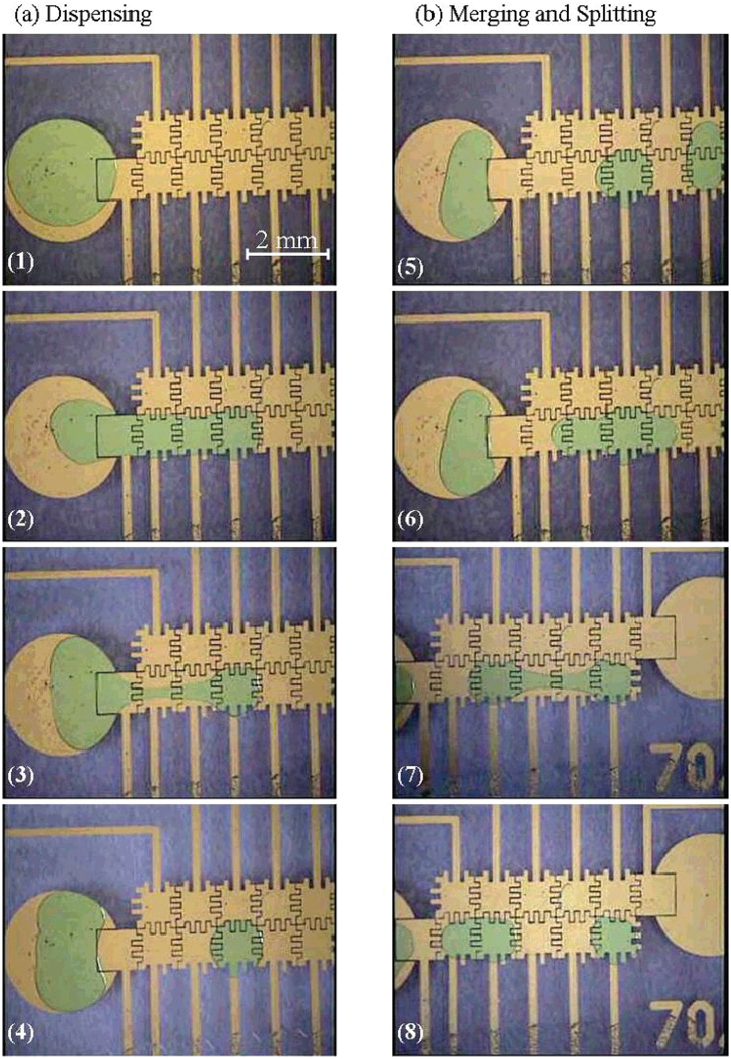
Video sequence depicting (
Dielectrophoretic manipulation
For manipulation and sorting of dielectric particles such as biological cells, dielectrophoresis has been widely used in microfluidic systems. 67 –69 When an electric field is applied to particles, a dipole is induced in the particles. 70 If the electric field is nonuniform, the particles will experience a force, which is not dependent on the polarity of the electric field but on internal properties of the particles compared to their surroundings. Therefore, using dielectrophoresis, biological cells can be manipulated, sorted, and separated by the electric field generated by the microelectrodes. An example of using dielectrophoresis as a selective filter was reported, and removal of PCR inhibitors was demonstrated in microfluidic system. 71 To increase the sensitivity of the PCR, removal of PCR inhibitors in sample is essential. By using dielectrophoresis, cells were captured by the electrodes in the fluidic chamber, whereas PCR inhibitors were washed by the buffer. After that, the dielectrophoretic force can be turned off and the cells released as a ready sample for the PCR.
Opto-electric manipulation
Electrokinetic force can be generated by microelectrodes, as discussed above. Moreover, an opto-electrically–induced electrokinetic scheme has also been reported to achieve particle or fluid manipulation in microfluidic systems. In 1970, pioneering work of optical manipulation was demonstrated by trapping the particles using optical tweezers. 72 Optical tweezers rely on optical pressure for particle manipulation. Recently, opto-electric manipulation was proposed to use light to activate and control electrokinetic manipulation instead of using microelectrodes. Critical reviews of opto-electric manipulation in microfluidic systems were reported. 73,74 The use of light provides a higher degree of freedom to construct the electric field through illumination displays such as an LCD screen. Opto-electric manipulation and concentration of colloidal particles were demonstrated. 75,76 Particle manipulation was performed between a parallel plate indium tin oxide electrode biased with an AC signal and illuminated with near-infrared (1064 nm) light. The technique of opto-electric manipulation is still in a developing stage and opens new portals of dynamic, multiscale, and high-throughput manipulation of biomolecules. It is expected that new applications using this technique will be demonstrated.
Magnetohydrodynamic (MHD) pumping
MHD pumping is based on the Lorentz force when an electric current is applied across a channel filled with conducting solution in the presence of a perpendicular magnetic field. 77 The Lorentz force is both perpendicular to the current in the channel and the magnetic field. Fluid flow is then induced along the channel direction. Continuous flow PCR was demonstrated using MHD pumping. 78 The reaction solution was passed through the required temperature zones such that the flow rate determined the temperature profile that the reactants experience. The reported MHD pumping can achieve a flow rate of 85 to 369 µm/s, with the electromagnet producing a magnetic flux density of 7.4 to 13.5 mT and peak channel current of 55 to 156 mA. However, the PCR using MHD pumping was confounded by electrolysis at elevated temperatures. The formation of bubbles may block the channel and could adversely affect the PCR process.
Electrochemical pumping
Electrochemical pumping uses the bubbles electrochemically generated by the electrolysis of water to drive the fluids. 79 Thus, the structure and operation of the electrochemical micropump are relatively simple, and it can be easily integrated with other microfluidic devices. A fully integrated microfluidic biochip for PCR amplification and DNA microarray detection was developed and is shown in Figure 6 . 80 Electrochemical pumps were actuated by electrolysis of saline solution using two platinum electrodes to generate gases when a DC current was applied. The steady flow rate was up to 0.8 mL/min with a power consumption of <150 mW. The analysis started with the preparation process of a whole blood sample, which included magnetic bead-based target cell capture, cell preconcentration and purification, and cell lysis, followed by PCR amplification and electrochemical DNA microarray-based detection.
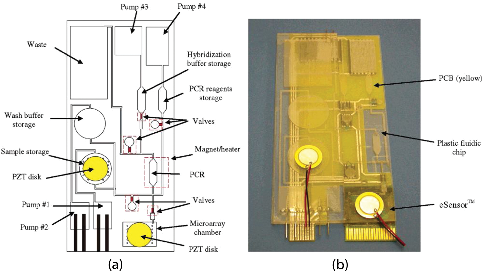
(
Capillary manipulation
Most of the mechanical and nonmechanical fluid manipulation approaches require external power sources (e.g., compressed air, rotational motion, and electrical potential or current) to drive the fluids. However, the requirement of the external instrumentation of on-chip fluid manipulation is one of the challenges in the development of the point-of-care devices. Capillary manipulation provides a power-free solution for the fluid manipulation on-chip.
Paper-based microfluidics, or microfluidic paper-based analytical devices, has attracted attention recently for a new class of diagnostic devices. 81 The basic functions required by the diagnostic device including sample manipulation and detection can be performed on a sheet of paper. Paper is inexpensive, thin, lightweight, available in a wide range of thicknesses, and disposable; thus, paper-based microfluidics is suitable for the development of diagnostic assays in developing countries and harsh environments. Moreover, paper can transport aqueous fluids by wicking due to capillary force, and passive pumping is realized. Also, well-defined pore sizes in paper can be manufactured that can separate suspended solids from samples before the bioassays. Paper is biocompatible with various biological samples and can be modified by a wide range of functional groups that can be covalently bound to proteins, DNA, or small molecules. 82 –84 Paper-based microfluidic devices can be fabricated by patterning sheets of paper into hydrophilic channels using different methods such as photolithography, 84 , 85 wax printing, 86 , 87 and plasma treatment. 88 The aqueous fluids can be transported along the channels by wicking in the hydrophilic fibers of paper. A paper-based microfluidic device has been demonstrated for the determination of glucose and protein simultaneously. 89 A single piece of paper was patterned by photolithography to realize the paper-based assays. The concentration of the glucose and protein was determined by matching the color intensity of the reacted sites and the printed color on label stock visually. However, differentiation of color intensity by the naked eye was complicated because of the influence of color perception from people and lighting. To pursue quantitative analysis based on paper-based microfluidic devices, a camera phone was used for image capturing of the reacted sites, and the color intensity was analyzed by the computer software. 90 The idea behind this was to provide a real-time and off-site diagnosis operated by unskilled personnel in the developing countries. More accurate results compared with visual inspection can be achieved. However, the color intensity of the digital image captured by the camera phone was affected by lighting conditions and the calibration curve of color from different camera phones. Therefore, electrochemical detection on paper-based microfluidic devices has been proposed for highly sensitive and selective analysis. 84 , 91 The determination of glucose, lactate, and uric acid in biological samples has been demonstrated. 84 The detection electrodes were screen printed by the carbon and Ag/AgCl ink onto a piece of paper. These examples are the recent developments of paper-based microfluidics. Paper-based microfluidic devices have a great potential to be a good alternative to traditional microfluidic systems for point-of-care diagnostic applications.
Biological Detection
Biological activities can be recognized by different signal transduction pathways performing on the microfluidic systems. A number of studies have reported various measurement techniques to achieve sensitive and specific biological detection. In this section, we roughly categorized the measurement techniques into labeled and label-free detection methods. For the labeled detection methods, we will focus on the discussion on fluorescent, electrochemical, and nanogold-based measurements. For the label-free detection methods, we will focus on the discussion on impedance, electrochemical, surface plasmon resonance (SPR)–based, and mass-sensitive measurements.
Labeled Detection Methods
Fluorescent measurement
Fluorescent measurement of biological activities is commonly used in conventional biological laboratory and microfluidic systems because of its high sensitivity and ease of conjugating fluorescent and enzyme-based labels to most biomolecules. Some of the commercially available fluorophore tages are Cy series (e.g., Cy5 and Cy3), fluorescein isothiocyanate (e.g., FITC), tetramethylrhodamine isothiocyanate, and Alexa series. Also, enzyme-labeled biomolecules manifest their signal through fluorescent product generated from enzyme reaction. Alkaline phosphatase (AP) and HRP are two of the most common enzyme labels. For example, laser-induced fluorescence (LIF) detection with a confocal microscope on microfluidic chips has been reported to measure Cy5 dye and Cy5-labeled immunoreagents. 92 The confocal epifluorescence setup is shown in Figure 7 . Samples and immunoreagents were mixed within 30 to 60 µm wide flow channels, allowed to react homogeneously, and then electrophoretically separated to determine the sample concentration. A 635 nm diode laser was used because it provides a convenient, compact, and rugged source for instruments that are possible for on-field applications. A detection limit of approximately 9 pM in a 20 µm deep channel was achieved with this diode laser. Moreover, a microfluidic system for flow cytometry and fluorescence-activated cell sorting has been reported based on gravity and electric force driving of cells. 93 A confocal LIF detector was employed for measurement of TO-PRO-3–stained DNA inside cells. Direct observing and manipulating of cells flowing in the flow cytometry channel was performed. However, the complicated confocal LIF optical systems may limit their wider applications. Many nonconfocal LIF detection systems with other optical arrangements (e.g., orthogonal 94 ) were also developed for coupling to microfluidic systems. A more integrated approach is to incorporate the optical lens into the microfluidic systems. A PDMS chip integrated with 2D optical lenses was fabricated to improve the performance of fluorescent spectroscopy. 95 Based on the design of the lens curvature radius, the properties of the light beam coming out from the fiber can be modified. The intensity of the fluorescence can be increased and a stronger response from the fluorescent dye can be induced, leading to about three times higher sensitivity than the setup with small curvature lenses. Alternatively, a miniaturized mosaic immunoassay was proposed based on patterning lines of antigens onto a surface by means of a microfluidic network. 96 Samples were delivered by the channels of a second microfluidic network across the pattern of immobilized antigens on the substrate surface. A mosaic of binding events can readily be measured in one screening using fluorescence. In summary, fluorescence measurement is the most widely used method because of its advantages of high sensitivity and a wealth of available fluorophores and labeling chemistries. However, fluorescent dyes are relatively costly, have limited shelf life, and are influenced by chemical factors (e.g., pH).
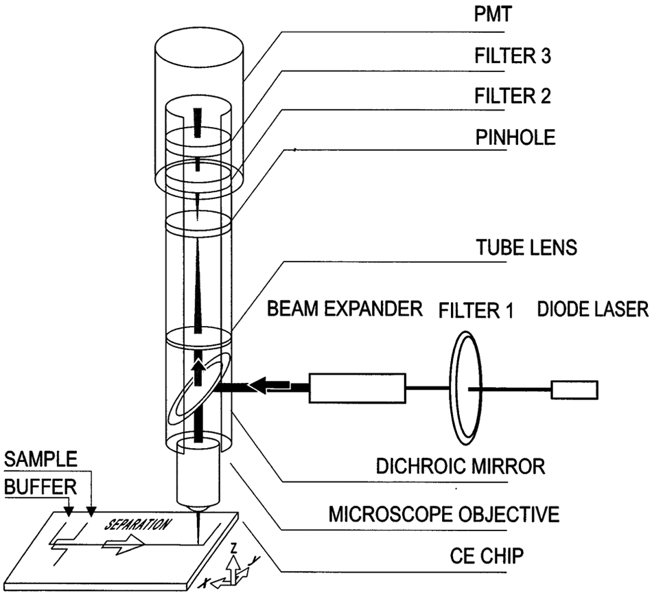
Confocal epifluorescence setup for Cy-5 detection on chip. Copyright 2000. Reprinted from ref. 92 with permission from Elsevier.
Electrochemical measurement
Electrochemical measurement uses electrodes to measure the current generated from the electrochemical activities while the potential of the electrodes are held at a specific value. The advantages of electrochemical measurement include easy miniaturization and low power requirements. That makes it suitable for on-chip biological detection integrated into microfluidic systems. Electrochemical measurement can be classified as both labeled and label-free detection. Here, labeled detection is first described. A typical example of an integrated microfluidic system was reported to determine the immunoassay results using electrochemical measurement. 97,98 An interdigitated array (IDA) electrode was used for the measurement electrode. AP and p-aminophenyl phosphate (PAPP) were used as an enzyme label for immunoassay and the enzyme substrate, respectively. The enzyme converted PAPP to the product (i.e., p-aminophenol [PAP]), which is oxidizable. PAP underwent a two-electron oxidation to p-quinoneimine (4-QI) when applying an oxidizing potential to the anode (i.e., one of the IDA electrodes). 4-QI converted back to PAP when diffused to an adjacent cathode held at reducing potential, undergoing a two-electron reduction. The immunoassay results can be measured by the current, and the measurement of the concentration of mouse IgG from 50 ng/mL to 100 ng/mL was demonstrated. 98
Nanogold-based measurement
Conventionally, nanogold (i.e., colloidal gold), is used in immunohistochemistry. Nanogold-labeled antibodies are bound to the surface antigens in biological tissues. The silver enhancement process can physically enlarge the particle size of nanogold. The silver-enhanced nanogold particles show dark brown under optical or electron microscope. 99,100 The antibodies can be indicated by nanogold, and the distribution over the tissue can be identified. For the diagnostic applications using nanogold, colorimetric and electrical DNA array detection have been demonstrated to determine the specific DNA sequence. 101 –105 DNA probes were first immobilized on the sites of the DNA array, followed by the application of the target DNA. Silver-enhanced nanogold particles were used to indicate the DNA hybridization on the sites. The hybridization results can be detected by either using a conventional flatbed scanner or measuring the conductivity changes across the microelectrodes. Figure 8 shows the protocol of the electrical detection of DNA hybridization using silver-enhanced nanogold particles. Similar methodology has also been applied for the immunoassay. Colorimetric and electrical immunoassays using antibody-nanogold conjugates and gold enhancement have been demonstrated. 106 –108 Moreover, nanogold particles have been applied to the electrochemical detection of protein binding activity. 109,110 The immunoassay of goat IgG was performed using donkey anti-goat IgG labeled by nanogold. The result of the immunoassay was determined by anodic stripping voltammetry through the oxidative gold metal dissolution in the acidic solution. The dynamic range for the assay was between 0.5 and 100 ng/mL. The most important advantage of using nanogold is that the nanogold particles are stable and can be stored for a very long period of time compared with the fluorescent dyes.
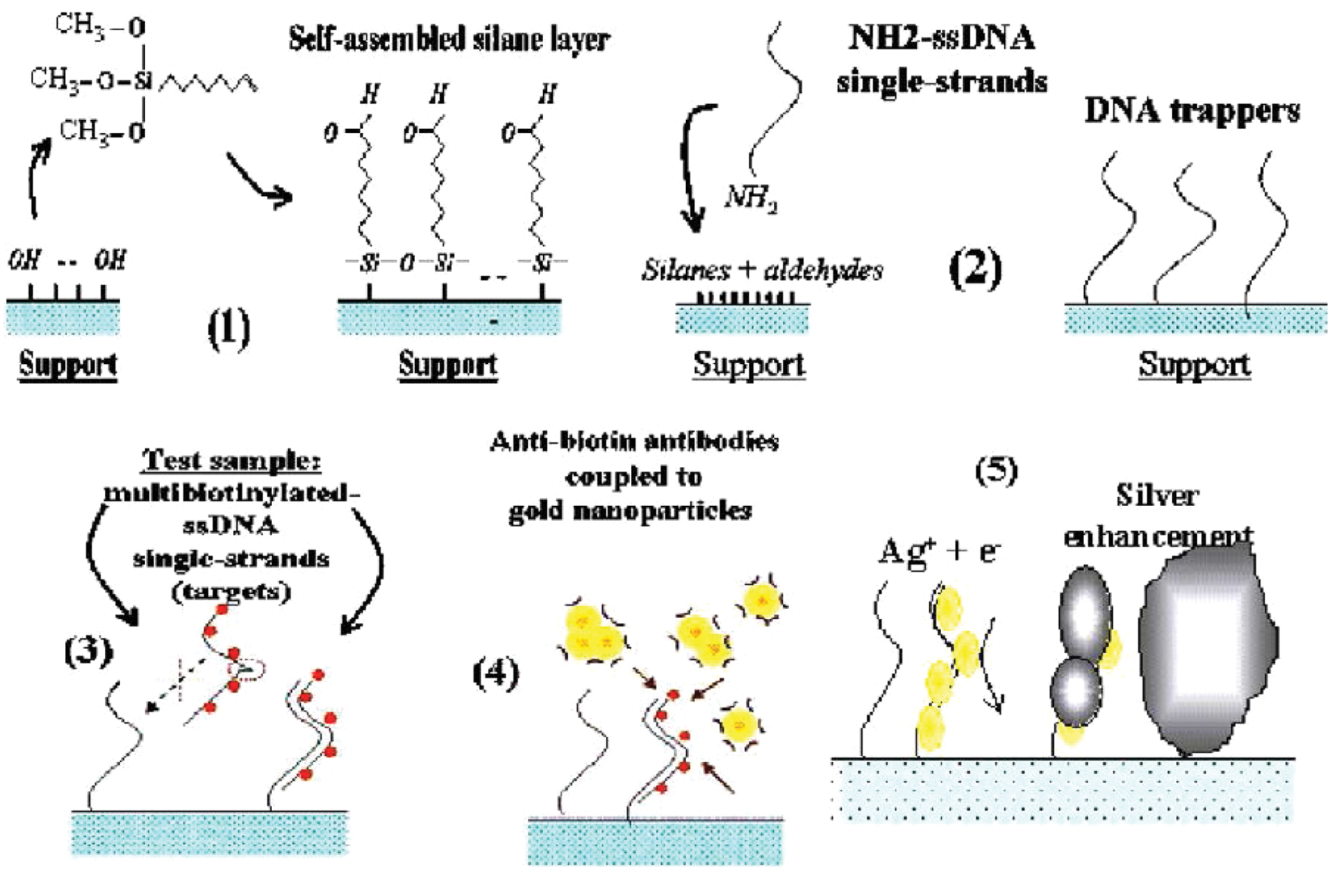
(1) Functionalization of the support, (2) DNA probes spotting and covalent bonding between the amino probes and the aldehydes, (3) hybridization of a second strand biotinylated DNA, (4) gold labeling with anti-biotin/gold particles, (5) amplification by silver precipitation. Copyright 2004. Reprinted from ref. 104 with permission from Elsevier.
Label-free Detection Methods
Impedance measurement
The working principle of impedance measurement is based on the microelectrodes fabricated on a substrate surface (e.g., glass or silicon) working as an electrical transducer. When analytes are presence on the microelectrodes, they cause the change of impedance measured across the microelectrodes. Because cells are sizable, cell analysis is suitable for using impedance measurement. A number of studies have been reported using impedance measurement to quantify the cell concentration. The detection of E. coli O157:H7 using an interdigitated microelectrode array was developed for monitoring the food samples. 111 –113 The detection of Salmonella has also been demonstrated by making use of the impedance properties of bacterial suspensions in deionized water. 114,115 The bacterial concentration could generate different electrical impedance spectral responses. Moreover, a microfluidic chip was developed for the detection and identification of bacteria. 116 Bacteria suspended in solution were recognized by the antibodies immobilized on the functionalized glass surface on the microfluidic chamber. The impedance within the chamber increased according to the bacterial concentration. Furthermore, adherent cells were cultured directly on a pair of interdigitated electrodes, and the impedance can represent the adhesive behavior of the cells, such as number, growth, and morphological behavior. 117 Measurement of 3D cell culture construct has also been reported to quantify the cell number. 34 Impedance measurement was also used to investigate the cell function during bacterial infection. Several virulence factors of the meningitis-causing pathogen were studied. 118 Alternatively, with the rapid development of nano fabrication techniques, nanoscale electrodes have been fabricated and used to determine the protein binding using the impedance measurement. 119 The impedance increased directly related to the protein concentration after protein binding at the electrode surface.
Electrochemical measurement
Electrochemical measurement can be also used in label-free detection. A typical example is to detect glucose concentration. 120 –122 An amperometric-type nonenzymatic glucose-sensing microfluidic system was reported, and the electrochemical measurement was based on a three-electrode setup. 120 The system was composed of a microfluidic channel network and a miniaturized electrochemical cell for nonenzymatic glucose sensing. Sample and buffer solution in the microfluidic system was transported by electro-osmotic flow. Three electrodes, which were nanoporous platinum as working and counter electrode and silver/silver chloride as reference electrode, were integrated into the electrochemical cell. Glucose concentration in buffer was determined by the direct oxidation of glucose. The sensitivity was 1.65 µA cm–2 mM–1 in the glucose concentration ranging from 1 to 10 mM.
Surface plasmon resonance
SPR provides a sensitive tool for real-time sensing of biomolecules without the use of label and becomes a key research tool for pharmaceutical development, food quality control, environmental monitoring, and clinical analyses. A number of commercial SPR imaging instruments have been launched, for example, Biacore Flexchip (GE Healthcare), SPRimager (GWC Technologies), and IBIS iSPR (IBIS Technologies). These benchtop instruments allow the detection of molecules of very different molecular weight simultaneously immobilized on the same sensor chip. Recently, a microfluidic system integrated with localized SPR was developed and is shown in Figure 9 . 123 The detection of protein binding was demonstrated using this integrated system. The immobilization of biotin on the sensor surface and the subsequent affinity binding of anti-biotin were quantitatively detected by the integrated system. The detection limit of 270 ng/mL of anti-biotin was achieved. Moreover, a microfluidic chip integrated with a 2D SPR phase imaging system was developed to detect the interaction of anti-rabbit IgG and IgG. 124 The microfluidic chip was composed of a fluidic transportation system for sample/reagent delivery and a temperature control system for reducing the thermal noise during SPR sensing. This automated system successfully detected IgG with a detection limit of 10 ng/mL. These demonstrations showed the feasibility of the integration of SPR-based biosensing with microfluidic technologies. It is expected that with the development of microfluidic technology, miniaturized and low-cost SPR instruments will be developed and provide a promising tool for practical diagnostic applications.
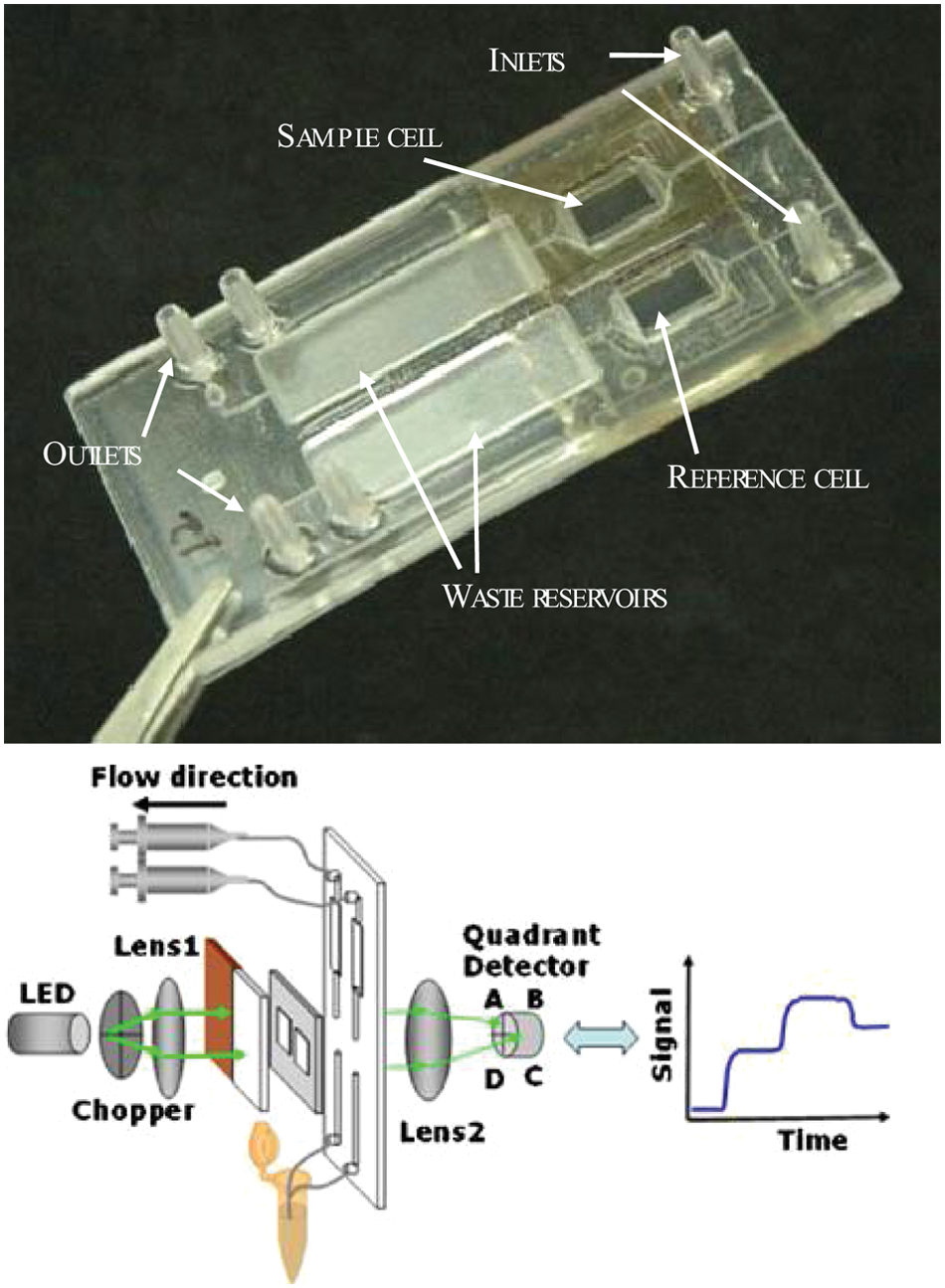
Photo and schematic of the microfluidic system integrated with localized surface plasmon resonance. Copyright 2004. Reprinted from ref. 123 with permission from Elsevier.
Mass-sensitive measurement
Surface acoustic wave (SAW) devices use horizontally polarized surface shear waves to monitor the absorbed mass (e.g., protein) on the surface directly. The mass adsorption can change the surface wave velocity to determine the biomolecular binding interactions in the sensing layer. An example of an SAW device for immunoassay is described for label-free detection in water. 125 The SAW device was coated with a thin parylene layer for creating a chemically homogeneous surface for biological sensing. Urease binding at the anti-urease–coated SAW device could be monitored in real time. Another example of the use of an SAW device is to detect cocaine molecules in gas. 126 Anti-benzoylecgonine antibodies were attached to the electrodes on the SAW device surface via a protein-A crosslinker. The cocaine vapor of ~1 ng at a flow rate of 180 cm3 min was used in the experiments. Real-time molecular recognition of cocaine molecules was achieved using the SAW device. Moreover, another mass-sensitive measurement for biomolecules can be achieved by quartz crystal microbalance (QCM). The principle is to measure the resonance frequency shift attributed to mass changes on a surface. The binding of immunoliposomes to antigen was demonstrated by the QCM device. 127 Antigen could be measured in the concentration range from 10−5 to 10−8 M. In summary, SAW and QCM devices provide a sensitive and cost-effective method for label-free detection of molecules in a water and gas environment.
Material and Manufacturing Techniques
In the beginning, development of microfluidic systems was based on the microelectronic manufacturing infrastructure. Silicon was used as a material for fabricating microfluidic systems for various applications. 128 –130 The well-established silicon fabrication process and extensive studies of silicon property make the growth of microfluidic systems fast. The fabrication process for silicon-based microfluidic systems involves substrate cleaning, photolithography, metal deposition, and wet/dry etching. 131,132 Normally, wet etching is a fabrication method for the microstructures with a low aspect ratio, whereas dry etching can fabricate relatively high-aspect-ratio microstructures. However, silicon substrate is relatively expensive and is not optically transparent. It may limit the biological applications using optical detection. Therefore, glass and polymer materials were introduced to fabricate the microfluidic systems. 133,134 Compared with silicon substrate, glass and polymer materials are less expensive and optically transparent. Polymer materials include PMMA, polystyrene, polycarbonate, and PDMS. Among these polymer materials, PDMS is one of the most commonly used materials for microfluidic systems in recent years because it is flexible, optically transparent, and biocompatible. The PDMS structure can be formed easily by molding. Microfluidic systems can also be formed by using two different types of materials, such as silicon/glass and glass/polymer. Moreover, to make functional microfluidic systems, adhesion between substrates is a problem of great practical concern. Silicon-silicon bonding (e.g., anodic bonding) may not be applicable for the polymer materials. Thermal compression, ultrasonic, or gluing by application of either epoxy or methanol may induce global and localized geometric deformation of the substrates or leave an interfacial layer with significant thickness variation. Therefore, special bonding processes for glass/polymer or polymer/polymer have been developed for fabricating the microfluidic systems. 135 –138
Conclusions
Excellent integration of microfluidic systems and diagnostic applications has been successfully demonstrated in the laboratory; however, microfluidic-based point-of-care devices have not made a great impact on the commercial market. One of the possible reasons is that these newly developed diagnostic protocols need to take time to compete with the existing methods that have been perfected over decades. However, there are still some challenges in the development of microfluidic technology, such as purification of raw samples, concentration of the analytes, dead volume in the fluid manipulation, and elimination of using external bulky and expensive detection systems. Although some uses of the fully integrated systems are being developed, fully sample-to-answer analysis systems are required for the commercial point-of-care devices, especially for untrained personnel. Moreover, the cost of the existing microfluidic systems is still relatively high because they are not massively produced. PDMS material has been widely used for microfluidic research because it is low cost and replicable. However, the fabrication process involves several hours in the curing process, that causes difficulties in mass production. Materials with similar properties and capabilities for mass production are required for practical applications. Besides the technology development, launching of in vitro diagnostic devices requires an approval in most of the countries. The related registration process is tedious, which makes it difficult to be considered and completed by research groups. These factors greatly hinder the development of commercial microfluidic-based diagnostic systems. However, recent research has opened the door for commercial applications. A commercial diagnostic product of the i-STAT system from Abbott Laboratories has been launched recently. This is an excellent example of the possibility of using microfluidic technology for real-time, portable, sensitive diagnostics.
Footnotes
Acknowledgements
The author would like to thank the National Science Council, Taiwan, for financial support (project NSC100-2221-E-182-022).
Declaration of Conflicting Interests
The author declared no potential conflicts of interest with respect to the research, authorship, and/or publication of this article.
Funding
The author received no financial support for the research, authorship, and/or publication of this article.