
Abstract
Community-acquired methicillin-resistant Staphylococcus aureus (CA-MRSA) is an important cause of invasive infections, including sepsis associated with myocardial dysfunction. Caspases 1 and 11, involved in activation of the inflammasome, have been shown to be critical in response to sepsis as well as myocardial dysfunction of numerous etiologies. We examined the survival, myocardial function, and production of inflammatory mediators in mice lacking caspases 1 and 11. Cas 1/11 KO mice demonstrated no significant difference in mortality or in cardiac shortening fraction relative to control mice. Cas 1/11 KO mice had significantly reduced upregulation of expression of tumor necrosis factor (TNF)-α and interleukin (IL)-6 in the heart relative to control mice after CA-MRSA infection, as well as reduced serum production of IL-1β, TNF-α, and IL-6, with no difference in IL-10 production. Other inflammatory mediators beyond IL-1β, TNF-α, and IL-6 may be involved in myocardial dysfunction in CA-MRSA sepsis.
Introduction
Staphylococcus aureus is an important cause of bacterial infections including invasive infections such as pneumonia, osteomyelitis, and sepsis. Methicillin-resistant S. aureus (MRSA) is a significant clinical problem, notably MRSA infections in previously healthy patients, referred to as community-associated MRSA (CA-MRSA). 1
Myocardial dysfunction is a common problem in sepsis and appears to result from direct effects of microbes and circulating inflammatory mediators on the cells of the myocardium, as well as secondary insults from vascular dysfunction. 2 The molecular mechanisms responsible for causing cellular injury and myocardial dysfunction have not been fully elucidated but likely include processes such as alterations in calcium channels, mitochondrial injury, oxidative stress, and protease activation. 2 The specific pathophysiology leading to cardiac dysfunction in CA-MRSA sepsis is not well defined despite its clinical importance, as severe hemodynamic dysfunction and cardiovascular collapse have been reported in CA-MRSA sepsis case series.3,4
Inflammatory caspases such as caspases 1 and 11 play a key role in activation of the inflammatory response, particularly by facilitating the production of mature interleukin (IL)-1β. 5 Mice with a deletion in the caspase 1 and caspase 11 genes have been shown to have reduced mortality in endotoxic shock and sepsis models, as well as improved cardiac function in heart failure and ischemia-reperfusion models.6–8 In this article we make use of caspase 1/11 knockout mice in a murine model of CA-MRSA sepsis that allows evaluation of myocardial depression using echocardiographic measures of shortening fractions, as well as gene expression changes in heart tissue during sepsis.
Methods
Mice
Control strain NOD/ShiLtJ mice were purchased from Jackson Laboratories (Bar Harbor, Maine). Caspase 1/11 knockout mice (NOD.129S2(B6)-Casp1tm1Sesh Casp4del/LtJ) came from a breeding colony maintained in the University of Texas Southwestern Medical Center animal facility, descended from mice originally purchased from Jackson Laboratories. Male mice of 6–10 weeks of age were used for experiments. All experiments were done according to protocol no. 2009-0344 approved by the Institutional Animal Care and Use Committee at the University of Texas Southwestern Medical Center. All animals were allowed free access to commercial mouse chow and tap water.
Infection model
CA-MRSA USA300 strain TCH1516 was obtained from ATCC (Manassas, VA) as a freeze-dried sample. Strain TCH1516, a sequenced USA300 CA-MRSA strain, was isolated from an adolescent patient with fatal sepsis. 9 Overnight bacterial cultures were inoculated from isolated colonies streaked on tryptic soy agar (TSA) plates (Becton Dickinson, Franklin Lakes, NJ), then diluted 1:100 in tryptic soy broth (TSB; Becton Dickinson)/0.25% glucose and grown at 37 C with shaking at 100 r/min. OD600 measurement was performed on a Spectronics (Westbury, NY) Genesys 5 spectrophotometer. At an OD600 of 0.75 (mid-logarithmic phase), bacterial cultures were centrifuged at 3000 r/min in a Beckman (Indianapolis, IN) swinging bucket rotor centrifuge and washed once in sterile phosphate-buffered saline (PBS) and then diluted in sterile PBS for injection of 100 µL total through the tail vein. An infectious inoculum of 5 × 10⁷ colony-forming unit (CFU) was used for infections, based on results from pilot experiments that showed this dose caused substantial mortality in infected control mice more than 48 h post-infection, but allowed the majority to survive to 48 h after infection (data not shown). For survival curves, mice were assessed every 3–4 h for first 24 h and then every 6 h for 24–72 h. Animals were euthanized if they were found to be moribund. At indicated time points, blood was collected from the retro-orbital vein, and the animals were sacrificed with CO2 inhalation. The hearts were immediately collected and flash frozen in liquid N2 and stored at −80°C until use.
Evaluation of myocardial function
Mice were prepared for echocardiography by removal of hair on chest using Nair (Church & Dwight Co., Inc., Ewing, NJ) under sedation with isoflurane. At indicated time points, animals were gently restrained without sedation and echocardiography was performed using an Acuson Sequoia 512 (Siemens, Malvern, NJ) echocardiogram machine using a 15L8 8–14 mHz probe. After obtaining short axis views, M-mode images directed across the left ventricle were taken (three per mouse). DICOM Viewer software was used to measure left ventricular end-diastolic (EDD) and end-systolic dimensions (ESD) for the first measurable M-mode image from each mouse. Three separate measurements of EDD and ESD were obtained from the first measurable M-mode image from each mouse and averaged. Shortening fraction (%) was calculated using the equation 100 × ((EDD − ESD)/EDD).
RNA isolation and quantitative polymerase chain reaction
RNA was isolated from mouse hearts using the Aurum™ Total RNA Fatty and Fibrous Tissue Kit (Bio-Rad, Hercules, CA). Frozen hearts were placed without thawing into glass dounces containing PureZOL (Bio-Rad) and then homogenized using glass pestles. RNA extraction was then conducted according to manufacturer’s instructions.
For quantitative polymerase chain reaction (PCR), complementary DNA (cDNA) was generated using the iScript™ cDNA Synthesis Kit (Bio-Rad) according to manufacturer’s instructions. Quantitative PCR was performed using the IQ SybrGreen Supermix (Bio-Rad). The primers used are described in Table 1. Primers were designed using the PrimerBlast program 10 and evaluated for secondary structure using the Mfold web server (http://mfold.rna.albany.edu/?q=mfold), with the exception of HPRT1, the primer for which was obtained from RTPrimerDB #45 (http://medgen.ugent.be/rtprimerdb/). Primer efficiencies were all between 90% and 105%. Reactions included 1 µg total cDNA and were run in duplicate on the MyQ Real-Time PCR Detection System (Bio-Rad). Results were analyzed using qbasePLUS software (Biogazelle, Zwijnaarde) with reference genes of hypoxanthine guanine phosphoribosyltransferase (HPRT) and ribosomal protein L32 (Rpl32).
List of primers used for qPCR.
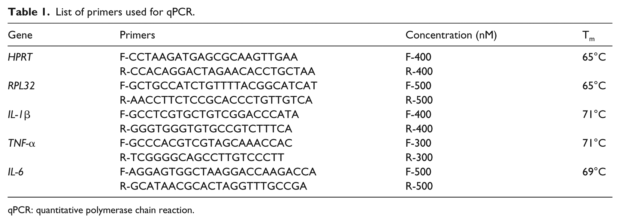
qPCR: quantitative polymerase chain reaction.
Serum cytokine quantification
Blood taken from mice was allowed to clot at room temperature for 30 min and then centrifuged in a tabletop mini-centrifuge at 13,200 r/min for 10 min at 4°C and serum was removed and stored at −80°C. Serum cytokines were measured using the Bio-Plex Pro (Bio-Rad) system. The Bio-Rad TH17 six-plex mouse panel was used for analysis.
Statistical analysis
The SAS system (version 9.4; SAS Institute, Cary, NC) was used in all statistical analyses. For survival analysis, the Cox proportional hazards model was used to examine the association between survival and genotypes or doses. In exploratory analysis, the interaction between genotypes and doses did not show statistical significance, so it was not included in the final model. Hazard ratios and its 95% confidence interval (CI) were reported. A P value of less than 0.05 was considered statistically significant. For echo data, multiple linear regression was applied with genotype, time, and their interaction as covariates. Time is treated as a categorical variable. Log-transformation with base 10 is applied to the outcome variables as the plotted outcome variables are skewed. Different time points were compared pairwise, and multiple comparisons were adjusted using Bonferroni’s method. Serum cytokine data and qPCR data are analyzed similarly with one exception. Due to the skewed distribution of data, log-transformation with base 10 is applied to the outcome variables of serum cytokine and qPCR data. The estimated mean at each time point and its 95% CIs were plotted by genotype for each outcome variable.
Results
We hypothesized that mice lacking caspase 1 and caspase 11 would have improved mortality and cardiac function in the setting of CA-MRSA sepsis due to decreased production of inflammatory mediators associated with impaired myocardial function. Therefore, we used caspase 1/11 knockout mice (Cas 1/11 KO) and the background control (NOD/ShiLtJ) mice to determine the effect of knockout of caspase 1 on mortality, cardiac function, and cardiac and systemic inflammatory responses. For a clinically relevant infection model, we used TCH1516, a USA300 strain of CA-MRSA isolated from an adolescent patient with a fatal case of sepsis. 9
We used our model to determine whether there was a significant difference in mortality between Cas 1/11 KO mice and the background control strain NOD/ShiLtJ. There was no significant difference between survival of Cas 1/11 KO and control mice (data not shown).
In order to examine the cardiac function, we evaluated infected mice by echocardiography to measure shortening fraction as an indicator of cardiac contractility. Figure 1 shows the shortening fraction for NOD/ShiLtJ and Cas 1/11 KO mice over a time course of 48 h. There was not a statistically significant difference in shortening fractions between NOD/ShiLtJ and Cas 1/11 KO at any time point after applying Bonferroni correction (significant P ⩽ 0.00833). This finding is consistent with the lack of difference in mortality.

CA-MRSA sepsis is associated with decreased cardiac contractility with later improvement over time. NOD/ShiLtJ and Cas 1/11 KO mice were infected with 5 × 10⁷ CFU of TCH1516 CA-MRSA and underwent echocardiography at indicated time points over 48 h. The graph shows the shortening fraction (%) at indicated time points. Data from Cas 1/11 KO mice are shown with square symbols and dashed red lines; NOD/ShiLtJ mice have data represented by solid blue lines and circular symbols. Statistical analysis by linear regression and differences between least squares means did not show a significant difference between NOD/ShiLtJ and Cas 1/11 KO mice after applying Bonferroni correction (significant P ⩽ 0.00833). Number of animals was n = 3–5 per time point for NOD/ShiLtJ mice and n = 8–14 for Cas 1/11 KO mice.
Inflammatory caspases such as caspase 1 and caspase 11 play an important role in triggering the inflammatory response to infection or other insult. We examined the expression of the key inflammatory mediators IL-1β, IL-6, and TNF-α in myocardial tissue over a time course using qPCR (Figure 2). Figure 2(a) shows that there was no significant difference in expression of IL-1β between control and Cas 1/11 KO mice at any time point after applying Bonferroni’s correction for multiple comparisons. Applying Bonferroni’s correction for multiple comparisons, a significant P value at each time point would be ⩽0.00833. There was a trend toward significant difference for IL-1β gene expression between control NOD/ShiLtJ mice and Cas 1/11 KO mice at 2 h (P = 0.0468), where the Cas 1/11 KO expression was higher, and at 12 h (P = 0.0316), where the control NOD/ShiLtJ were lower. IL-6 gene expression (Figure 2(b)) was significantly higher in control NOD/ShiLtJ mice compared to Cas 1/11 KO mice at 12 h (P = 0.0052). TNF-α gene expression (Figure 2(c)) was also significantly higher in control NOD/ShiLtJ mice at 12 h (P = 0.0054).

Divergent responses between control NOD/ShiLtJ and Cas 1/11 KO mice in upregulation of pro-inflammatory mediators in myocardium. NOD/ShiLtJ (blue lines and circles) and Cas 1/11 KO (red lines and squares) mice were infected with 5 × 10⁷ CFU of TCH1516 CA-MRSA and were sacrificed at specified time points. Hearts were collected at indicated time points for RNA isolation and quantitative PCR analysis for expression of (a) IL-1β, (b) IL-6, and (c) TNF-α. Gene expression was expressed as fold upregulation or downregulation relative to reference gene expression and was logarithmically transformed prior to analysis, and the estimated mean at each time point and its 95% confidence intervals were plotted. Statistical analysis by linear regression and differences between least squares means was performed. Applying Bonferroni’s correction for multiple comparisons, a significant P value at each time point would be ⩽0.00833. (b) IL-6 gene expression showed a statistically significant difference between control NOD/ShiLtJ mice and Cas 1/11 KO mice at 12 h (#P = 0.0052). There was a statistically significant difference in TNF-α gene expression between control NOD/ShiLtJ mice and Cas 1/11 KO mice at 12 h (*P = 0.0054) and a trend toward a statistically significant difference at 0 h (P = 0.0095). There was a trend toward significant difference for (a) IL-1β gene expression between control NOD/ShiLtJ mice and Cas 1/11 KO mice at 2 h (P = 0.0468) and at 12 h (P = 0.0316). There were 3–5 NOD/ShiLtJ mice per time point and 3–10 Cas 1/11 KO mice per time point.
Circulating serum inflammatory mediators play a significant role in the pathophysiology of sepsis and influence the inflammatory response in the myocardium.2,11 Therefore, we also examined the dynamics of the serum pro-inflammatory cytokines IL-1β, TNF-α, and IL-6, as well as the anti-inflammatory cytokine IL-10 (Figure 3). There was a significantly increased production of IL-1β by the control mice at 8 and 12 h (P < 0.001 for both) relative to Cas 1/11 KO mice (Figure 3(a)). This is expected, as caspase 1 is essential for efficient production of mature IL-1β. 7 However, there was also a marked difference between NOD/ShiLtJ and Cas 1/11 KO mice in the production of TNF-α and IL-6, neither of which require caspase 1 for activation. The most marked cytokine increase was in IL-6 in NOD/ShiLtJ mice, which was significantly relative to levels in Cas 1/11 KO mice at 8 h (P < 0.0001), 12 h (P < 0.0001), and 24 h (P = 0.004; Figure 3(b)). Even at baseline, the NOD/ShiLtJ mice had significantly increased levels of IL-6 relative to Cas 1/11 KO mice (P = 0.0053). IL-6 remained significantly elevated over the t = 0 time point at 48 h in NOD/ShiLtJ (P < 0.001) mice.

Serum pro-inflammatory cytokines are decreased in Cas 1/11 KO mice in response to CA-MRSA sepsis. Cas 1/11 KO and control NOD/ShiLtJ mice were infected with 5 × 10⁷ CFU of TCH1516 CA-MRSA and were sacrificed at indicated time points after blood was collected for serum, which was analyzed using the Bioplex system for quantitation of (a) IL-1β, (b) IL-6, (c) TNF-α, and (d) IL-10. The data were logarithmically transformed due to skewed distribution, and the estimated mean at each time point and its 95% confidence intervals were plotted. Data from Cas 1/11 KO mice are shown with square symbols and red lines; NOD/ShiLtJ mice have data represented by blue lines and circular symbols. Analysis by linear regression and differences between least squares means was performed. Applying Bonferroni’s correction for multiple comparisons, a significant P value at each time point would be P ⩽ 0.00833. There were statistically significant differences between Cas 1/11 KO and control NOD/ShiLtJ mice in production of IL-1β at 8 h (*P < 0.0001) and 12 h (**P < 0.0001). There was a significant difference between the two mice strains in production of IL-6 at 0 h (#P = 0.0053), 8 h (##P < 0.0001), 12 h (###P < 0.0001), and 24 h (####P = 0.004), and the difference approached significance at 2 h (P = 0.0363). For TNF-α, there was a significant difference between the mouse strains at 12 h (&P < 0.0001) and 24 h (&&P = 0.0041), and the difference approached significance at 8 h (P = 0.0114). There were 3–5 NOD/ShiLtJ mice per time point and 5–14 Cas 1/11 KO mice per time point.
Figure 3(c) shows that TNF-α was significantly increased in control NOD/ShiLtJ relative to Cas 1/11 KO mice at 12 h (P < 0.0001) and 24 h (P = 0.0041), and the difference approached significance at 8 h (P = 0.0114). The anti-inflammatory cytokine IL-10 (Figure 3(d)) did not show any significant differences between NOD/ShiLtJ and Cas 1/11 KO mice; both genotypes showed a peak at 12 h with a decrease afterwards. However, both genotypes of mice continued to have IL-10 levels significantly elevated above t = 0 levels (P = 0.0024 for Cas 1/11 KO mice and P = 0.0014 for NOD/ShiLtJ).
Discussion
Caspase 1/11 deletion and/or inhibition has proved beneficial in heart function in endotoxic/Gram-negative sepsis models as well as in other cardiomyopathy settings,6–8 so we hypothesized that caspase 1/11 knockout animals would have improved survival and myocardial function in the setting of CA-MRSA sepsis. Contrary to our hypothesis, though caspase 1/11 deficient mice did have reduction in production of IL-6 and TNF-α in the heart and IL-1β, TNF-α, and IL-6 in serum (Figures 2 and 3), they did not have a survival advantage over control mice and did not have significantly different shortening fraction (Figure 1).
There are several potential explanations for this lack of effect on survival and cardiac function in Cas 1/11 KO mice despite the reduced production of inflammatory mediators. One is that other inflammatory mediators are involved, which may include reactive oxygen/nitrogen species, matrix metalloproteinases, and heat shock proteins, as reported in other models.2,11,12 Second, as production of inflammatory mediators is essential for clearance of infection, too much of inflammatory mediators might cause excessive and detrimental inflammatory response, but too little would not successfully eliminate an infection. Another possibility is that genes which are not generally associated with inflammation are significantly affected in the heart in the setting of sepsis with deleterious effects on myocardial function. For example, Dos Santos and colleagues reported changes in septic mouse hearts including downregulation of metabolism-related genes and myosin heavy chain isotype switching in an iNOS-dependent manner. 12 Further studies should examine the role of other inflammatory mediators, the balance of pro- and anti-inflammatory mediators, and the role cardiac genes involved in critical cellular activities such as metabolism, structural protein synthesis, and intracellular signaling in this model.
Footnotes
Acknowledgements
The authors would like to acknowledge the technical assistance provided by Mohamed Elsheikh, who was supported by the Undergraduate Research Opportunities Program at the University of Minnesota. In addition, we would like to acknowledge Dr Elizabeth Braunlin for reading and commenting on this manuscript.
Authors’ note
Primary data and samples may be accessed on request to corresponding author Dr Janet R. Hume (
Declaration of conflicting interests
The author(s) declared no potential conflicts of interest with respect to the research, authorship, and/or publication of this article.
Funding
The author(s) disclosed receipt of the following financial support for the research, authorship, and/or publication of this article: This work was financially supported by the National Institutes of Health T32 Postgraduate Training Grant 5T32GM8593-15 (to Dr J. Hume) and National Institutes of Health Grant 5P50GM21681-44 (to Dr D. Carlson), as well as by start-up funds provided to Dr Hume via the Department of Pediatrics at the University of Minnesota Medical School.