
Abstract
Objective
This retrospective case series describes a hybrid fixation technique and determines the clinical outcomes, knee function, and activity level of patients at short-term follow-up.
Design
Seventeen patients (18 knees) with unstable osteochondritis dissecans (OCD) lesions involving the knee were treated with a hybrid fixation technique in which the salvageable fragment was fixed and osteochondral autograft transplantation system (OATS) was used for the unsalvageable fragment. Thirteen lesions involved the medial femoral condyle, 4 involved the lateral femoral condyle, and 1 involved the patella. Mean patient age was 17 years (range 12-28 years). All lesions were International Cartilage Repair Society (ICRS) grade III or IV. The patients were prospectively followed postoperatively. Outcome measures included the International Knee Documentation Committee (IKDC) score, Knee injury and Osteoarthritis Outcome Score (KOOS), and the Tegner activity scale.
Results
At mean follow-up of 36 months (range 24-67.2 months), the mean postoperative KOOS scores, given as mean (SD), were as follows: Quality of Life (QoL) 91.1 (17.0), Activities of Daily Living (ADL) 99.5 (1.5), Sport 94.5 (11.2), Pain 97.4 (5.8), and Symptoms 95.9 (6.5). Mean IKDC score was 96.2 (7.0). There was no significant difference between mean preinjury (7.95, SD = 1.1) and mean postoperative (7.45, SD = 1.5) Tegner scores (P = 0.363). The mean Magnetic Resonance Observation of Cartilage Repair Tissue (MOCART) score was 87.5 at a mean 7.8 months (range 3-18 months) postoperation. There were no reported complications.
Conclusion
The results of this case series suggest that patients with partially salvageable OCD lesions involving the knee can have positive short-term outcomes and can expect a low complication rate when treated with a hybrid technique of fixation with osteochondral autograft transfer.
Introduction
Osteochondritis dissecans (OCD) of the knee can cause significant pain and disability in both skeletally immature and mature patients. 1 These lesions are characterized by an osteochondral fragment that is no longer fixed to its osseous and cartilaginous surroundings. This may lead to an unstable fragment that becomes necrotic due to lack of local blood supply.2,3 Operative management is frequently needed to treat symptomatic lesions that have failed conservative management.
When approaching an unstable lesion at the time of surgery, the progeny fragment is determined to be salvageable—good bone for fixation and intact overlying articular cartilage, or unsalvageable—fragmented bone or damaged overlying articular cartilage. A salvageable fragment can be fixed with compression screws.4,5 An unsalvageable fragment can be excised, but restoration of the bone and cartilage leads to better long-term results with less risk of arthritis. 6 Many surgical techniques have been described, but consensus on ideal surgical treatment techniques are lacking. Isolated use of mosaicplasty has been shown to produce good outcomes,7-9 but is only indicated when the entire OCD fragment is unsalvageable, and concerns over donor site morbidity with multiple donor plugs remains. 10 Corticocancellous bone pegs have been associated with a high rate of loosening.11,12 Fixation of the OCD fragment using mosaicplasty has also been described with quality mid-term 13 and long-term 14 outcomes, but this may cause unnecessary disruption of the salvageable progeny fragment. In a small retrospective study of 7 patients, biologic fixation to completely salvage the fragment was shown to produce quality outcomes at minimum 7-month follow-up. 10 To our knowledge, there is a lack of longer term data evaluating the mid-term outcomes of patients with unstable OCD lesions treated with hybrid fixation to partially salvage the progeny fragment.
Therefore, the purpose of this retrospective case series study was to describe a hybrid fixation surgical technique to treat unstable OCD lesions of the knee and to analyze patient reported outcomes, knee function, and activity level of patients at short-term follow-up. It was hypothesized that the use of a hybrid fixation surgical technique for the treatment of unstable OCD lesions of the knee would result in good clinical outcomes and few complications at short-term follow-up.
Methods
Experienced surgeons performed all procedures at the Mayo Clinic, Rochester, Minnesota, and Osaka University Hospital, Osaka, Japan. All patients undergoing cartilage restoration procedures were included in a registry. The institutional review board (IRB ID No. 15-000601) at both centers approved the study. Informed consent was obtained from all study participants. All patients were evaluated preoperatively and postoperatively at minimum 2 years.
Inclusion Criteria
Patients were included in the study if they had (1) an unstable osteochondritis dissecans lesion involving the medial condyle or lateral condyle of the femur diagnosed by magnetic resonance imaging (MRI), (2) a surgical procedure using the novel hybrid fixation technique for treatment, and (3) a minimum 2-year follow-up. Patients were excluded if they had (1) an OCD lesion with no salvageable fragment, (2) an OCD lesion that was entirely salvageable, or (3) uncorrected malalignment, meniscal deficiency or ligament instability. Given the symptomatic and unstable, yet potentially salvageable, nature of the OCD lesions in this study, an attempt at nonoperative management was in many cases not attempted.
Patients
Seventeen patients (18 knees) with unstable OCD lesions of the knee who were treated with a hybrid fixation technique between the years 2002 to 2015 were included in the study (
Patient Characteristics.
MFC = medial femoral condyle; LFC = lateral femoral condyle.
Surgical Technique
All operations were performed by surgeons with extensive experience performing cartilage restoration procedures (AJK, BAL, YT, SH, and NN). Each procedure began with a diagnostic arthroscopy to further characterize the OCD lesion and identify associated pathology. A mini-medial parapatellar incision was made by extending the previously made anteromedial arthroscopy portal. For lesions involving the LFC, the incision was made laterally. Although all arthroscopic management of unstable OCD lesions has been described,
13
a mini-open technique was used in all instances for improved fixation visualization. The knee was then flexed to 90° in order to visualize the lesion (
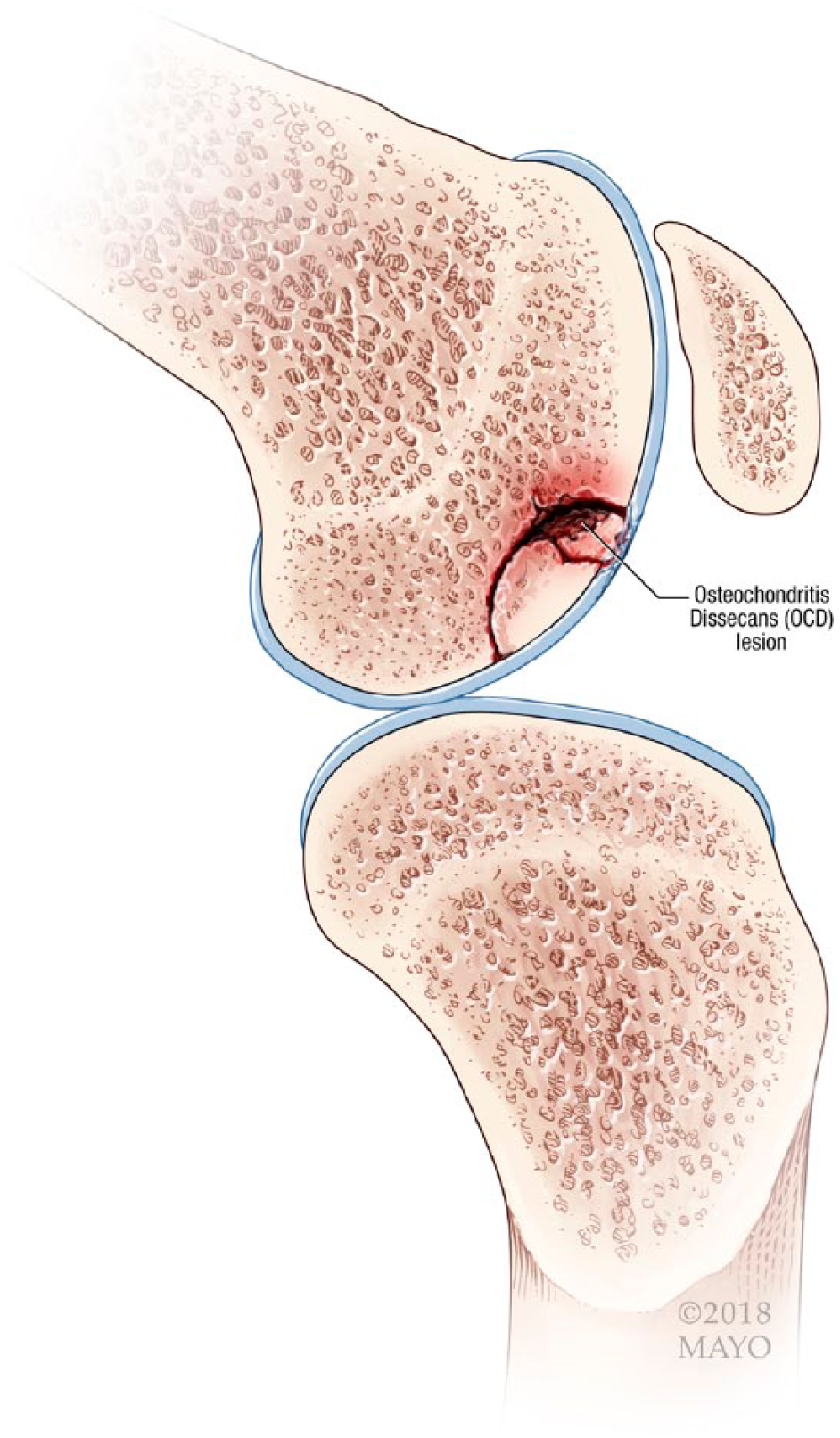
Example of an unstable osteochondritis dissecans lesion with a salvageable and unsalvageable fragment.
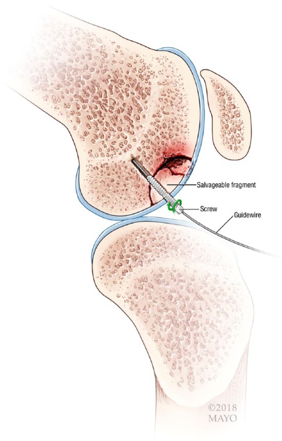
The salvageable fragment is fixated with a screw.
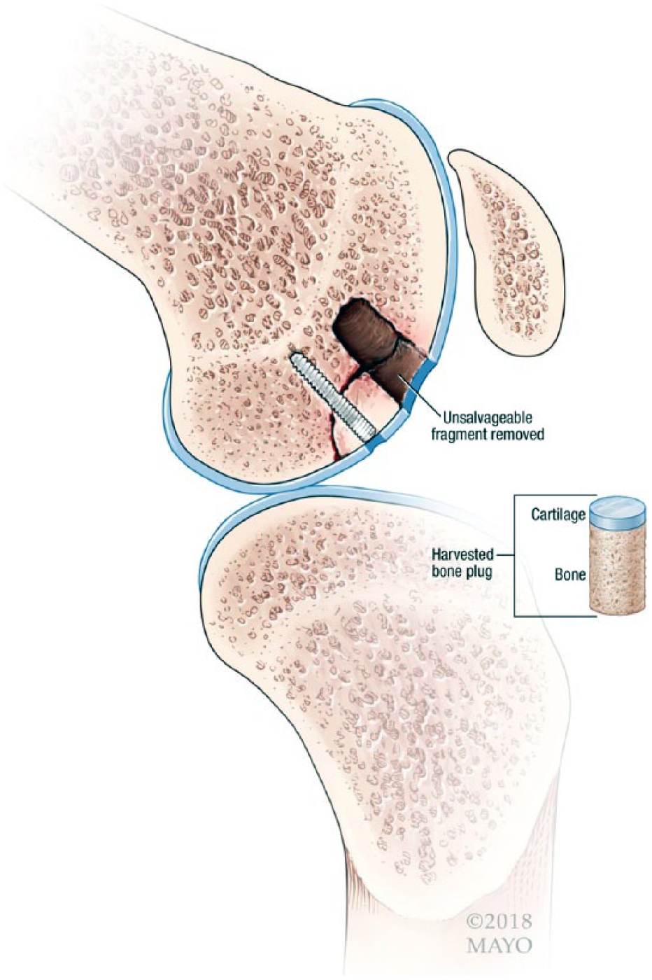
Unsalvageable portion is removed.
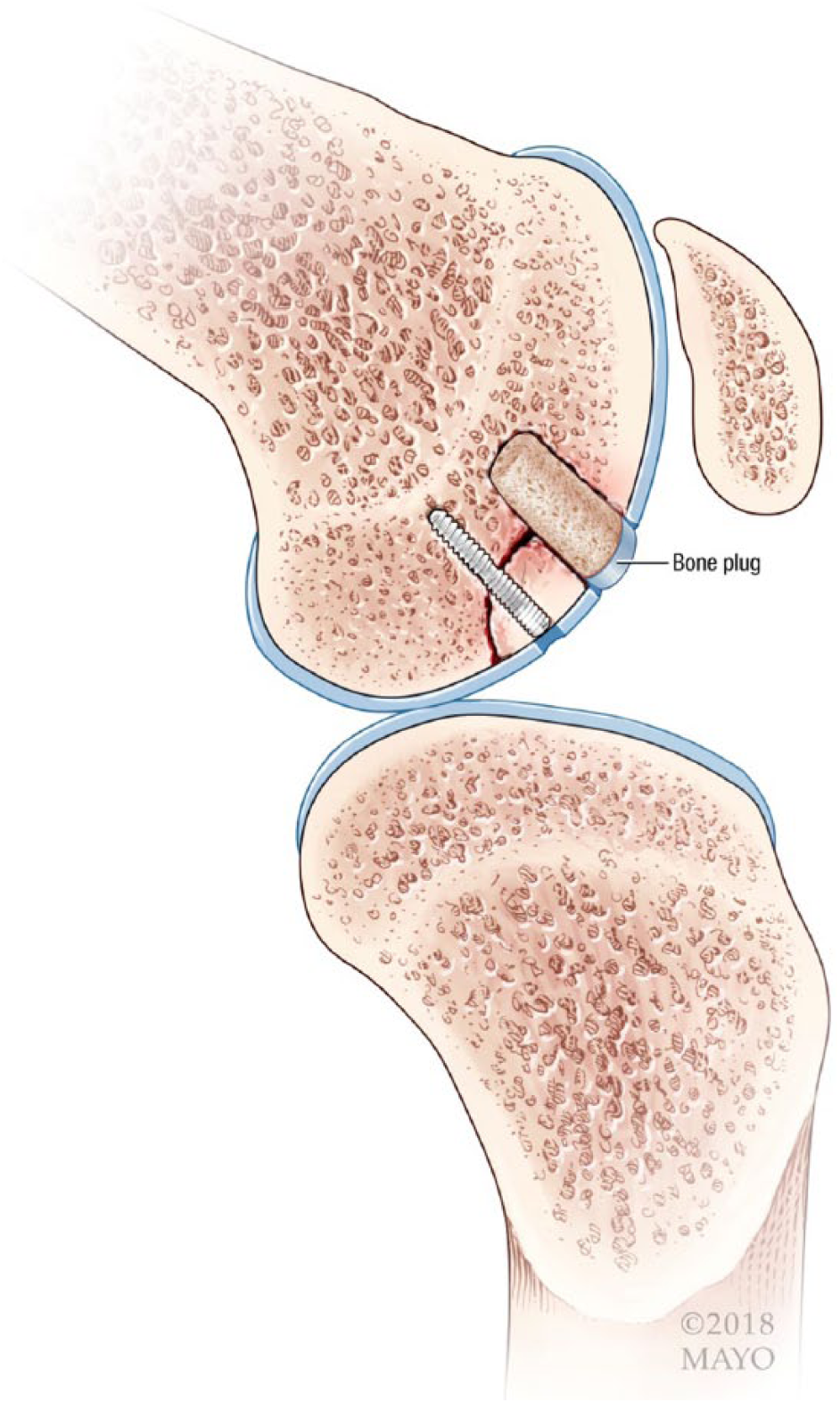
Osteochondral autograft transplantation system (OATS) plug is used to restore the articular surface and subchondral bone from where the unsalvageable fragment was removed.
For 6 weeks postoperatively, all patients remained partially weightbearing with the knee brace locked in full extension while ambulating. When not ambulating, immediate active and passive range of motion as tolerated was encouraged. Continuous passive motion machines were used starting at 0° to 30° and advanced as tolerated. Gradual weight bearing as tolerated was allowed after 6 weeks and the brace was discontinued. Typically, patients returned to all activities by 6 months after surgery.
Outcome Measures
The Knee injury and Osteoarthritis Outcome Score (KOOS) 17 Quality of Life (QoL) and Activities of Daily Living (ADL) subscales were used to assess health and well-being. KOOS has been shown to correlate with other measures of general health and well-being such as the Short Form–36 (SF-36) in patients with articular cartilage procedures. 18 Knee function was assessed using the International Knee Documentation Committee (IKDC) subjective knee score 19 and the KOOS Pain and Symptoms subscales. Each of these has been shown to be a valid measure of knee function following articular cartilage repair operations.20,21 The Tegner activity scale 22 and KOOS Sport (Sport/Rec) subscale were used to report patient activity level and sports-related disability. The Tegner activity level was assessed preinjury and postoperatively. Reoperation and complication rates were analyzed. Healing was assessed by MRI (5 knees) by using the Magnetic Resonance Observation of Cartilage Repair Tissue (MOCART) score,23,24 second-look arthroscopy (5 knees), MRI and arthroscopy (5 knees), computed tomography scan (3 knees), or clinical examination (1 knee).
Statistical Analysis
Descriptive analyses of the patient data were performed using means and ranges. Comparisons of preoperative versus postoperative clinical scores were conducted using Wilcoxon rank-sum tests. A 2-tailed t test was used for all statistical analysis with a critical α set to 0.05. Analysis was performed using SAS Statistical Discovery JMP version 7.0 (SAS, Inc. Cary, NC).
Results
Eighteen knees (17 patients) underwent hybrid fixation of an unstable OCD lesion of the knee between the years of 2002 and 2015. All patients had OCD lesions that were graded type III or IV based on the International Cartilage Repair Society (ICRS) scale of OCD lesions.
25
The mean follow-up was 36 months (range 24-67.2 months) with all patients reaching the minimum 2-years of follow up. All procedures were performed open, the salvageable fragment was stabilized with a pin, screw, or bone plug, and the unstable fragment was removed and replaced using an OATS technique (
Surgical Details.
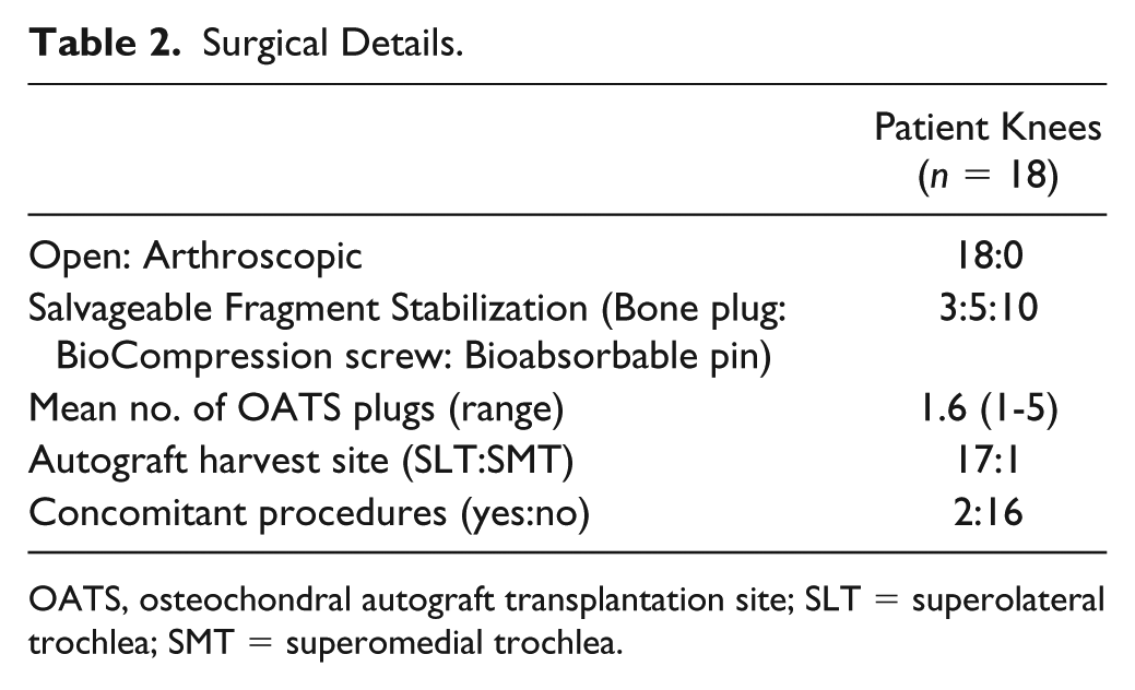
OATS, osteochondral autograft transplantation site; SLT = superolateral trochlea; SMT = superomedial trochlea.
Health Outcomes and Knee Function
The mean postoperative KOOS and IKDC scores are reported in
KOOS and IKDC Scores.
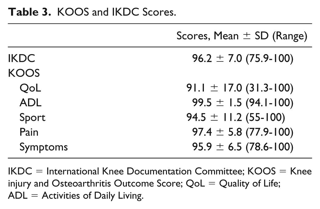
IKDC = International Knee Documentation Committee; KOOS = Knee injury and Osteoarthritis Outcome Score; QoL = Quality of Life; ADL = Activities of Daily Living.
Activity Level
All patients returned to sport. The mean postoperative KOOS Sport score was 94.5 (range 55-100). There was no significant difference between the mean preinjury (7.95; range 4-9) and mean postoperative (7.45; range 4-9) Tegner scores (P = 0.363) (
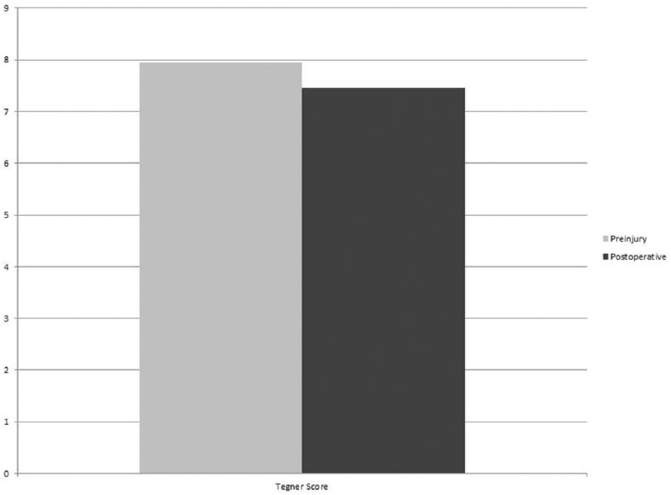
Preinjury and postoperative activity level.
Lesion Healing
Healing was assessed by MRI only in 5 knees, second-look arthroscopy and MRI in 5 knees, arthroscopy only in 5 knees, computed tomography scan in 3 knees, and clinically based on physical examination and rehabilitation clearance in 1 knee. The one patient in which healing was assessed clinically declined repeat imaging or arthroscopy. All were determined to be healed at a mean 5.6 months (range 4-7 months). Eight knees underwent repeat arthroscopy at a mean 5.8 months (range 4-7 months) to assess healing and 2 knees underwent planned repeat arthroscopy for screw removal at 5 months postoperatively. At that time, all lesions were stable to probing and the cartilage was completely filled (
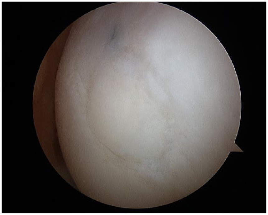
Osteochondritis dissecans lesion 5 months following treatment using the hybrid fixation technique.
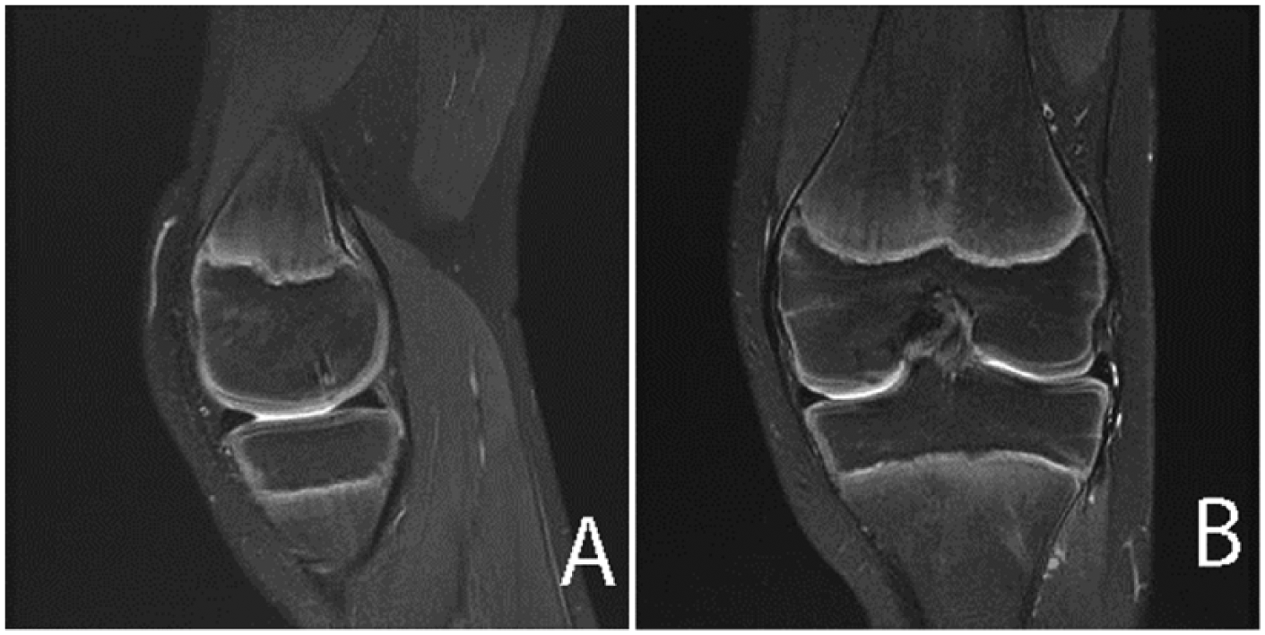
Magnetic resonance images demonstrating an osteochondritis dissecans lesion preoperatively (
Complications
There were no reported patient complications. Two knees (1 patient) underwent planned removal of the BioCompression screw used to stabilize the salvageable fragment. Otherwise, no reoperations were performed.
One patient accounted for the low KOOS Sport (55) and KOOS QoL (31.3) scores. In this patient, repeat MRI showed their lesion to be well healed without recurrence. It should be noted that this particular patient had progressive valgus malalignment and gained a significant amount of weight postoperatively.
Discussion
The main finding in the present study was that patients with unstable OCD lesions with both salvageable and unsalvageable fragments can have positive short-term outcomes and expect few complications following surgical treatment with a hybrid fixation technique which uses fragment fixation and OATS.
The technique described has the advantage of repairing an unstable OCD lesion in one surgical stage. It is indicated for lesions that have both a salvageable and unsalvageable fragment. The salvageable fragment is fixated to underlying healthy bone and the void left by the removed unsalvageable fragment is filled with an OATS plug(s). Lintz et al., 10 and Chadli et al. 26 more recently, described a “hybrid fixation” technique to stabilize the OCD fragment with both screws and OATS plugs. In this technique the entire fragment was salvageable, and the hybrid fixation was used to maintain the fixation benefits of the screw and improved revascularization benefits of the OATS plugs. Additionally, Miniaci et al. 13 and Berlet et al. 27 have described a technique in which the complete fragment was salvageable, and the lesion was fixated entirely with OATS plugs and no screws. In our technique, the osteochondral autograft plug is used to restore the bone and cartilage in a situation where a portion of the progeny fragment is fragmented and unsalvageable. Typically, this fragmented portion would lead to removal of the entire progeny fragment, with consideration of a second stage salvage procedure with an ACI sandwich technique 28 or the use of nonnative tissue in the form of osteochondral scaffolds 29 or osteochondral allograft transplant. 30 One stage autologous tissue with this technique may be considered an advantage for a young patient.
In the present study, KOOS and IKDC scores were used to assess health outcomes and knee function. The mean postoperative IKDC and KOOS scores were similar to that of prior studies, which evaluated patients following surgical treatment with other various techniques that salvaged the entire fragment.4,5,10,31 The technique described in this study is indicated when the entire fragment is unstable. Osteochondral allograft transplantation (OCA) and autologous chondrocyte implantation (ACI) are also options in this scenario and have shown positive results.32-34 However, ACI requires a second stage more costly procedure with extended rehabilitation time and allograft tissue for OCA is not always readily available. We believe that it makes sense to salvage autologous tissue in one surgical stage. Another viable option is the use of osteochondral scaffolds, which allow for a single-stage procedure. Perdisa et al. 29 reported mean IKDC scores of 82.2 and 90.1 at mean 2 and 5 years postoperatively, respectively, following the treatment of unstable OCD lesions with osteochondral scaffolds. These results are comparable to that of the present study. Unfortunately, this technique uses foreign material and does not preserve native tissue.
Activity level was assessed pre- and postoperatively using the Tegner activity scale. No significant difference was seen between preinjury and postoperative Tegner scores (P = 0.363). The return to preinjury activity level is consistent with that of other studies. Miniaci et al. 13 reported continued improvements in activity level as rated by a visual analog scale up to 18 months postoperatively following their described surgical technique. Lintz et al. 10 reported improved Hughston scores following a surgical procedure to fully salvage the progeny fragment with “hybrid fixation.” These are both very reasonable options for treatment of unstable OCD lesions but their described indications for use do not include instances in which the progeny fragment is only partially salvageable.
All knees except for one received either postoperative MRI or repeat arthroscopy. Lesion healing was adequately assessed by these measures. The 10 knees that underwent repeat arthroscopy demonstrated well healed lesions that were stable to probing. On MRI at a mean 7.8 months postoperatively, the mean MOCART score was 87.5. This mean MOCART score is slightly higher than that of a comparable study in which unstable OCD lesions were fixated with compression screws. In this study, the mean MOCART score was 72.0. 35 Additionally, Perdisa et al. 29 used MOCART to asses healing following the use of an osteochondral scaffold for treatment of unstable OCD lesions. They reported mean postoperative scores of 74.2 and 81.4 at 2 and 5 years, respectively. 29 Although Miniaci et al. 13 did not assess the lesions using the MOCART grading system, they noted healed lesions on MRI at 9 months postoperatively with only slight articular surface irregularity. Additionally, repeat arthroscopy on 2 patients showed smooth, stable lesions. 13
There were no reported complications at mid-term follow up. This may be due to the small number of patients included in this study, but it does suggest that this technique can be performed safely with a low risk for postoperative sequelae. Complications reported following other OCD lesion treatment options include graft failure with matrix-induced autologous chondrocyte implantation (MACI) use (low rate), 34 reoperation requiring revision with OCA, 32 and screw back-out with metal screw fixation. 5
The limitations of this study include the small sample size. This was due to the narrow indications for its use when both a salvageable and unsalvageable fragment make up the OCD lesion. The retrospective nature did not allow for assessment of pretreatment values and therefore true improvement cannot be assessed. Adult and juvenile OCD lesions were included. Assessment of lesion healing was heterogeneous. All patients achieved minimum 2-year follow-up, but the overall follow-up was short term. Patients were not randomized to this procedure and there was no control. Despite these limitations, this technique has not been previously described and provides a viable option for the treatment of unstable OCD lesions which have proven to be difficult to treat.
This case series introduces a surgical technique to treat unstable OCD lesions of the knee and suggests that patients can have positive short-term outcomes and expect few complications following its use. Longer term studies are needed to further compare its benefits to other reported surgical treatment options.
Footnotes
Acknowledgments and Funding
The author(s) received no financial support for the research, authorship, and/or publication of this article.
Declaration of Conflicting Interests
The author(s) declared the following potential conflicts of interest with respect to the research, authorship, and/or publication of this article: Bruce A. Levy receives or has received IP royalties and research support from Arthrex Inc., Biomet, Smith & Nephew, and Stryker; is a paid consultant for Arthrex, Inc. and Smith & Nephew; and is in the editorial or governing board of Arthroscopy: The Journal of Arthroscopic and Related Surgey, CORR, Journal of Knee Surgery, and Knee Surgery, Sports Traumatology, Arthroscopy. Norimasa Nakamura is a paid consultant for Biomet and Novartis; is on the board or is a committee member of the International Cartilage Repair Society and the International Society of Arthroscopy, Knee Surgery, and Orthopaedic Sports Medicine; and in on the editorial or governing board of Journal of Experimental Orthopaedics (Springer), Journal of Orthopaedic Science (Springer), and Cartilage (Sage). Aaron J. Krych receives or has received research support from Aesculap/B. Braun, Arthritis Foundation, Ceterix, and Histogenics; receives IP royalties from Arthrex Inc.; is a paid consultant for Arthrex Inc. and Vericel; is on the board or is a committee member of the International Cartilage Repair Society, International Society of Arthroscopy, Knee Surgery, and Orthopaedic Sports Medicine, Minnesota Orthopedic Society, and Musculoskeletal Transplantation Foundation; and is on the editorial or governing board of the American Journal of Sports Medicine.
Ethical Approval
The institutional review board (IRB ID No. 15-000601) at the Mayo Clinic, Rochester, Minnesota, and Osaka University Hospital, Osaka, Japan. approved the study.
Informed Consent
Written informed consent was obtained from all study participants.
Trial Registration
Not applicable.