
Abstract
Background
This study investigates the effect of Edgeworthia chrysantha Lindl. on the expression of brain-derived neurotrophic factor (BDNF) and its receptor tyrosine kinase B (TrkB) in the hippocampus and cortex of depression model rats.
Methods
A chronic unpredictable stress-induced depression model was used, and the behavior changes of animals as well as the expression levels of BDNF and TrkB were measured in all groups (normal control, model, venlafaxine hydrochloride, and Edgeworthia chrysantha Lindl. (100, 50, 25 mg·kg−1) dose groups).
Results
Our results revealed that the scores of horizontal and vertical movements in the open field experiment were significantly decreased in the model group compared to the normal control. Besides, the expressions of BDNF and TrkB in the hippocampus and cortex were significantly decreased in the model group compared to the normal control. On the other hand, treatment with Edgeworthia chrysantha Lindl significantly improved depressive behavior and increased the expression of BDNF and receptor TrkB in the hippocampus and cortex of rats compared to model group.
Conclusion
Edgeworthia chrysantha Lindl. demonstrates potential antidepressant effects, possibly mediated by the upregulation of BDNF and TrkB expression, highlighting its neuroprotective properties.
Introduction
Depression is a kind of neurological and mental disease caused by many factors and is accompanied by a serious suicidal tendency.1,2 The onset of depression is closely related to stress. 3 With the acceleration of people's work and lives and the intensification of various stress factors, the incidence of depression is increasing year by year, which brings a heavy burden to society and families.2,3 According to WHO statistics, depression affects as many as 300 million people globally and will top the global burden of disease by 2030. 4 Depression is the most common mental disorder in China, with a lifetime prevalence of 6.9%.5,6 114 800 people with depression die by suicide every year, resulting in a loss of 5 billion yuan. 7 Therefore, the treatment of depression is also getting more and more attention. The listed anti-depressants are mainly aimed at single-target treatment, which is not ideal and has more adverse effects.8,9
Finding safe and effective anti-depressant drugs from natural plants is one of the most important ways to develop anti-depressant drugs. Edgeworthia (Edgeworthia chrysantha Lindl.), known as the “Florilegium”, is a deciduous shrub of the genus Knotweed (Edgeworthia) in the family Thymelaeaceae. 10 The Edgeworthia chrysantha Lindl. is an integral part of the traditional Yao medicine system known as the “Five Tigers, Nine Cows, Eighteen Diamonds, and Seventy-Two Winds,” and it holds significant importance in the Yao region. Some scholars have found that the clove glycosides contained in Edgeworthia chrysantha Lindl. have other biological activities such as anti-fatigue, depression, improving blood circulation in the brain, sexual performance, inhibiting glucose metabolism, and so on. 11 The flowers of Edgeworthia chrysantha Lindl. has the effect of nourishing the heart and tranquilizing the mind, dispelling wind, and brightening the eyes, and it is the main treatment for green blindness, cataracts, excessive tearing, dreaming, deficiency of lymph, and loss of voice. In the Yao region, the flowers of Edgeworthia chrysantha Lindl. are commonly used to treat rheumatism and paralysis, women's postpartum disease, mental depression, infertility, high blood pressure, and other evidence. 12 The flowers of Edgeworthia chrysantha Lindl. contain many silver forging glycosides, zingiber officinale, nicotiana glycosides, rutin, and other flavonoids, which have strong antioxidant, free radical elimination, and anti-inflammatory effects and have a wide range of applications in the fields of pharmaceuticals, food, cosmetics, and other fields.
The signal axis, composed of brain-derived neurotrophic factor (BDNF) and its specific tropomyosin-related kinase receptor B (TrkB), is involved in regulating the development, cellular architecture, and functional status of the nervous system under physiological conditions. 13 In pathological conditions, it has the functions of anti-oxidation, anti-apoptosis, promoting the growth of neuronal axons and dendrites, enhancing synaptic plasticity, promoting nerve repair, etc, which has become a common molecular target for neuroprotection and nerve repair in recent years. 14
The present study hypothesizes that Edgeworthia chrysantha Lindl. exerts antidepressant effects by upregulating the expression of brain-derived neurotrophic factor (BDNF) and its receptor TrkB in the hippocampus and cortex, thereby mitigating depressive behaviors in a rat model of chronic unpredictable stress.
Materials and Methods
Animals
3-month-old healthy male Sprague-Dawley rats of clean grade, weighing (180-200) g, were provided by the Laboratory Animal Center of the Academy of Military Medical Sciences of the People's Liberation Army. License number: SCXK (Liao) 2015-0001.
Drugs and Reagents
Venlafaxine Hydrochloride Capsules (Chengdu Kanghong Pharmaceutical Group Co., Ltd); specification 75 mg per capsule, product lot number 06013. Polyclonal anti-BDNF antibody (Tested Reactivity: mouse, rat, human; Wuhan Sanying Biotechnology Co., LD, 25699-1-AP); polyclonal anti-TrkB antibody (Tested Reactivity: mouse, rat, human; abcam, ab18987); hematoxin (Sigma, H9627); neutral gum (Sinopsin Group, 10004160) was provided.
Extraction and Isolation of Edgeworthia chrysantha Lindl
The extraction of Edgeworthia chrysantha Lindl. were conducted by the Department of Chinese Medicine of Guangxi University of Traditional Chinese Medicine based on the literature method. 15 In brief, the plant-dried powder was extracted using ethanol (75%) at 25 ᵒC. After 4 times extraction, the total extract was prepared using a vacuum evaporator and fractionated using different solvents (Petroleum ether, chloroform, ethyl acetate, and n-butanol). Silica gel chromatography was used for separation and fractionation of chloroform fraction with increasing methanol amount to obtain the desired extract.
Instruments
RM 2016 rotary pathological micrograph (Leica Company, Germany); BX53 biological microscope (Olympus), continuous liquid separator (Eppendorf Company, Germany); JT-12J computerized biological tissue dehydrator (Wuhan Junjie Electronics Co., LD); BW-OF302 open field laboratory (Shanghai Soft Long Technology Development Co., LD); BW-MWM101 Morris water maze (Shanghai Soft Long Technology Development Co., LD).
Grouping and Administration
After 1 week of acclimatization feeding, rats were scored behaviorally with the open-field experiment, and rats with total scores of less than 30 or more than 120 points for horizontal and vertical movements within 3 min were excluded. 90 selected rats were randomly divided into 6 groups: normal control group, model group, venlafaxine hydrochloride group (12.5 mg·kg−1 venlafaxine hydrochloride), 16 high-dose group (100 mg·kg−1 of Edgeworthia chrysantha Lindl.), middle-dose group (50 mg·kg−1 of Edgeworthia chrysantha Lindl.), and low-dose group (25 mg·kg−1 of Edgeworthia chrysantha Lindl.). At the same time of modeling, the rats were given electric stimulation with the drug once a day for 21 days. The normal control and model groups were given equal volumes of distilled water.
Animal Model Building
In addition to normal feeding in the normal control group, the remaining group referred to the literature, including 9 stimuli of fasting for 24 h, water fasting for 24 h, overnight lighting, tail pinching for 1 min, shaking, heat stress, and swimming in cold water at 4 °C for 5 min, which were randomly arranged in 21 d. One stimulus was given every day, and the same stimulus could not appear consecutively, so the animals could not foresee the given stimulus, and each stimulus appeared 2 to 3 times.
Open Field Experiment
An experimental method to evaluate the autonomic action, autonomic inquiry behavior, and tension of laboratory animals in a specific environment. The open-field experimental video analysis system was used to test the 21 days of molding. The activity of the rats within 3 min was recorded while the surrounding environment was kept quiet. One point was scored for the number of times more than 50% of the rat's body entered a square, which was recorded as a horizontal movement, and one point was scored for the number of times both forelimbs of the rat left the bottom grid of the box, which was recorded as an upright movement. After each rat was tested, the open tank was cleaned with 75% alcohol before the next rat was tested. 17
Test of Learning and Memory Ability in Rats
The learning and memory ability of rats was tested with the BW-MWM 101 Morris water maze, which consists of a localization navigation test and a spatial exploration test, and the experiment was completed on the 21st day of modeling. 18
Place Navigation Test
It was used to measure the learning and memory acquisition abilities of water maze in rats. The rats were trained three days before the experiment, and the place navigation test was performed on the fourth day. From the water entry point of the diagonal image line where the platform is located, the rats were placed into the pool facing the pool wall, and the time required for the rats to find the platform within 120 s (ie escape latency) was recorded. If no platform is found within 120 s, the escape incubation period is marked as 120 s. 18
Space Exploration Experiment
The ability of the rats to retain the spatial location memory of the platform was tested. In the second part of the place navigation test, the platform was withdrawn, and the rats were placed in the pool from the diagonal entry point where the platform was located. The number of times the rats crossed the original platform location within 120 s was recorded. 19
Sugar Water Consumption Experiment
Tests were performed on the 21st day of molding. Before the experiment, the rats were trained to adapt to sugary drinking water by placing two water bottles in each cage at the same time. On the first day, two bottles are filled with 1% sucrose water. On the second day, 1 bottle is filled with 1% sucrose water, and 1 bottle is filled with pure water. On the third day, the rats were fasted and dehydrated. On the fourth day, the rats were given one bottle of sucrose water and one bottle of pure water for each cage. The consumption of sucrose water and pure water was measured after 1 h, and the sugar-water preference of rats was calculated in each group. Sugar water preference = 1% sugar water consumption/(1% sugar water consumption + pure water consumption) × 100%. 20
The Expressions of BNDF and TrkB in the Hippocampus and cortex Were Determined by Immunohistochemistry
On the 21st day of the experiment, after open-field behavior, the rats were anesthetized with pentobarbital sodium, fixed in the supine position, opened the chest cavity to expose the heart, inserted a needle into the left ventricular intubation at the apex of the heart, quickly injected normal saline, and cut open the right auricle. After the right auricle retained a clear colorless fluid, it was switched to 4% paraformaldehyde cardiac perfusion for approximately 300 ml. After the rats were stiffened, the brains were severed and removed, and the brain repair blocks were labeled and fixed in a 4% paraformaldehyde solution for more than 48 h. After gradient alcohol dehydration, transparency, and paraffin embedding, coronal sections were made, and the slices were about 4 μl thick. Slides were fished with slides coated with gelatin-complex alum. The primary antibody is rabbit anti-mouse BDNF polyclonal antibody or rabbit anti-mouse TrkB polyclonal antibody; the working concentration of BDNF is 1:100, and the working concentration of TrkB is 1:50. Secondary antibody: HRP-labeled goat anti-IgG. Follow the kit instructions strictly. Sections were deparaffinized, antigen repaired, endogenous peroxidase blocked, serum blocked, primary antibody added, enzyme-labeled secondary antibody added, DAB chromogen added, Harris hematoxylin re-stained, dehydrated, blocked, and photographed. 21 The stereotaxic map of the rat brain was compared with the Olympus BX53 biological microscope. The three sections of the same part of each animal were selected for each index, and the number of BDNF- or TrkB-positive cells in the CA3 region of the hippocampus was counted under a 400× light microscope. The average of the three sections was taken as the number of positive cells in the hippocampal CA3 region of that animal.
Statistical Analysis
Data were analyzed using SPSS 23.0 software, and values are expressed in mean ± Standard error of mean (SE). The normality of data was tested using the Shapiro-Wilk Test. A one-way ANOVA was used for comparison between the means of different groups followed by Tukey post-hoc test. P < .05 was considered statistically significant.
Results
Effect of Edgeworthia chrysantha Lindl. on Scores in an Open-field Experiment of Depressed Model Rats
As shown in Table 1, compared with the normal control group, the scores of horizontal movement and upright times of rats were decreased in the model group (P < .01). Compared with the model group, the scores of horizontal exercise and upright times were increased in the high, middle, and low dose groups and the venlafaxine hydrochloride group (P < .01). Compared with the high-dose and low-dose groups, the scores of horizontal movement and upright times of the rats were significantly increased in the middle-dose group (P < .01). (One animal in the model group died during the experiment.)
Comparison of Open-field Test Scores of Rats in Each Group.

Data are presented as mean ± SE. *P < .01, compared with the normal control group; #P < .01, compared with the model group; &P < .05, compared with high dose group.
Effects of Edgeworthia chrysantha Lindl. on Sugar and Water Consumption in Depressed Model Rats
As shown in Table 2, compared with the normal control group, the sugar water preference of the model group was decreased (P < .01). Compared with the model group, the sugar water preference of rats in the high, middle, and low-dose groups and the venlafaxine hydrochloride group was increased (P < .05, P < .01). Compared with the high-dose and low-dose groups, the sugar water preference of rats was higher in the middle-dose group (P < .05).
Comparison of Sugar-water Preference of Rats in Each Group.

Data are presented as mean ± SE. *P < .01, compared with the normal control group; #P < .01, $P < .05, compared with the model group; &P < .05, compared with high dose group.
Effects of Edgeworthia chrysantha Lindl. on Learning and Memory Ability of Depressed Model Rats
As shown in Table 3, compared with the normal control group, rats had an increased escape latency time and fewer crossings of the original platform in the model group (P < .01). Compared with the model group, the escape latency time of rats in high-dose, middle-dose, and low-dose groups and venlafaxine hydrochloride groups was decreased, and the number of times of crossing the original platform was increased (P < .05, P < .01). Compared with the high-dose and low-dose groups, the escape latency time of rats was reduced, and the number of times crossing the original platform was increased in the middle-dose group (P < .05).
Comparison of Learning Memory Ability Performance Scores in Each Group of Rats at Each Stage.

Data are presented as mean ± SE. *P < .01, compared with the normal control group; #P < .01, $P < .05, compared with the model group; &P < .05, compared with high dose group.
Effects of Edgeworthia chrysantha Lindl. on the Expression of BDNF in the Hippocampus and cortex of Depressed Rats
As shown in Table 4, Figures 1 and 2, under light microscopy, the positive cytoplasm of BDNF was stained a distinct brownish-yellow color. The positive cells were mainly concentrated in the CA3 region in the hippocampus and the prefrontal lobe in the cortex. In the normal control group, BDNF-positive cells were arranged neatly, with more positive cells and deeper cytoplasmic staining. Compared with the normal control group, the BDNF arrangement of rats in the model group was scattered, the cytoplasm staining was lighter, and the number was significantly reduced, which was significant (P < .01). Compared with the model group, the cytoplasmic staining of BDNF-positive cells in the hippocampus and cortex of rats in each administration group was enhanced, the arrangement was relatively concentrated, and the number of positive cells increased, which was significant (P < .01, P < .05).
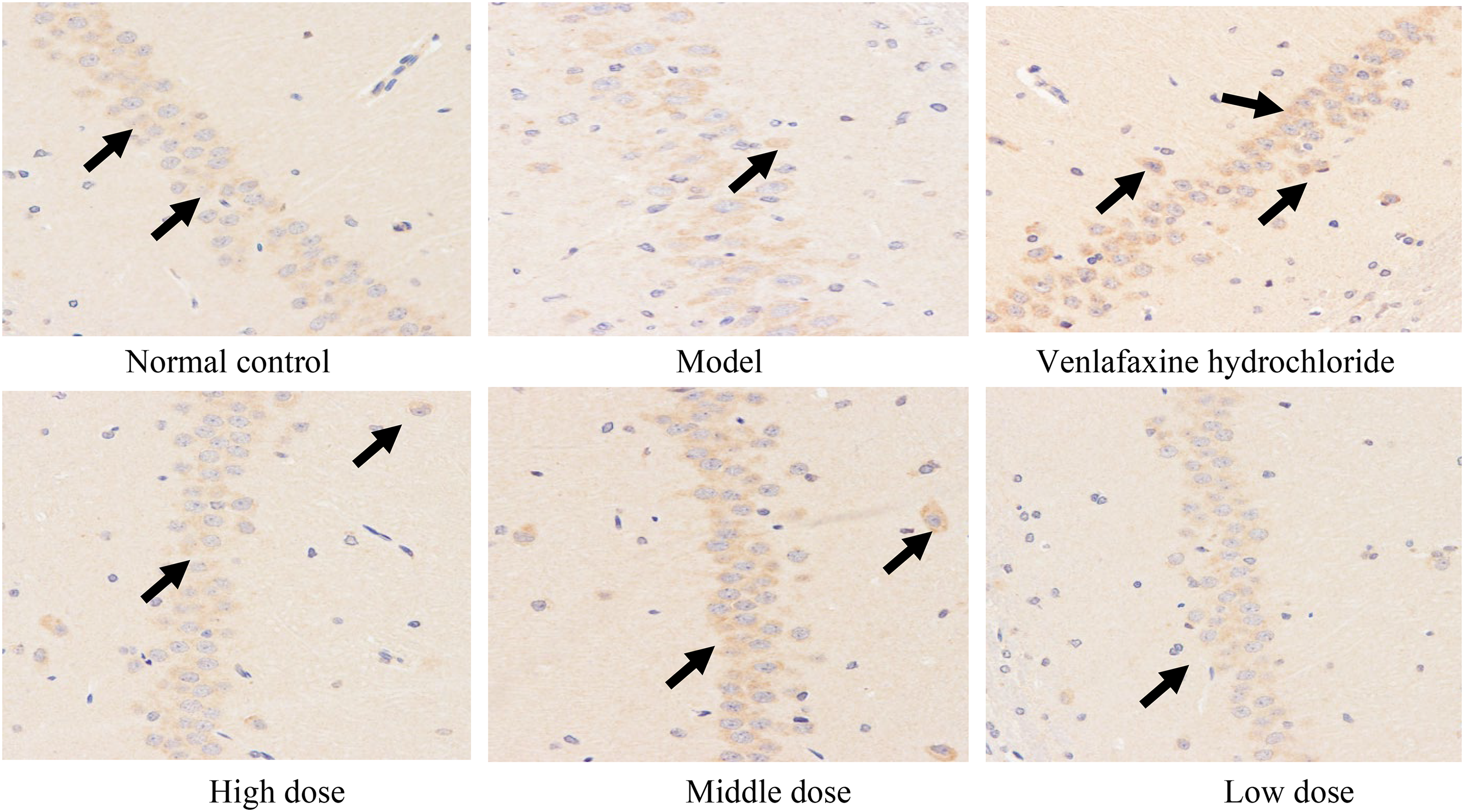
Expression of BDNF in the hippocampus was detected by immunohistochemistry (10 × 40).
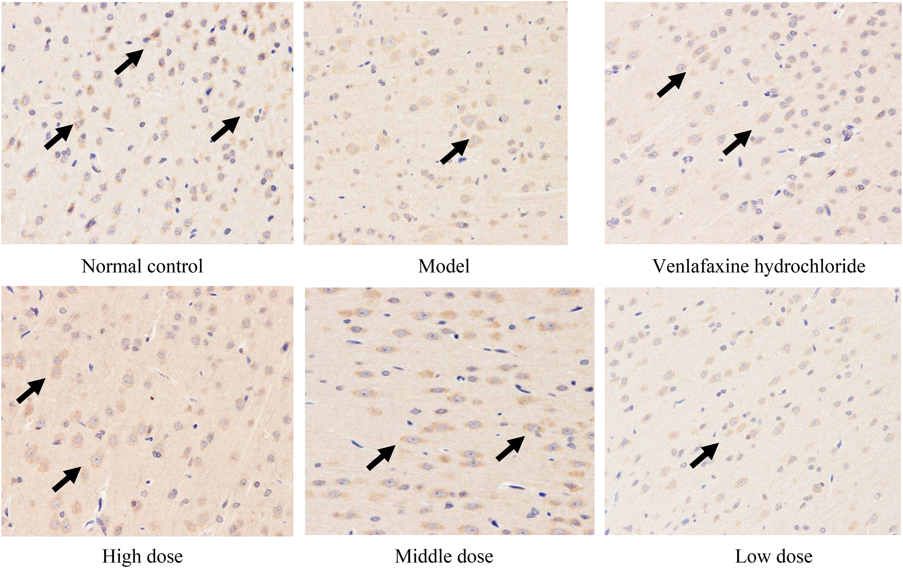
Expression of BDNF in the cortex was detected by immunohistochemistry (10 × 40).
Expression of BDNF in Hippocampus and cortex of Experimental Rats in Each Group.

Data are presented as mean ± SE. *P < .01, compared with the normal control group; #P < .01, $P < .05, compared with the model group; &P < .05, compared with high dose group.
Effects of Edgeworthia chrysantha Lindl. on TrkB Expression in the Hippocampus and cortex of Depressed Rats
As shown in Table 5, Figures 3 and 4, under the light microscope, the plasma of TrkB-positive cells was stained yellow-brown, and the nuclei were not colored. Positive cells in the hippocampus were mainly concentrated in the CA3 area and prefrontal cortical areas. TrkB-positive cells in the normal control group were neatly arranged, with more positive cells and deeper cytoplasmic staining. Compared with the normal control group, the TrkB-positive cells in the model group were more disorganized, with lighter cytoplasmic staining and a significantly lower number, which was significant (P < .01). Compared with the model group, the cytoplasmic staining of TrkB-positive cells in the hippocampus of rats was enhanced in each dosing group; the color was intense, the arrangement was relatively concentrated, and the number of positive cells was increased, which was significant (P < .01, P < .05).
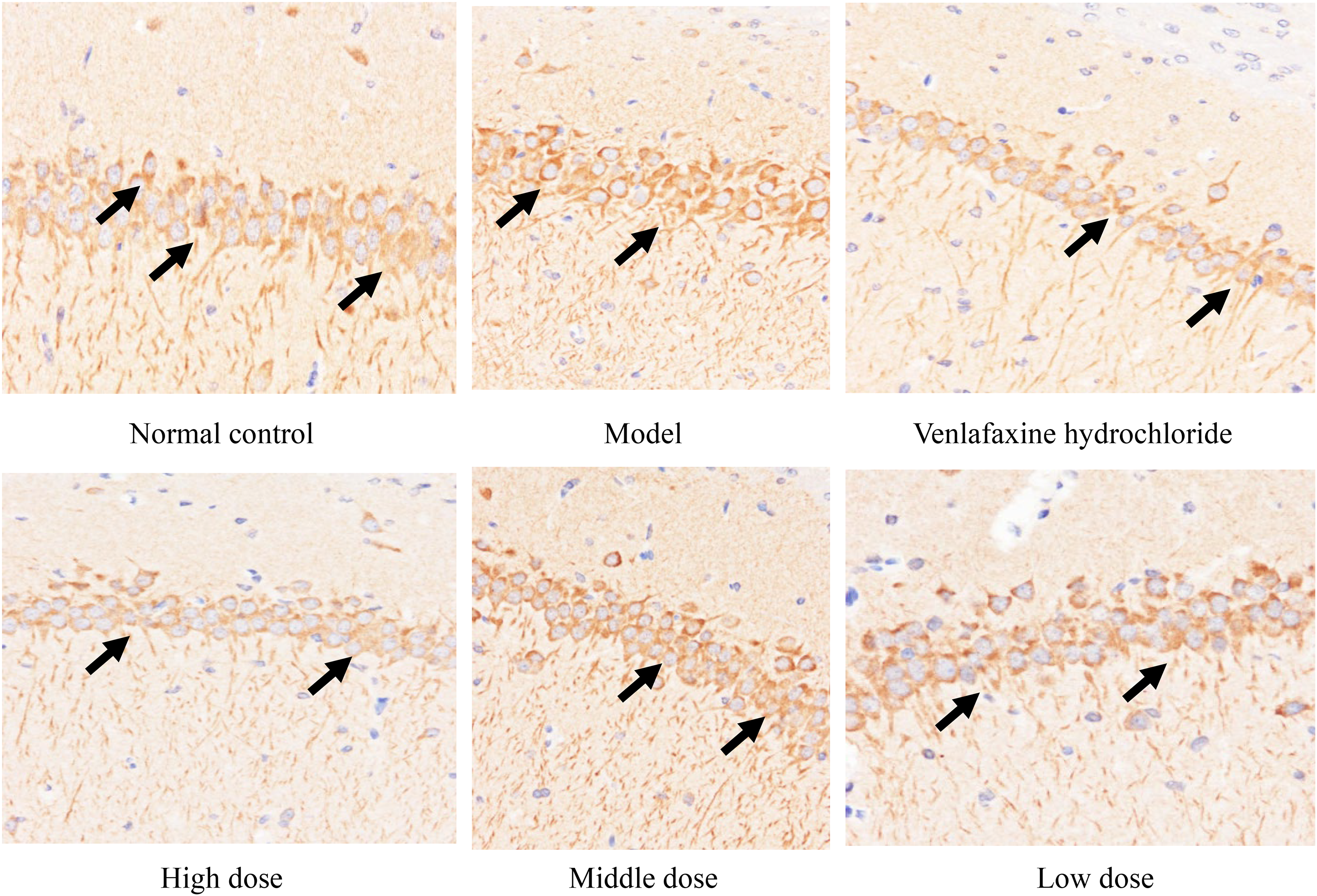
Expression of TrkB in the hippocampus was detected by immunohistochemistry (10 × 40).

Expression of TrkB in the cortex was detected by immunohistochemistry (10 × 40).
Expression of TrkB Protein in Hippocampus and cortex of Experimental Rats in Each Group.

Data are presented as mean ± SE. *P < .01, compared with the normal control group; #P < .01, $P < .05, compared with the model group; &P < .05, compared with high dose group.
Discussion
Long-term chronic low-intensity stress is one of the main causes of depression. The chronic unpredictable mild stress (CUMS) model induces depression by giving different stimuli to model animals every day, which is close to the cause of human depression by exposing animals to varied and unpredictable stressors daily, leading to behavioral and physiological changes associated with depressive disorders. 22 In the present study, we used established tests to evaluate depressive behaviors and the effects of Edgeworthia chrysantha Lindl. on these outcomes. The sucrose water depletion test is one of the main methods to quantitatively evaluate pleasure deficits in rodents which quantitatively assessed anhedonia, a core symptom of depression, as decreased sucrose consumption indicates reduced pleasure-seeking behavior.23,24 Besides, open-field experiment is one of the classical methods of animal behavior evaluation that evaluate hypoactivity and exploratory behavior, which are typically diminished in depression. 25 In a depressed state, animals generally show decreased active activity and exploratory behavior. Studies have shown that depression can lead to a significant decline in learning and memory.1,26 The Morris water maze is a good way to test animal spatial learning and reference memory. 27 Therefore, the present experiment was conducted to evaluate the success of the depressed rat model using the open field test, sugar-water consumption test, and Morris's water maze test and to observe, for the first time, the anti-depressant efficacy of Edgeworthia chrysantha Lindl. The results showed that after 21 days of modeling, compared with the normal control group, the score of the open field experiment was significantly reduced, the preference for sugar and water was decreased, and the ability to learn and remember was decreased in the model group, indicating the success of model preparation. Compared with the model group, the scores of the absenteeism experiment, sugar and water preference, and learning and memory ability of rats in the high, middle, and low dose groups were significantly improved, indicating Edgeworthia chrysantha Lindl. had anti-depressant effects and were more effective in the middle dose group.
It has been demonstrated that the hippocampus is a crossroads for the pathogenesis of depressive disorders, mediating functional and structural changes in the nervous system and that it is also closely related to higher cognitive functions such as emotion, learning, and memory.26,28,29 The hippocampal CA3 region is a subregion that is extremely sensitive to stress and is closely related to the onset and development of depression. 30 The prefrontal lobe has a role in depression that affects emotional, cognitive, and executive functions. Clinical and experimental studies have shown that damage to the prefrontal cortex can cause depressed mood and behavior in both humans and animals. 31 In the present study, we investigated the expression levels in the BDNF and TrkB in both CA3 of the hippocampus and brain cortex after treatment with Edgeworthia chrysantha Lindl.
BDNF is widely distributed in the central nervous system and has various pharmacological activities, such as improving neuronal pathology, promoting neuronal survival and differentiation, and preventing neuronal damage during the development of the nervous system, which can affect neuronal cell function through various signal transduction pathways. The expression of BDNF is of great significance in the study of the pathogenesis of depression. Its reduced expression is implicated in the pathophysiology of depression, and its upregulation is a hallmark of effective antidepressant treatments. 32 Our results revealed Edgeworthia chrysantha Lindl. significantly improvement of BDNF expression in the hippocampus and brain cortex of rats compared to the model group. In parallel, Martínez-Turrillas suggests that decreased levels of BDNF expression are involved in some of the pathophysiological processes of depression and may be one of the fundamental pathological changes in depression and that modulation of BDNF levels may enable the treatment of depression. 33 The improved mechanism of an electroconvulsive anti-depressant may be to improve the expression of BDNF, restore neuronal plasticity, promote neuronal survival, and alleviate depressive symptoms. 34
TrkB is a specific high-affinity receptor for BDNF, which is widely distributed in the central nervous system and has important effects on neuronal survival, growth, and differentiation. The biological effect of BDNF is dependent on binding to TrkB. Some findings have shown that BDNF acting in conjunction with its receptor TrkB increases synaptic plasticity, promotes axonal and dendritic growth, and increases synaptic terminal density. In the present study, the administration of Edgeworthia chrysantha Lindl. significantly enhanced TrkB protein expression in both the hippocampus and brain cortex of rats compared to the model group. This result is in line with the study conducted by Cai, Li 35 who concluded that neuromodulators could improve depressive symptoms and enhance the learning and memory ability of rats in the post-stroke depression model, and its mechanism may be related to the increase of hippocampal BDNF and TrkB content and the promotion of hippocampal synaptophysin I, SYNA, and other synaptic growth-related proteins, to enhance the plasticity of hippocampal neurons in post-stroke depression model rats. Our results showed increased TrkB expression in rats treated with Edgeworthia chrysantha Lindl., supporting its role in enhancing synaptic plasticity and promoting neuronal survival. Thus, the ability of Edgeworthia chrysantha Lindl. to enhance BDNF/TrkB signaling likely underpins its efficacy in reversing the behavioral and cognitive deficits observed in depressed rats.
However, there are some limitations to this study that need to be addressed in subsequent investigations. The results may not be as broadly applicable if rats are excluded based on behavioral scoring standards, which could induce selection bias. Furthermore, each group had a comparatively small sample size, which might have decreased the statistical ability to identify minor effects. While adequate for acute effects, the study's 21-day duration limits the assessment of long-term safety and efficacy. Furthermore, our knowledge of the fundamental mechanisms by which Edgeworthia chrysantha Lindl. works is limited by the lack of mechanistic research. Larger sample numbers, longer treatment durations, and more expansive inclusion criteria should all be included in future research to overcome these constraints.
Conclusions
The results of the present study showed that Edgeworthia chrysantha Lindl. significantly improved behavioral functions and increased BDNF and its receptor TrkB expressions in the hippocampus and cortex of rats in a dose-dependent manner. Increasing the expression of BDNF and TrkB in the hippocampus and cortex, regulating neuronal plasticity in the hippocampus and cortical areas, and protecting hippocampal and cortical neurons may be one of the anti-depressant pharmacological mechanisms of Edgeworthia chrysantha Lindl.
Footnotes
Acknowledgments
The authors thank the foundation project; Guangxi Natural Science Foundation Project (2022JJA141296); and the First-class Discipline Construction Project of Guangxi University of Traditional Chinese Medicine (2019XK070, 2018XK058) for supporting this work.
Author Contributions
Y.L., S.W., Y.Z., and Q.F. preparing the conception of the study; S.M., and L.Y., conducted the experimental work; Z.H. collected and analyzed data. All authors shared in writing this paper. All authors read and approved the final manuscript.
Data Availability
Data and material are available from the corresponding author upon reasonable request.
Declaration of Conflicting Interests
The authors declared no potential conflicts of interest with respect to the research, authorship, and/or publication of this article.
Ethical Approval
All the experimental procedures were approved by the animal ethical committee of Laboratory Animal Center of the Academy of Military Medical Sciences of the People's Liberation Army. License number: SCXK (Liao) 2015-0001.
Funding
The authors disclosed receipt of the following financial support for the research, authorship, and/or publication of this article: This work was supported by the Natural Science Foundation of Guangxi Province (No.2023GXNSFAA026504), Guangxi New Medical Science Research and Practice Project (No.XYK2022007,XYK202419), the Guangxi Multidisciplinary Innovation Grant in Traditional Chinese Medicine (No. GZKJ2304), NATCM's Project of High-level Construction of Key TCM Disciplines/Medicine for ethnicminorities (Zhuang medicine), (No.zyyzdxk-2023164), Guangxi Higher Education Key Laboratory for the Research of Durelated Diseases in Zhuang Medicine (Gui Jiao Ke Yan (2022) No.10).