
Abstract
Objective
To investigate the effects of phillyrin on D-galactose (D-gal)-induced aging in the brain of mice and mechanism.
Methods
Institute of Cancer Research mice were intraperitoneally injected with D-gal 150 mg/kg to induce the aging model, and the mice were treated with phillyrin 5, 15, and 45 mg/kg once a day for 8 weeks.
Results
The memory impairment induced by D-gal in brain aging was ameliorated by phillyrin treatment, and the oxidative stress in the aging brain and serum was inhibited, as evidenced by increased superoxide dismutase, glutathione peroxidase enzyme activities, and decreased malondialdehyde levels. Additionally, the hippocampus damage has also been well improved by phillyrin. Moreover, the protein expressions of nuclear factor erythroid-2-related factor 2 (Nrf2), heme oxygenase-1 (HO-1), and NADPH quinone oxidoreductase 1 (NQO1) were markedly up-regulated and the expression of Keap-1 was down-regulated in the phillyrin group than that in the aging model group.
Conclusion
These findings suggest that phillyrin is effective in the prevention of age-related memory impairment, and the attenuation oxidative stress by activating the Nrf2/HO-1 signaling pathway in the hippocampus may be one of the mechanisms.
Introduction
Aging is the most basic life characteristic of the organism with age increasing, the functional state of the organism decreases, and the stability of the internal environment and stress capacity are weakened, which is an irreversible phenomenon of gradual structural and functional degradation.1,2 In the process of aging, the progression of aging-related diseases will be greatly accelerated, such as neurodegenerative diseases, cardiovascular diseases, motor system diseases, and various cancers.3,4 In addition, as an increasing number of older people worldwide, the aging of China has also shown a deepening trend year by year. According to statistics, there are 190 million people aged over 65 in China, which accounts for 13.5% of the total population. 5 Such a large and fast-developing elderly population inevitably bring great pressure and severe challenge to the social development of our country. Therefore, it is essential to study how to delay aging or reduce the occurrence of age-related diseases. Owing to the aging is a complicated and multifactorial biological process, our understanding of the mechanism of aging is limited to date. However, studies have shown the increased oxidative stress in aging is the most representative factor. 6 With the growth of age, the ability to scavenge free radicals in the body declines, causing an increase in reactive oxygen species (ROS). The accumulating ROS can lead to the destruction of cell membrane structure and the disorder of organelle function, and then cause damage to macromolecules. 7 Thus the effective inhibition of increasing ROS is expected to moderate senescence, reducing its harmful effects on human health. 8
Chronic administration of D-gal to mice has long been a widely accepted model for mimicking aging, 9 attributed to the fact that excess D-gal promotes the production of ROS in the body, reduces the activity of antioxidant enzymes, and accelerates oxidative damage. 10 In addition, the characteristics of aging induced by D-gal such as impaired neurological structure and function, and reduced cognitive and motor abilities are resemble the natural aging process, 11 therefore, this model of aging has been widely used for the anti-aging effects and mechanisms of natural antioxidant substances.
Previous studies have shown that the nuclear factor erythroid-2-related factor 2 (Nrf2) signaling pathway is the critical antioxidant response signaling mechanism that takes a key role in the defense against oxidative stress. 12 In normal circumstances, Nrf2 and Kelch-like ECH-associated protein 1 (Keap-1) polymerize in the cytoplasm. 13 However, when the cell redox balance of the organism is broken, Nrf2–Keap-1 complex dissociates from the cytoplasm and subsequently Nrf2 enters the nucleus and activates the expression of multiple downstream target proteins and genes, such as heme oxygenase-1 (HO-1), NADPH quinone oxidoreductase 1 (NQO1), superoxide dismutase (SOD), glutathione peroxidase (GSH-Px), and catalase (CAT). 14 In this regard, activation of the Nrf2 pathway can serve as a feasible target for anti-aging. 15
Phillyrin is a double epoxy lignan glycoside compound with the molecular formula C27H34O11 and a relative molecular mass of 534.55 (Figure 1), well it is also one of the main components of Forsythia suspensa, a traditional medicine in China. 16 The previous researches have reported that phillyrin shows lots of pharmacological effects, such as anti-inflammatory, antiviral, anti-diabetic, and antioxidative.17–20 Phillyrin could reduce injury in intracerebral hemorrhagic mice via activating the Nrf2 signaling pathway, 21 another study found that phillyrin plays a neuroprotective role on cerebral ischemia/reperfusion injury in rats, 20 which are attributed to its antioxidant effect. However, to our knowledge, although numerous evidence suggests that phillyrin has antioxidant and anti-aging effects and could attenuate aging, the specific underlying mechanism about it has still remained unclear.

The structure of phillyrin.
Thus, the aim of the current study is to explore whether phillyrin can protect D-gal-induced aging mice from oxidation stress damage by improving antioxidant capacity, correlated with the Nrf2 signal pathway.
Materials and Methods
Animals and Treatments
The Institute of Cancer Research (ICR) mice were obtained from the Shanxi Medical University (License SYXK (Jin): 2019-0004). All mice were housed at an animal laboratory of the Center for Disease Control and Prevention in Shanxi Province (23 ± 2 °C, 12 h light/dark cycle) (License: SYXK (Jin): 2020-0005) with ad libitum food and water access. With 7 days of acclimatization feeding, 50 ICR mice were randomly assigned to five groups: the control group, the aging model group (the D-gal model group), the phillyrin low-dose group (5 mg/kg), the phillyrin medium-dose group (15 mg/kg), and phillyrin high-dose group (45 mg/kg). Except for the control group, the other four groups of mice were injected intraperitoneally with D-gal at a dose of 150 mg/kg/d solubilized in 0.9% normal saline (NS) for 8 weeks. At the same time, the mice in the phillyrin group were administered with phillyrin (5, 15, or 45 mg/kg/day, dissolved in 1% methylcellulose) intragastrically via gavage, 16 the doses of phillyrin (5, 15, or 45 mg/kg/day) used in this study were determined based on previous study. 22 And the untreated group and D-gal model group were also given an equal volume of 1% methylcellulose by gavage. Phillyrin (purity ≥ 98% according to the HPLC) and D-gal were purchased from Weikeqi Biological Technology Co., Ltd (Ichuan) and Beijing Solarbio Science and Technology Company (Beijing) respectively.
Body Weight and Brain Index Measurement
The weight of mice was measured and recorded weekly using a fixed electronic balance. At the end of the behavioral test in the eighth week, mice were anesthetized and sacrificed after fasting for 12 h, and the brain of the mice were separated and weighed one after another. The brain index was measured as brain weight/body weight (mg/g).
Morris Water Maze (MWM) Test
To assess the spatial cognitive ability of mice, the MWM test was performed in the eighth week, and the whole water maze test includes positioning navigation trail and spatial probe test.23,24 In the positioning navigation trail, mice were put into the water facing the wall of the pool at a regular time period every day, and the time it takes to search for and climb onto a platform was recorded as the escape latency. Each mouse received daily training from four different quadrants for five consecutive days. If the mice could find the platform within 60 s and they were kept on the platform for 10 s. However, if the mice failed to find the platform within the allotted time, they were directed to the platform, and the escape latency was recorded as 60 s.
A spatial probe test was used to evaluate the ability of spatial location memory retention in mice. After the end of the positioning navigation trail, the platform was removed and the mice were placed into the pool facing the pool wall at any entry point for a duration of 60 s. During this period of time, the swimming trajectories of mice were recorded. Additionally, the crossing times of the platform and the percentage of time spent in the target quadrant were also recorded.
Histological Examination of Mice Brain
Three mice brains from each group were stored in 10% formaldehyde solution for 48 h, and the brains were encapsulated in paraffin and made into 4 to 5 μm thick sections. Finally, hematoxylin and eosin (HE) staining was used to evaluate the histopathological changes in the brain.
Measurement of SOD, GSH-Px, and Malondialdehyde (MDA) Levels in Serum and Brain Tissue
At the end of the water maze test, mice were anesthetized with 1% sodium pentobarbital for blood collection from the inner canthus after fasting for 12 h. The collected blood samples were placed in a 4 °C refrigerator for 2 h, and the serums were separated by centrifugation at 3000 r/min for 10 min. The brain tissues of mice were added with normal saline at the ratio of weight (g): volume (mL) = 1:9 to make 10% tissue homogenates. After centrifugation (4 °C, 3500 r/min, 10 min), the supernatant was taken. Finally, SOD and GSH-Px enzyme activities and MDA levels in the serum and brain tissues of mice were measured according to the kit instruction (Jiancheng Institute of Biological Engineering, Nanjing).
Western Blot Analysis
The brain tissue was homogenized by adding radioimmunoprecipitation assay (RIPA) lysis buffer and phenylmethylsulphonyl fluoride (PMSF) in appropriate proportions and incubated for 30 min on ice. After centrifugation at a low temperature of 10 000 r/min for 10 min, the supernatant was taken and the protein content was determined using the BCA kit. The samples of the same amounts of protein (25 μg) were separated by sodium dodecyl sulfate-polyacrylamide gel electrophoresis (SDS-PAGE) and transferred to polyvinylidene fluoride (PVDF) membranes. The PVDF membranes were blocked with PBST blocking solution containing 5% skimmed milk powder at room temperature for 1 h, and then the membranes were incubated with primary antibodies against β-actin (dilution ratio of 1:2000, BM0627, BOSTER), GAPDH (dilution ratio of 1:2000, GB15002, Servicebio), Nrf2 (dilution ratio of 1:2000, GTX103322, GeneTex), HO-1 (dilution ratio of 1:1000, ab52947, Abcam), Keap-1 (dilution ratio of 1:3000, A00514-3, BOSTER) and HO-1 (dilution ratio of 1:3000, GB11282, Servicebio) at 4 °C refrigerator overnight, followed by incubation with secondary antibodies (dilution ratio of 1: 5000, BA1056, BOSTER) at room temperature for 1 h on the next day. Finally, protein bands were detected using an enhanced chemiluminescence reagent, and the band gray value was quantified using the Image J software.
Statistical Analysis
The data statistical analysis was performed using SPSS 26.0 software and GraphPad Prism 8.0. Data from the experiment were presented as the mean ± SEM and different groups’ statistical analysis was determined by one-way analysis of variance (one-way ANOVA) with post hoc comparisons of least significant difference (LSD). Differences were regarded as statistically significant when the P value was lower than .05.
Result
Effect of Phillyrin on Body Weight and Organ Index
In present experiment, the body weights of the mice were weighed and recorded once a week. No statistical changes were found in the original weight of all groups at the start of the experiment. In contrast, after 8 weeks of D-gal administration, the mean weight gain of mice in the model group was dramatically lower compared to the control group (P < .01), medium-dose and high-dose addition of phillyrin markedly antagonized this decline (P < .05 or P < .01) (Table 1). Additionally, we also calculated the brain index of the mice, we found the brain index of the model treatment group were markedly reduced compared to that of the control group (P < .01), conversely, the medium-dose and high-dose phillyrin inhibited these decrease caused by D-gal (P < .05) (Table 1).
Effect of Phillyrin on Body Weight and Drain Index.
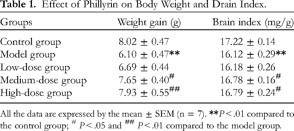
All the data are expressed by the mean ± SEM (n = 7).
Effect of Phillyrin on Memory Impairments in D-Gal-Induced Mice
The MWM test was conducted to evaluate the influence of phillyrin on the spatial cognitive competencies in mice induced by D-gal. During five consecutive days of positioning navigation trials, the results showed that the time it took all five groups of mice to find the submerged escape platform decreased with the training days. However, the model group had a greatly longer escape latency on the fourth and fifth days of training relative to the control group (P < .05 or P < .01), and phillyrin addition markedly shortened escape latency (P < .05) (Figure 2A). Simultaneously, in the spatial probe test, mice in the D-gal-treated group traversed the platform significantly less than the control group, and it significantly elevated in the medium-dose and high-dose phillyrin groups than that in the D-gal treatment group. In addition, the percentage of time spent in the target quadrant of the model group was significantly less than in the control group, however, the medium-dose and high-dose phillyrin groups notably ameliorated this shortening situation (Figure 2B and C). As shown in Figure 2D, we also recorded the swimming trajectories of each group of mice, the swimming track of mice in the model group were obviously random and had no clear direction to find the platform in comparison with the control group, whereas those in the phillyrin group could found the target platform more quickly than that in the model group.

Effect of phillyrin on spatial learning and memory impairment in the MWM test (A-D). (A) Escape latency of positioning navigation trials. (B) The percentage of time spent in the target quadrant. (C) The crossing number of the platform. (D) Swimming trajectory of all groups of mice. All data were expressed as mean ± SEM (n = 7).
Effects of Phillyrin on Oxidative Stress in Serum and Brain Tissue
To investigate the protective effect of phillyrin against D-gal-induced oxidative stress in aging mice, the changes in oxidative stress markers such as SOD, GSH-PX, and MDA were observed. The changes in MDA levels in serum and brain tissues were similar, and MDA levels were markedly higher in the model group than that in the untreated group (P < .05), the administration of phillyrin caused a decrease in the MDA levels relative to the D-gal alone induced aging mice (P < .05, P < .01) (Figure 3C and F). In addition, in the brain tissue, the SOD activity of the model group markedly reduced relative to the control group (P < .05), and the SOD activity in each phillyrin group increased. However, there were no significant statistical differences compared to the model group. Further, for the GSH-PX, the activity in the model group was markedly decreased (P < .01), while the high-dose phillyrin group exhibited a notable effect in increasing the GSH-PX activity in brain tissue (P < .05). In serum, the results indicated a dramatic reduction in the activities of SOD and GSH-PX of the D-gal-treated aging group relative to the control group (P < .01, P < .05), and the medium-dose or high-dose phillyrin groups attenuated this reduction (P < .05).

Effect of phillyrin treatment on antioxidant enzymes and MDA in serum and brain tissue. (A) to (C) The activities of SOD, GSH-Px, and levels of MDA in the brain tissue. (D) to (F) the activities of SOD, GSH-Px, and levels of MDA in the serum. All values were presented as mean ± SEM from each group (n = 7).
Effects of Phillyrin on the Expression of Nrf2/HO-1 Signaling Pathway Components in Brain Tissue of Aging Mice Induced by D-Gal
In order to study whether Nrf2/HO-1, an oxidative stress response-related signaling pathway in the organism, takes a crucial role in the mechanisms of action of phillyrin in D-gal-induced aging, the expression levels of Nrf2, Keap-1, HO-1, and NQO1 in the hippocampus of each group were explored by western blot analysis. We observed that the Keap-1 protein expression levels were notably higher in the D-gal model group than in the control group (P < .01), while following 8 weeks with the administration of phillyrin, the expression levels were decreased (P < .01). In addition, the expression levels of Nrf2 in the model group reduced compared to the control group (P < .05), whereas the expression levels were higher in the phillyrin medium or high-dose treatment group (P < .01). Agreement with the above Nrf2 expression level results, the relative levels of HO-1, NQO1 in the model group were decreased in comparison with that in the control group (P < .01, P < .05), Conversely, addition the phillyrin attenuated the downregulation of those protein levels (P < .05, P < .01) (Figure 4).

Effect of phillyrin treatment on the expression of Nrf2/HO-1 signaling pathway components in brain tissue. Data are presented as mean ± SEM from each group (n = 3).
The Effects of Phillyrin on Brain Hippocampus Histology in D-Gal-Induced Aging Mice
HE staining was performed to study the effect of phillyrin on the morphology of brain tissue. The results revealed histopathological changes of phillyrin in hippocampal CA3 of mice. The cell morphology of CA3 in the control group was mostly normal and the structure was intact and clear, further, these nuclei were large and round, with clear nucleoli and no apparent apoptosis. In contrast, the model group exhibited the cell's shape was irregular and the structure was loosely, accompanied by nuclear pyknosis, while these adverse changes in pathology were significantly lower following phillyrin pretreatment (Figure 5).

Effect of phillyrin treatment on hippocampal CA3 histopathological alterations (hematoxylin and eosin [HE] staining, magnification 200 ×). (A) Control group, (B) model group, (C) low-dose group (5 mg/kg), (D) medium-dose group (15 mg/kg), and (E) high-dose group (45 mg/kg).
Discussion
Aging is a biological process that human beings cannot avoid. Further, as the rapid population aging, age-related degenerative diseases like Alzheimer's disease and Parkinson's disease are increasing, 25 these will undoubtedly bring great challenges to our quality of life and economy. In addition, our country, China, with an increasing trend of the elderly population, is one of the most rapidly aging countries in the word. 26 Therefore, how to delay aging scientifically is becoming an urgent issue. In recent years, Chinese medicine, owing to the natural herb, has received extensive attention for the treatment of anti-aging and aging-related diseases. 27 Phillyrin, an active substance of traditional Chinese medicine Forsythia suspensa, is often used as a quality control index to evaluate Forsythia suspensa. 28 It has also been used for some treatments, such as antioxidant, anti-inflammatory, antiviral, and so on. The present research was explored to investigate the anti-aging activity of phillyrin and its underlying mechanism. Firstly, we established the mice aging model by intraperitoneal administration of D-gal (150 mg/kg) for 8 weeks. D-gal is a reducing sugar of the organism, 29 it is converted into hydrogen peroxide (H2O2) and aldehydes by galactose oxidase as its levels exceeds the normal range, promoting the production of ROS and eventually leading to aging.30,31 Thus, D-gal intervention has been successfully applied as one of the methods for establishing an aging animal model. In the present experiment, body weight and organ index were measured as physical indicators to investigate the effects of phillyrin on protection against D-Gal-induced aging. The results showed that the weight gain of the model group was lower than the control group, which was consistent with previously reports. 28 It is widely believed that senescence could lead to organ atrophy in the body, just as expected, the results showed that the brain index in the aging model group decreased significantly compared to the control group. Further, the brain index and weight gain of the mice increased after treatment with phillyrin. The above results fully suggested that phillyrin could increase these decrease induced by D-gal.
The water maze test is a common behavioral research method for testing the cognitive abilities of animals. 32 It is well known that the learning and memory ability of the body declines to vary degrees with aging, owing to oxidative stress and neurodegenerative changes in brain tissue. 33 So in the water maze experiment, the present results showed that although with the extension of training time, it took longer time to find the underwater platform for the D-gal treatment group mice, which was in accordance with previous studies, such as the learning deficits with the impairment of MWM performance in D-gal induced senescence. 34 The results indicated we successfully established the aging model. Further, in the spatial probe test, the crossing times of the platform and the percentage of time spent in the target quadrant of the model aging group were markedly higher than the control group, however, addition with phillyrin of medium-dose or high-dose could reverse these situations. These findings indicated that phillyrin could attenuate memory damage and improve cognitive function. Based on the above experiments, HE staining on the brain tissue of mice was carried out, and we observed the morphological changes of organs, tissues, and cells to confirm whether the body had pathological damage. 35 And the pathological results of brain tissue also proved that the CA3 region of hippocampus structure in the aging model mice were disordered and the nucleus were pyknotic. After treatment with phillyrin, these changes were significantly alleviated. In conclusion, phillyrin could attenuate brain damage and improve cognitive ability in aging mice. As such, phillyrin could be a viable anti-aging pharmaceutical compound.
Oxidative stress is critical in aging. With the occurrence of aging, the balance of the oxidation and anti-oxidation system was broken, leading to the increase of free radicals, which subsequently lead to cell damage, tissue, and organ disorders. 36 Previous studies have shown that antioxidant enzymes can prevent cells against damage caused by free radicals. 37 SOD and GSH-Px are crucial antioxidant enzymes in the oxidative defense system, SOD can remove excessive free radicals in the body and protect cells from damage, and is the first gate of defense for the antioxidant defense system. 38 GSH-Px could catalyze the decomposition of hydrogen peroxide in the body and specifically convert reduced glutathione (GSH) to oxidized glutathione (GSSG), thereby protecting the structural and functional integrity of cell membranes. 39 MDA is the product of lipid peroxidation, which can lead to cell membrane damage. So the level of MDA could indicate the severity of free radical damage to the body's cells. 40 We found significant reductions in the serum and brain tissue SOD, and GSH-Px activities in the aging model mice, which suggested oxidative stress and associated damage caused in the body induced by D-gal. Whereas the phillyrin treatment significantly improved the activities of SOD and GSH-Px as well as decreased the content of MDA in the serum and brain tissue compared to the model aging group, showing that phillyrin could enhance the anti-oxidative capacity of the organism. Also, our results were in agreement with previous studies of the antioxidant effect of phillyrin, for example, Yan et al. revealed that the phillyrin treatment had significantly higher SOD, GSH-Px, and T-AOC activities and lower MDA content in the serum and brain compared with the normal mice. 19 So our study further confirms that phillyrin can attenuate D-gal-induced aging by regulating SOD, GSH-px, and MDA contents.
To further explore the mechanisms of anti-aging by phillyrin, we have performed a WB assay. Results showed the decreased expression of Nrf2 and the increase of Keap-1 in the D-gal-induced aging group. Similar results were observed in Chen's study, that patchouli alcohol inhibits oxidative stress induced by the D-gal and improves the quality of aging cartilage. 41 Previous researches have shown the Nrf2 signaling pathway is one of the most markable endogenous anti-oxidative stress pathways. Nrf2 is an important transcriptional regulator and improving its activity could alleviate senescence. 42 For example, Chen et al. suggested that Leonurine has an anti-aging effect and could be related to the up-regulation of the Nrf2 signaling pathway. 15 Under normal circumstances, Nrf2 is anchored by Keap-1 in the cytoplasm, which promotes Nrf2 ubiquitination and proteasomal degradation. However, if the body is subjected to oxidative stress, the interaction between Nrf2 and Keap-1 will be disrupted and Nrf2 translocates to the nucleus and combines with ARE to regulate the genes expression of downstream antioxidant enzymes such as HO-1, NQO1, and so on, which take a significant part in regulating the oxidative response of the organism.43,44 For example, trehalose promoted the expression level of Nrf2 by blocking the binding of Keap1 to Nrf2 in the liver of D-gal-induced aging mice, 6 the result was in consistent with our study, we observed that phillyrin activated the expression of Nrf2 and reduced level of its negative regulator Keap-1 compared to the model mice. Further, downstream Nrf2 pathway members HO-1 and NQO1 expression increased by phillyrin treatment. All in all, the above results suggest that the protective effect of phillyrin against aging is partially attributable to the activation Nrf2 signaling pathway and inhibition of D-gal-induced oxidative stress. However, there are limitations in our research, for example, as a nuclear transcription factor, Nrf2, it would be interesting to perform cellular fractionation to obtain differential abundance between nuclei and cytosol. Additionally, it would provide a piece of evidence for the conclusion that phillyrin facilitated ROS clearance by performing a ROS staining assay. Furthermore, Bach1, as a transcription factor, is also a negative regulator of Nrf2, 45 Bach1 can repress the expression of antioxidant genes by binding to ARE, whereas Nrf2 activates the expression of antioxidant genes at the same loci after being exposed to stress responses, 46 for which it is necessary to closely follow the changes in Bach1 in our future experiments.
Conclusion
Overall, we confirmed that phillyrin treatment could improve cognitive activity and reduce D-gal-induced oxidative stress in mice. Furthermore, the underlying mechanism of phillyrin in aging mice may be through activation of the Nrf2/HO-1 signaling pathway to alleviate oxidative stress. The above results indicate that phillyrin is a promising agent that could provide a strategy for reducing aging.
Footnotes
Acknowledgments
We thank Haifeng Zhao for providing writing assistance and for providing language help (Department of Nutrition and Food Hygiene, School of Public Health, Shanxi Medical University, Taiyuan 030001, Shanxi, PR China). In this study, Xuemin Li was responsible for the design of the experiment and the writing of the article, Ling Wang was responsible for the conduct of the experiment. Other authors (Chenyang Li, Ruotao Tian, Yongfei Bai, Xiaohong Zhang, Shuqin Li) have contributed their own efforts to animal behavior testing, data analysis, and cartography. Here, we would like to express our gratitude. This work was supported by the Fundamental Research Funds from the Center for Disease Control and Prevention in Shanxi Province (No. 2021020).
Data Availability Statement
The data presented in this study are available on request from the corresponding author.
Declaration of Conflicting Interests
The authors declared no potential conflicts of interest with respect to the research, authorship, and/or publication of this article.
Funding
The authors received no financial support for the research, authorship, and/or publication of this article.
Institutional Review Board Statement
This study was approved by the Ethics Committee of the Center for Disease Control and Prevention in Shanxi Province (Approval ID: 2021003).
Informed Consent Statement
Not applicable.