
Abstract
Schisandra sphenanthera has been used as a Chinese traditional medical herb with its restorative and antioxidant functions for hundreds of years. Anwulignan is a monomeric compound isolated from the total lignans of S. sphenanthera. Our previous study showed that anwulignan has a strong antioxidant activity in vitro. However, it is unclear if anwulignan possesses any function in improving learning and memory. In this study, we examined the effect of anwulignan on the learning and memory impairment induced by
Nowadays, intensive efforts have been spent on the development of medicine and food supplement to prevent patients from suffering learning and memory impairment, which is attributed to oxidative damages of neurons. 1 The dried ripe fruit of Schisandra sphenanthera Rehd. Et Wils (Schisandraceae) with its restorative and antioxidant functions has been widely used as a traditional medicine. 2 We wondered if S. sphenanthera or its ingredient has any effect on learning and memory impairment. Anwulignan 3 is a main monomeric compound isolated from Schisandraceae total lignans. Our previous studies showed that anwulignan has a strong antioxidant activity in vitro. In this study, we investigated if anwulignan could reduce learning and memory impairment in a mouse model.
To examine the effects of anwulignan on learning and memory impairments, the step-through test and Morris water maze were performed for the behavioral activities of mice. Step-through test is an animal model developed to detect the learning and memory ability of small animals.
4
Rodents are nocturnal animals that like a dark place and avoid a bright place. When they are given electrical stimulation in the dark place, they are forced to escape to a bright place and acquire the memory. The latency and error times can be used as indices to judge the memory strength. Institute of Cancer Research (ICR) mice were treated with 220 mg kg−1

Effects of anwulignan on the learning and memory of
The Morris water maze is a classic test for the spatial memory, which consists of a navigation trial and a probe trial. Using this test, we examined anwulignan for its effect on spatial memory in mice. The result of the navigation trial is shown in Figure 1C. During the first 3 days, the escape latencies of all groups appeared to decrease but the changes were statistically insignificant. However, on the fourth and fifth days,
In the

Pathological changes of the hippocampal tissue of mice under an optical microscope (HE ×200). Black arrows: damaged neurons (the nucleus was round or pyramidal; the nuclear chromatin was concentrated and deeply stained; the cytoplasm was red stained).
In order to determine the antioxidation property of anwulignan, superoxide dismutase (SOD) activity and malondialdehyde (MDA) content were measured. As shown in Figure 3, in comparison with the control group, SOD activities were significantly reduced (P < 0.01) and MDA levels were significantly increased (P < 0.01) in the blood and the hippocampal tissue of mice in the model group. In comparison with the model group, in anwulignan groups, SOD activities were significantly increased both in the blood (at 2 and 4 mg kg−1, P < 0.05 for both) and in the hippocampus of mice (at 2 and 4 mg kg−1, P < 0.05 and P < 0.01) and MDA levels were significantly decreased in both the blood and the hippocampus (at 2 and 4 mg kg−1, P < 0.05 and P < 0.01, respectively). These results showed that anwulignan has an antioxidant capacity.

Effects of anwulignan on SOD activities and MDA levels in the blood and hippocampal tissue of the mice (mean ± SD, n = 15). (a) SOD activities; (b) MDA levels. *P < 0.05, **P < 0.01, as compared to the model group; ## P < 0.01, as compared to the control group. SOD, superoxide dismutase; MDA, malondialdehyde.
It has been known that p38 mitogen-activated protein kinase (MAPK)-nuclear factor [erythroid-derived 2]-like 2 (Nrf2)-heme oxygenase 1 (HO-1) signaling pathway plays an important role in the protection of oxidative stress injury. 6 It is unclear whether anwulignan has any effect on this pathway. Therefore, we examined the expression of p38 MAPK, Nrf2, and HO-1 in the hippocampus of mice by reverse transcription polymerase chain reaction (RT-PCR) and western blot.
As shown in Figure 4, the expression of Nrf2 and HO-1 in the hippocampus of mice in the model group as well as all anwulignan groups was significantly upregulated (P < 0.05 or P < 0.01) in comparison to that in the control group. P38 MAPK remained unchanged. Interestingly, the expression of Nrf2 and HO-1 genes in the hippocampus of anwulignan group (only at 4 mg kg−1) was further increased (P < 0.05) in comparison to that in the model group. It is very fascinating that both
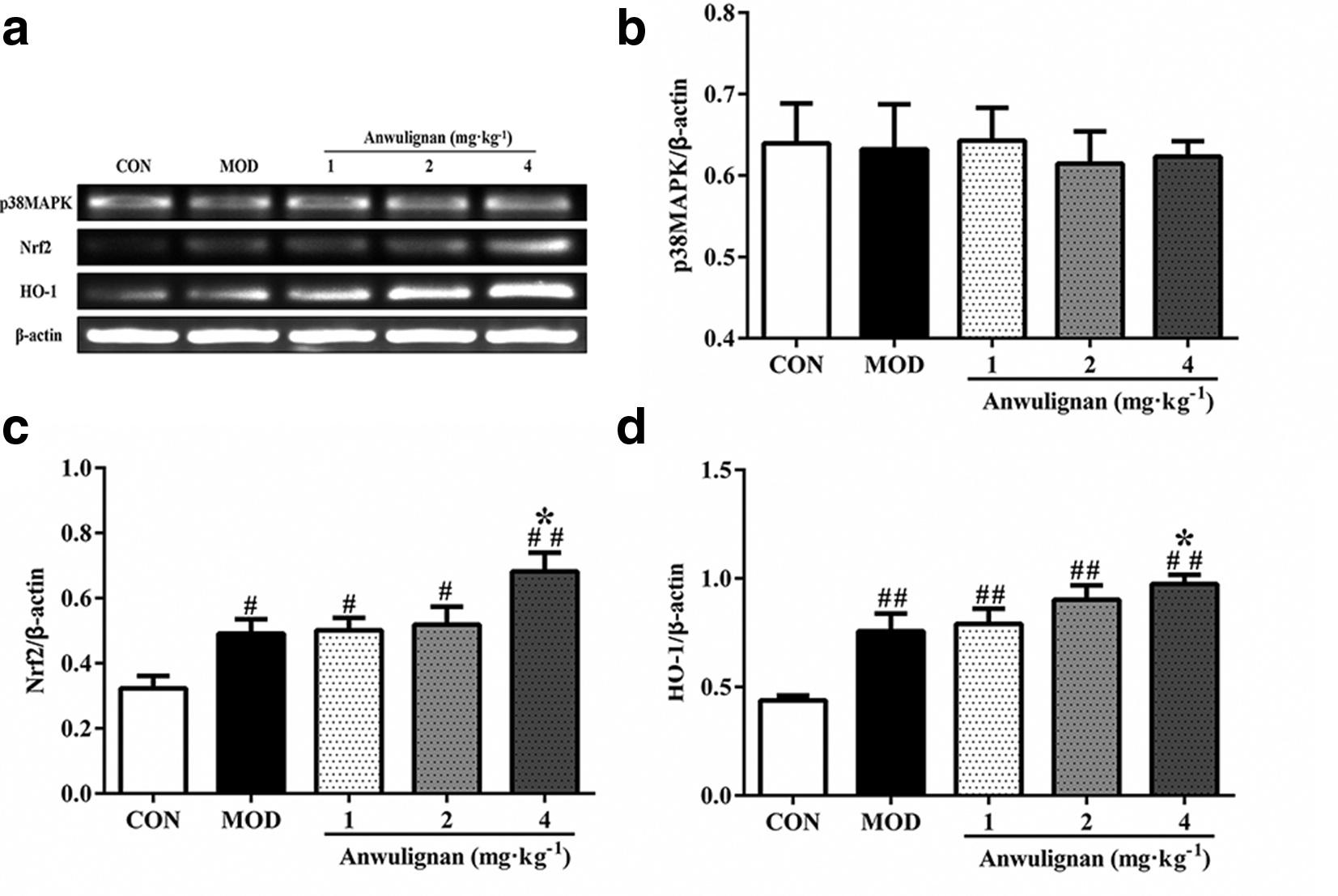
Effects of anwulignan on the expression of p38 MAPK, Nrf2, and HO- 1 mRNA in the hippocampus of the mice (RT-PCR; mean ± SD, n = 3). (a) Agarose gel electrophoresis image of p38 MAPK, Nrf2, HO-1, and β-actin. (b) Column chart of p38 MAPK/β-actin. (c) Column chart of Nrf2/β-actin. (d) Column chart of HO-1/β-actin. # P < 0.05, ## P < 0.01, as compared to the control group; *P < 0.05, as compared to the model group. MAPK, mitogen-activated protein kinase; Nrf2, nuclear factor [erythroid-derived 2]-like 2; HO-1, heme oxygenase 1 RT-PCR, reverse transcription polymerase chain reaction.
The western blot results are illustrated in Figure 5. The phosphorylation of p38 MAPK, Nrf2 ,and HO-1, but not p38 MAPK, was significantly upregulated (P < 0.05 or P < 0.01) in the hippocampus of mice in the model group as well as all anwulignan groups in comparison to those in the control group. Meanwhile, anwulignan at 2 and/or 4 mg kg−1 increased these expressions significantly in comparison to the model group (P < 0.01 for all). The protein levels of these components were consistent with those in the mRNA levels. The expression of p38 MAPK in both mRNA and protein levels was found not to be changed significantly, but the phosphorylation of p38 MAPK (the ratio of p-p38 MAPK/p38 MAPK) was significantly increased (P < 0. 01 for all) in the model group (compared with the control group) and in anwulignan groups (at 2 and/or 4 mg kg−1, in comparison with the model group).
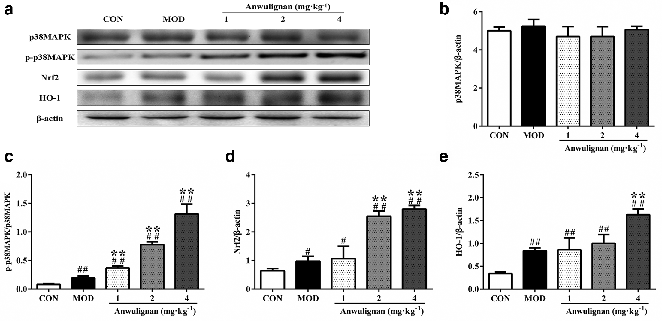
Effects of anwulignan on the expression of p38 MAPK, p-p38 MAPK, Nrf2, and HO-1 proteins in the hippocampus of the mice (western blot; mean±SD, n = 3). (a) Electrophoresis image of p38 MAPK, p-p38 MAPK, Nrf2, HO-1, and β-actin. (b-e) Column charts of p38 MAPK/β-actin, p-p38 MAPK/p38 MAPK, Nrf2/β-actin, and HO-1/β-actin. # P < 0.05, ## P < 0.01, as compared to the control group; *P < 0.05, **P < 0.01, as compared to the model group. MAPK, mitogen-activated protein kinase; Nrf2, nuclear factor [erythroid-derived 2]-like 2; HO-1, heme oxygenase 1.
According to the reports, accumulation of free radicals may partly contribute to the learning and memory impairment.
7
The hippocampus takes on an important role of declarative memory, cognition and location navigation, and information processing.
9
There is an evidence showing that the destruction of bilateral hippocampi causes an obvious decline of learning and memory, and the long time oversupply of
In this study, we specially measured the activation of p38 MAPK-Nrf2-HO-1 pathway. p38 MAPK is a class of MAPKs that are responsive to stress stimuli.
11
Oxidative damage can activate the phosphorylation of p38 MAPK (p-p38 MAPK),
12
then, p-p38 MAPK stimulates Nrf2 to be dissociated from Keap1. Nrf2 is a basic leucine zipper protein that regulates the expression of antioxidant proteins against oxidative damage triggered by injury.
13
After the dissociation, Nrf2 enters the nucleus, binds to antioxidative response element, and promotes the expression of HO-1.
14
HO-1, the final and key element of this pathway, is an inducible isoform of hemeoxygenase, which is a powerful free radical scavenger in the body.
15
Our results showed that the intracellular p38 MAPK-Nrf2-HO-1 signaling pathway was activated by not only anwulignan, but also
Experimental
Animals
Male ICR mice, weighing 20 ± 2 g, were provided by the Experimental Animal Research Center of Jilin University with the license number of SCXK (Ji): 2016-0005. The mice were raised in separate cages and in a diurnal cycle of 12 hours:12 hours and were fed based on the AIN93G diet. The animal experiments were approved by the Institutional Animal Care and Use Committee (IACUC) (2017-03) of Beihua University.
Animal Administration and Experimental Protocol
ICR mice were randomly divided into 5 groups: control group (distilled water orally and normal saline subcutaneously), model group (distilled water orally and 220 mg kg−1
Step-Through Test
On the first day, the mice were placed in a light compartment. They could go into the dark compartment and return to the light compartment through a door between the compartments freely in a DT-200 step-through (Chengdu Taimeng Software Co., Ltd. China) apparatus for 3 minutes of familiarization. Then, on the second day, the mice were again placed in the light compartment with the door opened, but the power was turned on. When the mice stepped into the dark compartment, they would suffer an electric shock and return to the light compartment. The test lasted for 5 minutes. The latency (the time for the mice to first step into the dark compartment) and error time (the total time for mice to step into the dark compartment) were recorded. A retention test was conducted 24 hours later, and the mice went through the same process. 16
Morris Water Maze Test
A MT-200 Morris water maze (Chengdu Taimeng Software Co., Ltd. China) video tracking test system was used for this test. The pool was filled with water (23 ± 1°C). The platform was placed in the center of the first quadrant and 2 cm below the water surface. This test lasted for 6 days. The first 5 days were for the navigation trials, and the last day was for a probe trial. In the navigation trial, mice were trained twice a day. In the probe trial, the platform was removed, and the mice were allowed to swim for 120 seconds and the swimming paths were recorded and analyzed to obtain the numbers of the mice crossing the platform (the crossing numbers) and the duration the mice stayed in the first quadrant (the staying time), in which the platform had been placed. The performance on the fifth and sixth days was regarded as the final result of the navigation trial and probe trial and used to assess the space memory of mice. 17
Assay of SOD and MDA in the Hippocampus and Blood
The detection of SOD and MDA was conducted following a specific protocol. Fifteen mice from each group in step-through test were sacrificed with anesthesia using ether at 30 minutes after the last administration. Then their serum samples were collected and hippocampal tissues were taken. SOD activities in the serum and hippocampal tissue were assayed by WST-1 method and the MDA levels were measured by thiobarbituric acid method according to the kit instructions.
Histological Analysis
After completing Morris water maze test, three mice in random were anesthetized with ether and sacrificed by decapitation to obtain the hippocampus samples. The hippocampus was fixed with 10% formalin, embedded with paraffin, and sliced into pieces of 5 to 10 μm thickness. The slices were stained by H&E (hematoxylin-eosin) staining and examined under an OLYMPUS optical microscope (OLYMPUS company, Japan) for pathological changes and photographed.
Reverse Transcription PCR
After Morris water maze test, three mice in random were anesthetized with ether and sacrificed by decapitation to obtain the hippocampus samples. Total RNA was isolated and cDNA was synthesized using a reverse transcription reaction kit. RT-PCR amplification reaction was performed according to the kit instructions with specific primers designed with primer software 6 and synthesized by Beijing DingguoChangsheng Biotechnology Company (Beijing, China). β-actin was taken as the internal reference. The primers are shown in Table 1.
Primer Sequence.

MAPK, mitogen-activated protein kinase; Nrf2, nuclear factor [erythroid-derived 2]-like 2; HO-1, heme oxygenase-1.
The PCR amplification condition consists of pre-denaturation at 94°C for 3 minutes, denaturation at 94°C for 30 seconds, annealing for 30 seconds (annealing temperature: 55°C for p38 MAPK, 58°C for Nrf2 and HO-1, 63.5°C for β-actin), and extension at 72°C for 30 seconds, with 30 cycles; then extension at 72°C for 7 minutes to make unsynthesized DNA molecules to complete synthesis, and finally the products are stored at 4°C. About 10 µL PCR products was electrophoresed on agarose gel, in which the results were visualized and photographed using Tanon 1600 Gel Image system (Shanghai Tianneng Technology Co., Ltd. Shanghai, China).The density of the photographs was scanned by the gel imaging system, and quantitated for p38 MAPK, Nrf2, and HO-1 mRNA amount.
Western Blot
The hippocampus samples were treated with lysis buffer for ice cracking for 1 hour, and then the lysates were centrifuged at 16 000 × g to obtain the supernatant. The protein concentration was determined by bicinchoninic acid method, and 10% sodium dodecyl sulfate-polyacrylamide gel electrophoresis was used to separate Nrf2, HO-1, p38 MAPK, and p-p38 MAPK. The proteins were transferred onto polyvinylidene fluoride (PVDF) membrane for 2 hours, and then blocked with the blocking buffer (tris-buffered saline-T buffer containing 5% skim milk powder) for 1 hour and the primary antibodies of Nrf2 (1:1000), HO-1 (1:1000), p38 MAPK (1:1000), and p-p38 MAPK (1:1000) were added onto the membrane, respectively. PVDF membrane was incubated at 4°C overnight . Then the second antibody (horseradish peroxidase goat anti-rabbit IgG antibody) (1:2000) was added and incubated for 2 hours. Finally, the membranes were developed with ElectroChemi-Luminescence (ECL) color solution.
Statistical Analysis
The experimental data were presented in mean ± SD. Statistical analysis was performed by one-way analysis of variance, followed by Tukey’s test for multiple comparisons using SPSS software (SPSS Inc., Chicago, IL,USA). Differences were considered to be significant when P value was less than 0.05.
Footnotes
Declaration of Conflicting Interests
The author(s) declared no potential conflicts of interest with respect to the research, authorship, and/or publication of this article.
Funding
The author(s) disclosed receipt of the following financial support for the research, authorship, and/or publication of this article: This project was supported by the Grants from Jilin Provincial Science and Technology Department (20170309006YY, 20150311047YY), the Education Department of Jilin Province (JJKH20180376KJ), and the Health Department of Jilin Province (2018J089).