
Abstract
Primary lung cancer, a high-mortality disease, is divided into small cell lung cancer (SCLC) and non-SCLC. The 5-year survival rate of lung cancer is ∼15%. Berberine (C20H18NO4), obtained from, for example, Coptis chinesis, C. deltoidea, and C. teewall (Figure 1), is a traditional Chinese and Mongolian medicine for treating inflammatory diarrhea.1–3 Berberine has shown anti-inflammatory, antidiabetic, and antitumor effects,4–6 can inhibit cancer proliferation and tumor metastasis, and induce tumor cell differentiation and apoptosis. Micro RNAs (miRNAs), a group of highly conserved endogenous noncoding small RNA molecules with a length of 18-23 bases,7,8 help regulate various biological processes, including malignant tumors. miRNAs bind to the 3′-UTR region of the target gene, form a silencing complex, and inhibit the protein translation of the target gene. miRNA molecules are extensively involved in tumor cell proliferation, apoptosis, invasion, and metastasis and also regulate tumor cell autophagy and apoptosis. 9 miR-144 can increase anemia and reduce glutathione regeneration and antioxidant capacity. 10 miR-144 is poorly expressed in liver, lung, and prostate cancer cells.11–13 miR-144 appears closely related to human cancer,10,13–15 and upregulating its expression may promote the proliferation of cervical cancer cells.
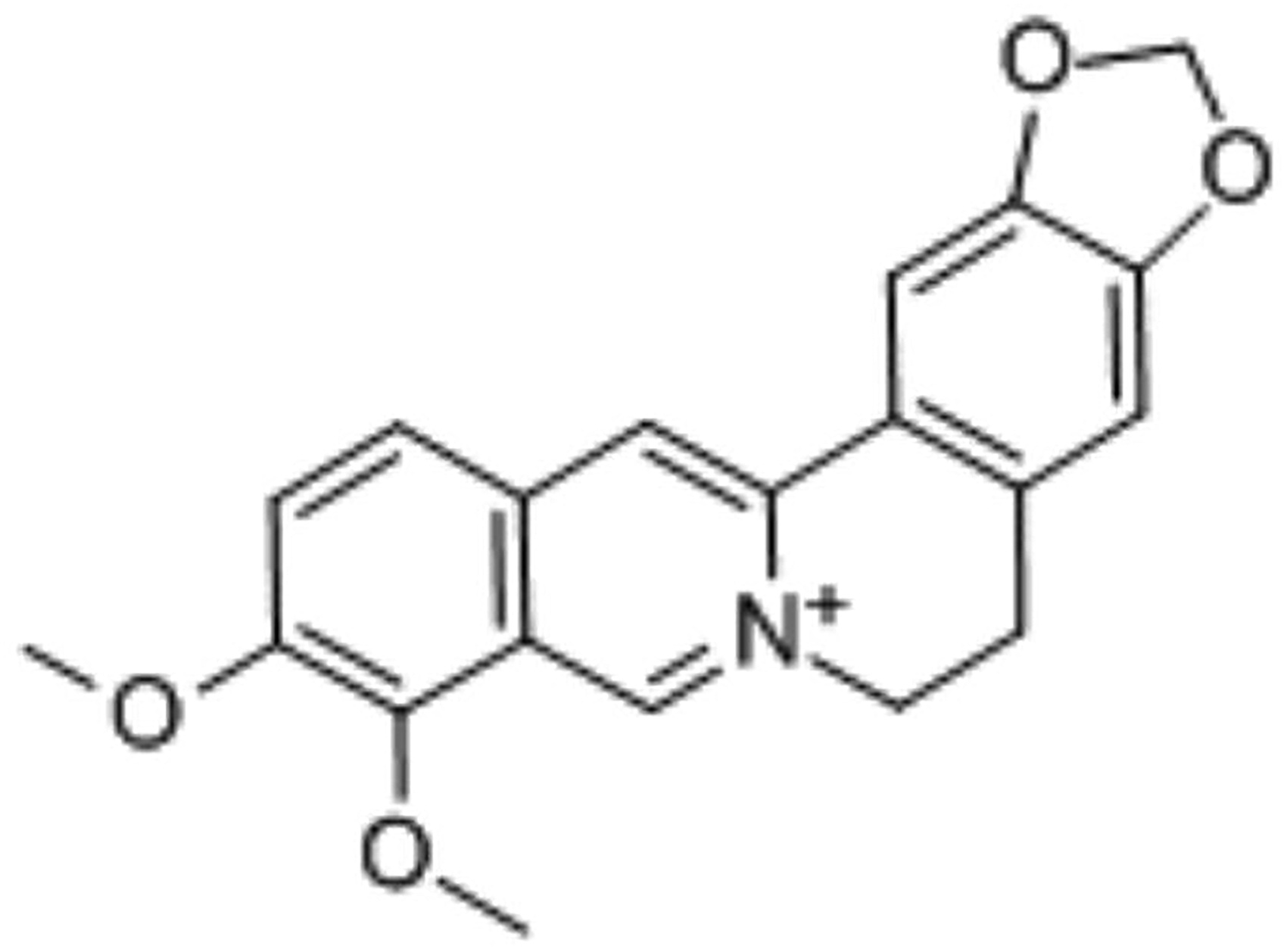
The structure of berberine.
We have used several botanical medicines to treat adenocarcinomic human alveolar basal epithelial cells (hereafter referred to as A549 cells) and experienced promising outcomes with Huanglian. These experiences led us to investigate the effects of berberine on A549 lung cancer cells. Additionally, we explored changes in miR-144 expression and the apoptosis and autophagy pathways.
Materials and Methods
Berberine (purity >98%) was purchased from the Chinese National Institute (Beijing, China). A549 cells (Chinese Academy of Sciences, China) were cultured at 37 °C in 5% CO2.
Cell Proliferation
After berberine treatment, cell proliferation was assessed using a cell counting kit-8 (CCK-8) (K1018, Apexbio) kit. Cells (1 × 10⁴ cells per well) were plated in 96-well plates (100 μL/well), and 10 μL of CCK-8 solution was added at 24, 48, and 72 h for 2 h at 37 °C. The absorbance values at 450 nm were then measured using an enzyme marker. The cell growth rate was calculated using the following equation: % growth rate = [(mean experimental absorbance-mean blank absorbance)/(mean control absorbance-mean blank absorbance) × 100; each test was repeated 5 times.
Determination of miRNA Levels With qRT-PCR
Total miRNA, isolated from the A549 cells, was reversely transcribed into complementary DNA using the TIANScript RT kit (Tiangen Biotech, China). Then, the SYBR Premix Ex Taq TM II kit (Takara) was used to determine relative gene expression levels and enrichment. The miRNA was quantified with a SYBR-Green PrimeScript miRNA RT-PCR kit (Takara).
Western Blotting
Total cellular proteins were extracted using a high-performance radioimmunoprecipitation analysis lysis buffer (C0481, Sigma-Aldrich) containing 1% protease inhibitor and 1% phosphatase inhibitor (Beyotime). The protein concentration of each sample was determined using a bicinchoninic acid kit (23227, Thermo Fisher Scientific). The proteins were separated by polyacrylamide gel electrophoresis and transferred to polyvinylidene fluoride membranes (Millipore), which were blocked with 5% bovine serum albumin at room temperature for 1 h. Primary antibodies were incubated with membranes overnight at 4 °C. Next, the membranes were incubated with horseradish peroxide–labeled goat antirabbit immunoglobulin G (ab205718, Abcam) dilutions at room temperature for 1.5 h. After incubation, the membranes were developed using developing liquid (NCI4106, Pierce). Proteins were quantified using Clinx Chemi Analysis software (ChemiScope 6000).
Cell Transfection
The miR-144 inhibitor (siRNA-GUACAUCAUCUAUACUGUA) was purchased from GenePharma. Cell transfection was conducted using Lipofectamine 3000 (Thermo Fisher Scientific). After 48 h, the cells were harvested for experimental analysis.
Statistical Analysis
Statistical analyses of data were performed using Graphpad Prism 9.0 (GraphPad Software). Where appropriate, data are expressed as mean ± standard deviation. Independent sample t tests were used for between-group comparisons; one-way analysis of variance (ANOVA) was used for multiple-group comparisons, followed by Tukey's post hoc tests. Finally, between-group comparisons at different time points were performed using 2-way ANOVA. A P < .05 indicated statistical significance.
Results
The Effects of Berberine on A549 Proliferation
CCK-8 was used to explore the effects of berberine function on A549 proliferation. Different berberine concentrations were added to the A549 cells at 12, 24, 36, and 48 h. A549 proliferation was reduced with increasing berberine concentrations (Figure 2). The optimal growth percentage was 36 h.

A549 cell growth percentage following exposure to berberine (0, 20, 40 µg/mL) for 12, 24, 36, and 48 h. Each experiment was performed 5 times.
Berberine Upregulated miR-144 Expression
The miR-144 expression was detected by quantitative PCR (Figure 3). miR-144 expression increased with increasing berberine concentrations.

The upregulation of miR-144 following berberine (0, 20, and 40 µg/mL) administration in A549 cells after 36 h. *P < .05 versus 0 group; #P < .05 versus 20 µg/mL group. Each experiment was performed 5 times.
Berberine Affects Apoptosis and Autophagy
To detect the influence of berberine on the apoptosis pathway, the expression of caspase-3, caspase-3 cleaved, Bcl-2, and Bax was detected (Figure 4A). We found that caspase-3/caspase-3 cleaved and Bcl-2/bax (Figure 4B and C) gradually decreased as the berberine concentration increased. Berberine also affected the apoptosis and autophagy pathways. Here, we explored the expression of beclin-1, LC3I, and LC3II (Figure 4D) and observed that each gradually rose with increasing berberine concentrations (0-40 µg/mL) (Figure 4E and F).

Effects of berberine on apoptosis (caspase-3, caspase-3 cleaved, Bcl-2, and Bax) and autophagy (beclin-1, LC3I, and LC3II) by berberine (0, 20, and 40 µg/mL). (A) Representative western blots showing apoptosis pathway proteins: caspase-3, caspase-3 cleaved, Bcl-2, Bax, and GAPDH protein detection. (B and C) Histograms summarizing results from (A) for caspase-3/caspase-3 cleaved and Bcl-2/bax, respectively. (D) Representative western blots showing autophagy pathway proteins: beclin-1, LC3I, LC3II, and GAPDH protein detection. (E and F) Histograms summarizing results from (D) for beclin-1, LC3I, and LC3II, respectively. *P < .05 versus 0 group; #P < .05 versus 20 µg/mL group. Each experiment was performed 5 times.
miR-144 Function in A549 Cells After Berberine Administration
We transfected a miR-144 inhibitor into A549 cells to explore the function of miR-144. The expressions of caspase-3/caspase-3 cleaved and Bcl-2/bax (Figure 5A) were higher in the miR-144 inhibitor group and miR-144 inhibitor + berberine group (Figure 5B and C). Beclin-1, LC3I, and LC3II were inhibited when miR-144 was silenced (Figure 5D). The expression of beclin-1, LC3I, and LC3II in the miR-144 inhibitor transferred groups was low (Figure 5C and D). A549 proliferation in the miR-144 inhibitor + berberine group with 20 µg/mL was higher than in the other groups. There were no significant differences between the miR-144 inhibitor group and the miR-144 inhibitor + berberine group (Figure 6).

Effects of berberine (20 µg/mL) on A54g after miR-144 inhibition. (A) Representative western blots showing apoptosis pathway proteins: caspase-3, caspase-3 cleaved, Bcl-2, Bax, and GAPDH. (B and C) Histograms summarizing results (A) for caspase-3/caspase-3 cleaved and Bcl-2/bax, respectively. (D) Representative western blots showing autophagy pathway proteins: beclin-1, LC3I, LC3II, and GAPDH. (E and F) Histograms summarizing results (D) for beclin-1, LC3I, and LC3II, respectively. *P < .05 versus 0 group; #P < .05 versus 20 µg/mL group. Each experiment was performed 5 times.
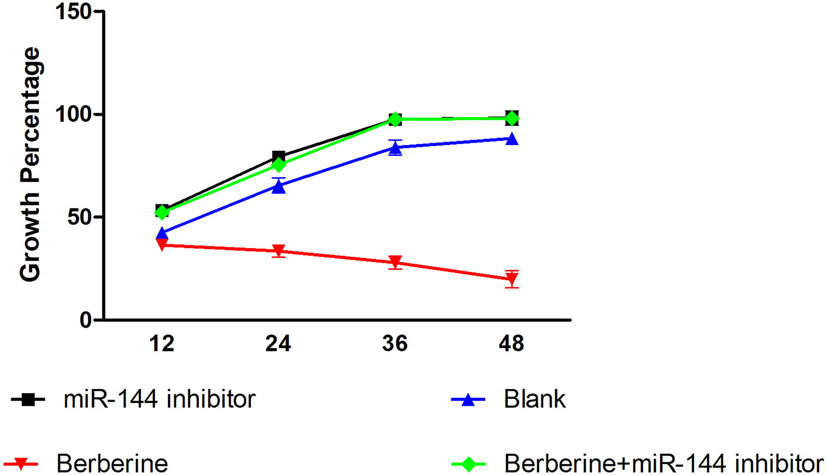
Effects of berberine (20 µg/mL) on A54g proliferation after miR-144 inhibition for 12, 24, 36, and 48 h.
Discussion
Berberine, an isoquinoline alkaloid extracted from several medicinal plant species,16,17 demonstrates various pharmacological effects. 5 We found that this alkaloid suppressed A549 proliferation through apoptosis and autophagy. Berberine promoted A549 cell apoptosis, mainly by inhibiting caspase-3 cleaved and Bcl-2 expression and promoting Bax expression. Thus, the expressions of caspase-3/caspase-3 cleaved and Bcl-2/bax were inhibited. Autophagy is a catabolic process correlated with various physiological procedures and pathological conditions. In the autophagic process, LC3 I—expressed in soluble form within the cytoplasm—transforms into LC3 II, which localizes on the autophagosome membrane.18–20 Beclin-1 is a protein that mediates autophagy.21,22 Interestingly, berberine also promoted A549 autophagy by increasing the expressions of beclin-1, LC3I, and LC3II.
miR-144 is downregulated in lung and hepatic breast cancer cells. In lung cancer, miR-144 is significantly downregulated. 10 We found that berberine upregulated miR-144 expression. Furthermore, A549 apoptosis and autophagy were enhanced after berberine administration. However, when miR-144 was inhibited, the miR-144 expression, apoptosis, and autophagy of A549 were reduced, with or without berberine (Figure 7). In future studies, a mouse model will be explored. In conclusion, berberine inhibited A549 proliferation and promoted apoptosis and autophagy through miR-144.

The mechanism of action of berberine.
Footnotes
Authors’ Note
The datasets used and/or analyzed during the current study are available from the corresponding author on reasonable request.
Declaration of Conflicting Interests
The author(s) declared no potential conflicts of interest with respect to the research, authorship, and/or publication of this article.
Funding
The author(s) received no financial support for the research, authorship, and/or publication of this article.