
Abstract
The skin plays various crucial biological functions and gradually degenerates due to the process of skin aging. Hence, the development of new ingredients for delaying or even reversing skin aging is an essential subject that needs to be addressed. Our screening effort for anti-skin aging ingredients has led to the discovery that Sideritis scardica extract (SSE) possesses collagenase inhibition, advanced glycation end product (AGE) formation inhibition, antioxidative, and antiallergic activities, as well as ultraviolet B (UVB)-induced matrix metalloproteinase-1 (MMP-1) expression inhibition activity. However, the effect of S. scardica, commonly known as a mountain herb, on skin aging is unknown and needs additional study. Further studies on SSE have led to the isolation of 3 flavonoids with an uncommon 8-hydroxyl moiety, isoscutellarein (
Introduction
The skin is the largest organ of the human body and acts as an anatomical barrier against both physical damage and pathogens. The importance of skin is not limited to the role of a barrier but also includes crucial thermoregulation and neurosensory functions. 1 Although the skin is essential for human physical and physiological functioning, it is not impervious to the effect of degeneration over the lifespan of a human being. Aged skin has a thinner epidermal layer, more pigmentation, reduced elasticity, increased wrinkles, and low collagen turnover. 2 The aging of skin can also impede its biological roles due to the loss of its structural integrity, which subsequently leads to the manifestation of various dermatological and even systemic diseases.2–5 The cause of skin aging can be divided into 2 separate processes, intrinsic and extrinsic.6,7 Intrinsic skin aging is the dilapidation of skin condition over time and is accompanied by biological aging. In contrast, extrinsic skin aging is caused by environmental factors such as ultraviolet (UV) light, physiochemical factors such as advanced glycation end products (AGEs), reactive oxygen species (ROS), and allergens, as well as physical damage.2,6,7
Exposure to UV radiation can cause skin aging via the degradation of collagenase, which then negatively affects collagen turnover in the skin. 2 Considering that collagen is an important component of the extracellular matrix that forms the epidermal layer of the skin, degradation of collagen can adversely affect the skin's physical integrity. 8 The degradation of collagen by UV is catalyzed by matrix metalloproteinase-1 (MMP-1), which is a type of collagenase.9–11 Both UV A (UVA; 320-400 nm) and UV B (UVB; 280-320 nm) are known to elevate MMP-1 expression.12–14 Although the exact mechanism by which MMP-1 is induced after exposure to UV radiation is unknown, studies have shown that MMP-1 gene expression is modulated by the p38 mitogen-activated protein kinase (MAPK) pathway in response to exposure to UV. 15 Another study has suggested that MMP-1 secretion is mediated by the protein kinase B (PKB/Akt) pathway. 16 In addition, agents such as AGEs, ROS, and allergens cause physiochemical damage to skin.17–23 AGEs are formed through the Maillard reaction, which is a nonenzymatic reaction between sugar and protein, 24 and the amount of AGEs in the body increases with aging and is a side effect of diabetes.17–19 The accumulation of AGEs in the cutaneous layer is one of the features of skin aging, as the crosslinking of AGEs such as pentosidine and carboxymethyl-lysine to collagen, elastin, and fibronectin decreases skin elasticity, tissue permeability, and repair mechanisms.25,26 It is also noteworthy that AGEs can also trigger an allergic reaction by binding to AGE-binding receptors that are present in mast cells, monocytes, and macrophages, which leads to the degranulation process that releases chemokines such as histamine.23,27 Furthermore, physiochemical damage resulting from ROS is also a known cause of skin aging.20,21 ROS can be derived from various sources, such as exposure to UVA, which is one of the primary causes of ROS generation in skin.21,28 Additionally, ROS are also generated in skin as a byproduct of enzymatic reactions, such as from the mitochondrial electron transport chain, nicotinamide adenine dinucleotide phosphate oxidases, xanthine oxidoreductase, or nonenzymatic reactions that form AGEs. 20 Irrespective of the sources of ROS, oxidative damage to skin cells can contribute to epidermal diseases such as psoriasis and skin cancer. 21
Sideritis scardica, commonly known as mountain herb, ironwort, or Mursalski, belongs to the Lamiaceae family. 29 The genus in which S. scardica is classified is comprised of plants that are xerophytic and usually grow in high-altitude areas of more than 1000 meters above sea level. S. scardica is endemic to the Mediterranean region, where it is known to be distributed in Albania, Bulgaria, Greece, and the Republic of Macedonia.29–31 In the Mediterranean region, it is traditionally consumed in the form of a tea that is locally known as mountain tea, Olympus tea, Pirin tea, or Mursalski tea. Traditionally, it is used for the treatment of inflammation, common cold, bronchitis, gastrointestinal disorder, pain, and rheumatic relief, as well as for reducing stress and anxiety, and some of these traditional claims have been verified scientifically.29,31,32 Scientific studies conducted on S. scardica have also indicated that it has antiobesity and insulin resistance prevention properties. 33 Additionally, promising effects of S. scardica on memory and cognitive functions in the elderly, as well as its antiseptic properties, have also been reported.32,34,35 However, the effect of S. scardica on skin aging was not known at the time of the preparation of this article. Phytochemical studies have revealed that the major classes of compounds found in S. scardica include mono-, di-, tri-, and sesquiterpenoids, fatty acids, esters, and flavonoids.29,32
Considering the crucial biological roles that are played by the skin, the development of new ingredients for delaying or even reversing skin aging is a subject that needs to be addressed, especially in aging societies. Furthermore, as world life expectancy continues to increase, anti-skin aging agents can also be viewed as an approach to improve the quality of life. For this reason, we explored S. scardica and its isolated compounds for their collagenase inhibition, AGE formation inhibition, anti-oxidative, and anti-allergic activities, as well for their effects on UV-induced MMP-1 expression. We also discuss the features of the isolated compounds that could lead to the observed anti-skin aging activities and the plausible mechanism for eliciting anti-skin aging activities in this paper.
Results
Bioactivities of SSE
In line with our search for ingredients that possess the potential to delay or reverse skin aging, we screened for ingredients with collagenase inhibition, AGE formation inhibition, and anti-oxidative properties. The screening results revealed that SSE dose-dependently inhibited the enzymatic activity of Clostridium histolyticum-derived collagenase from 62.5 to 500 μg/mL (Figure 1a). Apart from inhibiting collagenase activity, SSE also inhibited AGE formation in a dose-dependent manner (Figure 1b). However, the oxygen radical absorbance capacity (ORAC) value of SSE at 2912 ± 1153 µmol of Trolox equivalent (TE)/g also suggested that SSE possesses antioxidative activity (Table 1). In addition to the above-mentioned enzyme-based screening, cell-based screening also indicated that SSE possesses anti-allergic activity. The anti-degranulation activity on RHL-2H3 cell lines, which were used to evaluate SSE anti-allergic activity, showed that SSE inhibits the degranulation process in a dose-dependent manner (Figure 1c).

Bioactivities of SSE. (a) Collagenase inhibitory activity of 62.5 to 500 μg/mL of SSE on bacterial-derived collagenase. GSE was used as positive control. Each value represents the mean ± SD of 3 independent experiments. **P < .01 indicates significant compared to positive control calculated using Student's t-test. (b) AGEs formation inhibitory activity of 25 to 100 μg/mL of SSE. AG was used as a positive control. Each value represents the mean ± SD of 3 independent experiments. *P < .05 indicates significant compared to positive control calculated using Student’s t-test. (c) Anti-degranulation activity of 25 to 100 μg/mL of SSE on RBL-2H3 cells. WRT was used as a positive control. Each value represents the mean ± SD of 3 independent experiments. *P < .05 indicates significant compared to positive control calculated using Student's t-test. (d) Immunoblot analyses of MMP-1 expression on cell lysate harvested at 24, 48, and 72 h after irradiation with 5 mJ/cm2 of UVB. The expressions of MMP-1 relative to GAPDH were shown in the graph and expressed as means ± SEM. (e) The effect of 3 to 300 μg/mL of SSE on the expression of UVB-induced MMP-1 expression. The expressions of MMP-1 relative to GAPDH were shown in the graph and expressed as means ± SEM. *P < .05, **P < .01, and ***P < .0001 indicate significance compared to the control calculated using Dunnett's multiple comparisons test.
ORAC Values of SSE,
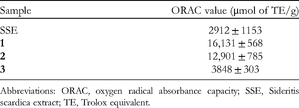
Abbreviations: ORAC, oxygen radical absorbance capacity; SSE, Sideritis scardica extract; TE, Trolox equivalent.
Next, we investigated the effect of SSE on the expression of MMP-1, which is a type of secretory collagenase that catalyzes the degradation of extracellular matrix collagen, 11 using UVB-irradiated normal human dermal fibroblasts (NHDF). Immunoblotting showed that UVB-induced MMP-1 expression increased in a time-dependent fashion (Figure 1d). Irradiation of NHDF with 5 mJ/cm2 UVB did not affect cell viability, while a slight decrease in cell viability was observed for 10 mJ/cm2 (data not shown). Therefore, we evaluated the effect of SSE on UVB-induced MMP-1 expression in NHDF at 72 h after irradiation with 5 mJ/cm2 UVB. Immunoblotting revealed that SSE inhibited UVB-induced MMP-1 expression in a dose-dependent manner, in which concentrations from 10 to 300 μg/mL showed significant inhibition activity (Figure 1e). Note that SSE did not significantly affect NHDF cell viability at concentrations up to 300 µg/mL (data not shown).
Structural Elucidation of Isolated Compounds from SSE
In continuation of the above findings, we proceeded to identify the bioactive compounds related to the above-mentioned activities. SSE was acid-hydrolyzed and further partitioned with EtOAc. Further separation and purification with repeated column chromatography and high-performance liquid chromatography (HPLC) of the EtOAc fraction resulted in the isolation of 3 flavonoids,

Structures and heteronuclear multiple-bond correlations (HMBCs) of
Bioactivities of the Isolated Compounds
The flavonoids that were isolated from SSE were evaluated for their collagenase inhibition, AGE formation inhibition, and anti-oxidative activities. All 3 compounds showed dose-dependent collagenase inhibition activity, in which

Bioactivities of
Regarding cell-based evaluations, the evaluation of the anti-degranulation activity of RBL-2H3 cell lines revealed that
Discussion
In view of the importance of skin to our well-being, the damaging effect of UV, AGEs, ROS, and allergens that lead to skin aging should not be taken lightly. Consequently, the development of ingredients that exhibit collagenase inhibition, AGEs formation inhibition, antiallergic, antioxidative, and UVB-induced MMP-1 expression inhibition activities would be essential for delaying or reversing skin aging to impart a younger-looking skin and even prevent skin diseases.
In our screening effort to search for an ingredient with anti-skin aging activities, we discovered that SSE possesses all of the above-mentioned properties. Interestingly, to our knowledge, S. scardica is not known to have anti-skin aging activities. Considering this gap in knowledge, we proceeded to further fractionate and purify the chemical components from SSE, which subsequently led to the elucidation of 3 flavonoids,
Considering that
In addition, analyses of the cell-based antiallergic activity revealed that the order of potency of
Taken together, all of the above observations suggested that the collagenase inhibition activity of
Conclusion
In summary, this study revealed that SSE showed dose-dependent anticollagenase, AGE formation inhibition, antioxidative, antiallergic, and UVB-induced MMP-1 expression inhibition activities. Subsequently, the 3 flavonoids isolated from SSE,
Materials and Methods
General Experimental Procedures
The 1H- and 13C-NMR spectra were measured on a JEOL ECP-600 spectrometer with tetramethylsilane as the internal reference. For isolation, a YMC multipreparative HPLC system (LC-Forte/R) was used. For analysis, a HITACHI ELITE LaChrom system was used. Absorbance and fluorescent analyses for the bioassay were measured with a Tecan infinite 200 microplate reader (Tecan, Japan Co., Ltd) and UV/VIS spectrometer, UV-1850 (Shimadzu).
Plant Material
S. scardica from Bulgaria was purchased through TAKA CORPORATION in 2020, and a voucher specimen was deposited at the herbal museum of Tokiwa Phytochemical Co., Ltd.
Preparation of SSE
The dried aerial parts of S. scardica (4 kg) were macerated and extracted twice with 50 wt% EtOH (88 L) under reflux for 1 h. The extracted solution was filtered, which was followed by treatment with activated carbon for 1 h. The filtrate was then filtered, and dextrin, emulsifier, and water were added, followed by the concentration of the mixture. The concentrate was then sterilized at 90 °C for 30 min and spray-dried to obtain the powdered SSE (1.4 kg).
Isolation of Flavonoids
Sixty grams of the obtained SSE was dissolved in 500 mL of MeOH, and then 200 mL of water was added. Acid hydrolysis was then performed by adding 180 mL of concentrated HCl and stirring at 75 °C for 2 h. The solution was reduced to approximately 1 L under vacuum at 40 °C. This step was followed by partitioning once with 500 mL of EtOAc and twice with 250 mL of EtOAc. The collected EtOAc fraction was filtered and evaporated under vacuum to obtain the EtOAc extract. The EtOAc extract was then further partitioned with a silica gel column (Wakogel® C-200, Wako) and eluted with a gradient of n-hexane-EtOAc in ratios of 8:2, 6:4, 4:6, and 2:8, and EtOAc to give 20 fractions. Further purification of fraction no. 9 with preparative HPLC (Cosmosil AR-II 5 μm, 28 mm i.d. × 250 mm, 22% MeCN + 0.1% trifluoroacetic acid (TFA), detector: 280 nm, flow rate: 10-25 mL/min) yielded
Cell Culture and Maintenance
The RBL-2H3 rat basophilic leukemia cells (Japanese Collection of Research Bioresources Cell Bank) were maintained in Dulbecco's modified Eagle's medium (Wako) supplemented with 10% fetal bovine serum (FBS; Cell Culture Bioscience) and 1% penicillin/streptomycin (Wako). Neonatal NHDF (TaKaRa BIO) were maintained in minimum essential media alpha (Wako) supplemented with 10% FBS (Biosera) at 37 °C in humidified 5% CO2.
Determination of Collagenase Inhibition Activity
C. histolyticum-derived collagenase (Wako) was used to evaluate collagenase inhibition activity. The enzyme reaction was carried out in Tris/HCl buffer, pH 7.1 (with 20 mM CaCl2), where PZ-peptide (BAC) was used as a substrate. The reaction mixture of each sample was incubated at 37 °C for 30 min. After incubation, 25 mM citric acid was added to terminate the reaction. EtOAc was added, followed by vigorous mixing and centrifugation at 1600 rpm for 10 min to extract the reaction end product. The upper layer obtained from centrifugation was collected, and sodium sulfate was added and incubated for 30 min for dehydration. The absorbance of the solution was then measured at 320 nm. Grape seed extract (GSE) was used as a positive control.
Determination of AGE Formation Inhibition Activity
The AGEs formation inhibition activity was evaluated under the following conditions. A reaction solution containing 40 μL of 8 mg/mL HSA (Sigma), 20 μL of 20 mol/L glucose, and 20 μL of each sample in 100 μL of phosphate buffer saline, pH 7.4 (PBS) was incubated at 60 °C for 40 h. The fluorescence of AGE production was measured at excitation (Ex) 370 nm/emission (Em) 440 nm. Aminoguanidine (AG) was used as a positive control.
Determination of Anti-oxidative Activity
The antioxidative activity of the samples was measured by comparison of the ORAC value. 43 In brief, the sample and (±)-6-hydroxy-2,5,7,8-tetramethylchroman-2-carboxylic acid (Trolox) (Sigma) as a standard were diluted in phosphate buffer, and fluorescein (Sigma) and 2,2'-azobis(2-amidinopropane) dihydrochloride (Wako) were added to the solution. The time-course changes in fluorescence intensity (Ex 485 nm/Em 530 nm) were measured, and the area under the curve of each sample was calculated and converted to a TE.
Determination of Anti-allergic Activity
The antiallergic activity of each sample was evaluated via the antidegranulation activity of HSA-induced RBL-2H3 cells. Briefly, 2.5 × 10⁵ cells/mL were seeded into a 24-well plate and incubated at 37 °C overnight 1 day prior to the experiment. The following day, the growth medium from the 24-well plate was removed, and each well was washed twice with PBS. Anti-dinitrophenol-immunoglobulin E (anti-DNP-IgE; Sigma) dissolved in the growth medium was added to each well except the control well and incubated for 2 h at 37 °C. After incubation, the growth medium containing anti-DNP-IgE was removed, and each well was washed twice with MT buffer. The sample was added and incubated for an additional 10 min. This was followed by the addition of DNP-labeled HSA (Sigma) to each well and incubation for another 30 min. After that, the 24-well plate was placed on ice to stop the reaction. The supernatant from each well was collected, and then 0.1% Triton was added to each well, followed by homogenization for 10 s. p-Nitrophenyl-2-acetoamido-2-deoxy-β-D-glucopyranoside (Wako) was added to each well to initiate the reaction and incubated for 25 min at 37 °C. After incubation, 2 M glycine buffer was added to stop the reaction. The absorbance of the supernatant and cell lysate was measured at 405 nm. The amount of degranulation was calculated from the absorbance of the supernatant in relation to the cell lysate. Wortmannin (WRT) was used as a positive control.
UV Induction of MMP-1
NHDF was washed with PBS twice and irradiated with 5 mJ/cm2 UVB in a thin layer of PBS using a UVB lamp (302 nm, Analytik Jena Japan). Subsequently, UVB-irradiated cells were seeded into a 24-well plate. After 24 h, the cells were incubated with serum-free media in the presence or absence of SSE or the flavonoids isolated from SSE.
Immunoblotting
The cells were lysed using NuPAGE LDS sample buffer (1×) containing 100 mM dithiothreitol (DTT). Samples were boiled at 95 °C for 5 min and then subjected to sodium dodecyl sulfate–polyacrylamide gel electrophoresis. The separated proteins were transferred to polyvinylidene difluoride (PVDF) membranes (Merck KGaA). The PVDF membranes were incubated in 5% nonfat dry milk (Yukijirushi) in Tris-buffered saline with Tween 20 and then incubated with anti-MMP-1 antibody (Proteintech Japan) for 90 min at room temperature or overnight at 4 °C. Bound antibodies were detected using horseradish peroxidase-conjugated secondary antibodies (Proteintech Japan). For the detection of glyceraldehyde 3-phosphate dehydrogenase (GAPDH), the PVDF membranes were incubated with an horseradish peroxidase-conjugated anti-GAPDH antibody (FUJIFILM Wako Pure Chemical Corporation) for 30 min. The bands were detected using an enhanced chemiluminescent system (FUJIFILM Wako Pure Chemical Corporation).
Supplemental Material
sj-docx-1-npx-10.1177_1934578X221094910 - Supplemental material for Anti-skin Aging Activities of Sideritis scardica and 3 Flavonoids With an Uncommon 8-Hydroxyl Moiety
Supplemental material, sj-docx-1-npx-10.1177_1934578X221094910 for Anti-skin Aging Activities of Sideritis scardica and 3 Flavonoids With an Uncommon 8-Hydroxyl Moiety by Fumiaki Sato, Chin Piow Wong, Kaito Furuya, Chikako Kuzu, Ryosuke Kimura, Tomoha Udo, Haruno Honda and Jinwei Yang in Natural Product Communications
Footnotes
Acknowledgments
We are grateful to Kota Hiratsuka, Ayaka Watanabe, and Mayu Teruo for technical assistance.
Declaration of Conflicting Interests
The authors declared no potential conflicts of interest with respect to the research, authorship, and/or publication of this article.
Funding
The authors received no financial support for the research, authorship, and/or publication of this article.
Ethical Approval
Not applicable, because this article does not contain any studies with human or animal subjects.
Informed Consent
Not applicable, because this article does not contain any studies with human or animal subjects.
Supplemental material
Supplemental material for this article is available online.
References
Supplementary Material
Please find the following supplemental material available below.
For Open Access articles published under a Creative Commons License, all supplemental material carries the same license as the article it is associated with.
For non-Open Access articles published, all supplemental material carries a non-exclusive license, and permission requests for re-use of supplemental material or any part of supplemental material shall be sent directly to the copyright owner as specified in the copyright notice associated with the article.