
Abstract
Acute liver injury is a serious threat to human health. Complementary therapy including a traditional Chinese herb has been used for the prevention and treatment of liver injuries. Schisandrae sphenantherae fructus (Schisandra) is the mature dry fruit of Schisandra sphenanthera Rehd. et Wils. Wuzhi capsule, a preparation containing Schisandra and its main component anwulignan, is used to treat hepatitis and hepatic insufficiency caused by viruses and drugs in the clinic. However, to date, there has been little study to reveal the effect of anwulignan in the protection of the liver. Therefore, in this study, we hypothesized that anwulignan could protect carbon tetrachloride (CCl4)-induced acute liver injury in mice. Anwulignan was shown to reduce significantly the liver index, decrease liver histopathological injury, decrease the serum level of aspartate aminotransferase and alanine aminotransferase, increase the activities of superoxide dismutase (SOD) and glutathione peroxidase, reduce liver malondialdehyde content, and downregulate the expression levels of interleukin (IL)-6, IL-1β, and tumor necrosis factor-α in the liver tissue, as well as the protein expression levels of receptor-interacting serine/threonine-protein kinase 1 (RIPK1), RIPK3, and phosphorylated mixed lineage kinase domain-like protein. All these results suggest that anwulignan can alleviate the CCl4-induced acute liver injury in mice, which may be related to its antioxidant, anti-inflammation, and inhibition of liver cell necroptosis effects.
Acute liver injury is a serious threat to human health, which is usually caused by hepatitis viruses, drug overdose, and chemical toxins. 1 -3 Complementary therapy, including the use of traditional Chinese herbs, has been used for the prevention and treatment of liver injuries. Schisandrae Sphenantherae Fructus (Schisandra), the mature dry fruit of Schisandra sphenanthera Rehd. et Wils, is permitted to be used in healthcare food in China because of its nourishing and strengthening functions. 4 Schisandra is widely used not only in China but also in Japan and Korea as a component of healthcare foods and as a drug. 5 Wuzhi Capsule, a preparation containing the ethanol extract of Schisandra, has been used for the treatment of hepatitis and hepatic insufficiency caused by viruses and drugs in the clinic. 6 -8 Anwulignan is 1 of the main components of the Wuzhi Capsule. 9 Anwulignan (C20H24O4: 4 -[(2S,3R)-4-(1,3-benzodioxol-5-yl)-2,3-dimethylbutyl]-2-methoxyphenol) is a representative monomer lignan in Schisandra, with a unique chemical structure, as shown in Figure 1. However, to date, there is no study to reveal the effect of anwulignan in the protection of the liver, except for our 1 previous study showing its hepatoprotective effect in D-galactose-induced subacute aging mice. 10 We wondered if anwulignan could protect against acute liver injury caused by chemicals and noticed that carbon tetrachloride (CCl4), a common chemical toxin, reportedly, can induce acute liver injury characterized by oxidative stress, inflammatory response, and cell necrosis. 11 -13 Therefore, in this study, we established a CCl4-induced acute liver injury mouse model to observe the protective effect of anwulignan and reveal its underlying mechanisms in antioxidation, anti-inflammation, and antinecroptosis. This research will provide an experimental basis for the further development and utilization of anwulignan.
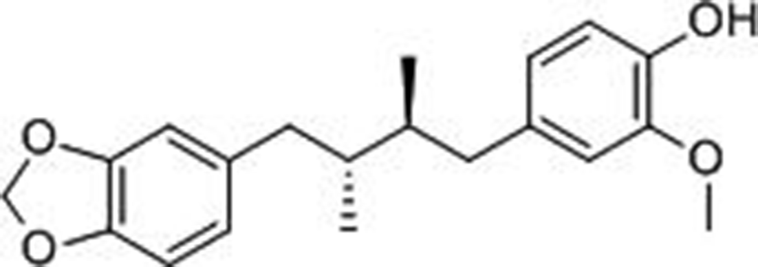
Chemical structure of anwulignan.
Results and Discussion
Liver index, serum alanine aminotransferase (ALT) and aspartate aminotransferase (AST) activities, and histopathological evaluation are all important indexes to judge the degree of liver injury. 14,15 All these parameters in the model (MOD) group were significantly higher than those in the control (CON) group (P < 0.01), but significantly lower in the anwulignan-MOD group than those in the MOD group (P < 0.01 or P < 0.05; Figure 2(A–C)).

Effects of anwulignan on the liver index and serum AST and ALT activity in CCl4-induced liver injury in mice. (A) Liver index; (B) serum AST activity; and (C) serum ALT activity. Compared with CON group, ## P < 0.01; compared with MOD group, * P < 0.05, ** P < 0.01; (A–C) (mean ± SD, n = 10). (D). Hemotoxylin and eosin staining of liver tissue (400×, n = 3). The green arrows indicate nuclear fragmentation, and the red arrows indicate inflammatory cells around the liver lobules. ALT, alanine aminotransferase; AST, aspartate aminotransferase; CCl4, carbon tetrachloride; CON, control; MOD, model.
Hematoxylin and eosin (HE) staining showed that the morphology of hepatocytes was normal, the structure of hepatic lobules was complete, and the central vein and hepatic sinuses were normal in the CON group. However, in the MOD group, there were deeply stained karyopyknosis of hepatocytes in the middle of the hepatic lobules, cytoplasmic pink staining, unclear structure of hepatic sinuses, nuclear fragmentation (as shown by green arrows in Figure 2(D)) and narrowing of hepatic sinuses, eosinophilic changes of hepatocytes around the hepatic lobules, and infiltration of inflammatory cells around the liver lobules (as shown by red arrows in Figure 2(D)). However, all of these histopathological changes were apparently alleviated in the anwulignan-MOD group, with a normal cell arrangement, and without eosinophil change and inflammatory cell accumulation (Figure 2(D)). The above results suggest that anwulignan alleviates the CCl4-induced liver injury in mice.
Oxidative stress injury is 1 of the main mechanisms of CCl4-induced acute liver injury in mice, 16 and superoxide dismutase (SOD), glutathione peroxidase (GSH-Px), and malondialdehyde (MDA) are important indicators to judge the degree of hepatocyte injury. 17,18 Therefore, in this study, we first detected these 3 indicators in the liver tissue. The results showed that the activities of SOD and GSH-Px were lower significantly (P < 0.01), and the content of MDA was higher (P < 0.01) in the MOD group than those in the CON group. However, anwulignan increased the activities of SOD and GSH-Px significantly (P < 0.01 or P < 0.05), and decreased the content of MDA (P < 0.01) (Figure 3), suggesting that anwulignan alleviates the CCl4-induced liver injury through its antioxidant effect.

Effects of anwulignan on SOD and GSH-Px activities and MDA content in liver tissue of mice. (A) SOD activities; (B) MDA contents; and (C) GSH-Px activities. Compared with CON group, ## P < 0.01; compared with MOD group,*P < 0.05, **P < 0.01 (mean ± SD, n = 10). CON, control; GSH-Px, glutathione peroxidase; MDA, malondialdehyde; MOD, model; SOD, superoxide dismutase.
The inflammatory response is 1 of the main factors that affect liver function in an acute liver injury. 19,20 Interleukin (IL)-6 and IL-1β are important inflammatory cytokines, and their levels directly reflect the degree of inflammation. 21 In this study, reverse transcription-polymerase chain reaction (RT-PCR) was used to detect the expression levels of IL-6 and IL-1β mRNA in liver tissue. As shown in Figure 4(A), the expression levels of liver IL-6 and IL-1β messenger ribonucleic acid (RNA) in the MOD group increased significantly (P < 0.01) compared with those in the CON group. However, anwulignan, decreased their expression levels significantly (P < 0.01 or P < 0.05). Tumor necrosis factor-alpha (TNF-α), mainly produced by macrophages and monocytes, is a proinflammatory cytokine that initiates necroptosis. 22,23 In this study, liver TNF-α was detected by RT-PCR, Western blot, and immunohistochemistry, and serum TNF-α by enzyme-linked immunosorbent assay (ELISA). As shown in Figure 4(D–I), in the MOD group, both liver TNF-α and serum TNF-α increased significantly (P < 0.01) in comparison with the CON group. However, anwulignan decreased the levels of both liver TNF-α and serum TNF-α significantly (P < 0.05). All the above results suggest that the protective effect of anwulignan on the CCl4-induced acute liver injury may be related to the inhibition of IL-6, IL-1β, and TNF-α.

Effects of anwulignan on inflammatory factors in the liver tissue of mice. (A) Image of RT-PCR detection; (B) IL-6 mRNA expression levels detected by RT-PCR; (C) IL-1β mRNA expression level detected by RT-PCR; (D) TNF-α mRNA expression level detected by RT-PCR; (E) image of Western blot detection; (F) TNF-α protein expression level detected by Western blot; (G) image of immunohistochemical staining; (H) TNF-α protein expression level detected by immunohistochemistry; and (I) TNF-α protein expression level detected by ELISA. Compared with CON group, # P < 0.05, ## P < 0.01; compared with MOD group, *P < 0.05, **P < 0.01 (mean ± SD; A-H: n = 3; I: n = 10). CON, control; ELISA, enzyme-linked immunosorbent assay; IL, interleukin; mRNA, messenger ribonucleic acid; MOD, model; RT-PCR, reverse transcription-polymerase chain reaction; TNF-α, tumor necrosis factor-alpha.
Programmed necrosis (necroptosis) was first proposed by Degterev et al. It is a new type of cell death with typical necrotic morphology and regulated by signal molecules. 24 The receptor-interacting serine/threonine-protein kinase 1 (RIPK1)/RIPK3/mixed lineage kinase domain-like protein (MLKL) pathway has been considered as the main regulatory pathway in necroptosis. 25 In this study, we detected the expression levels of RIPK1, RIPK3, and MLKL mRNA and proteins in the liver tissue by RT-PCR and Western blot, respectively. The detection of mRNA showed that, compared with those in the CON group, the expression levels of RIPK1 and RIPK3 mRNA increased significantly in the MOD group (P < 0.01 or P < 0.05). However, anwulignan decreased their expression levels significantly (P < 0.05; Figure 5(A)). Consistent with the above mRNA results, the expression levels of RIPK1 and RIPK3 proteins increased significantly in the MOD group, and anwulignan also decreased the expression of p-MLKL protein significantly compared with the MOD group (P < 0.01 or P < 0.05). These results suggest that anwulignan alleviates CCl4-induced liver necroptosis in mice, which is related to the regulation of the expression levels of RIPK1/RIPK3/MLKL pathway-related proteins.
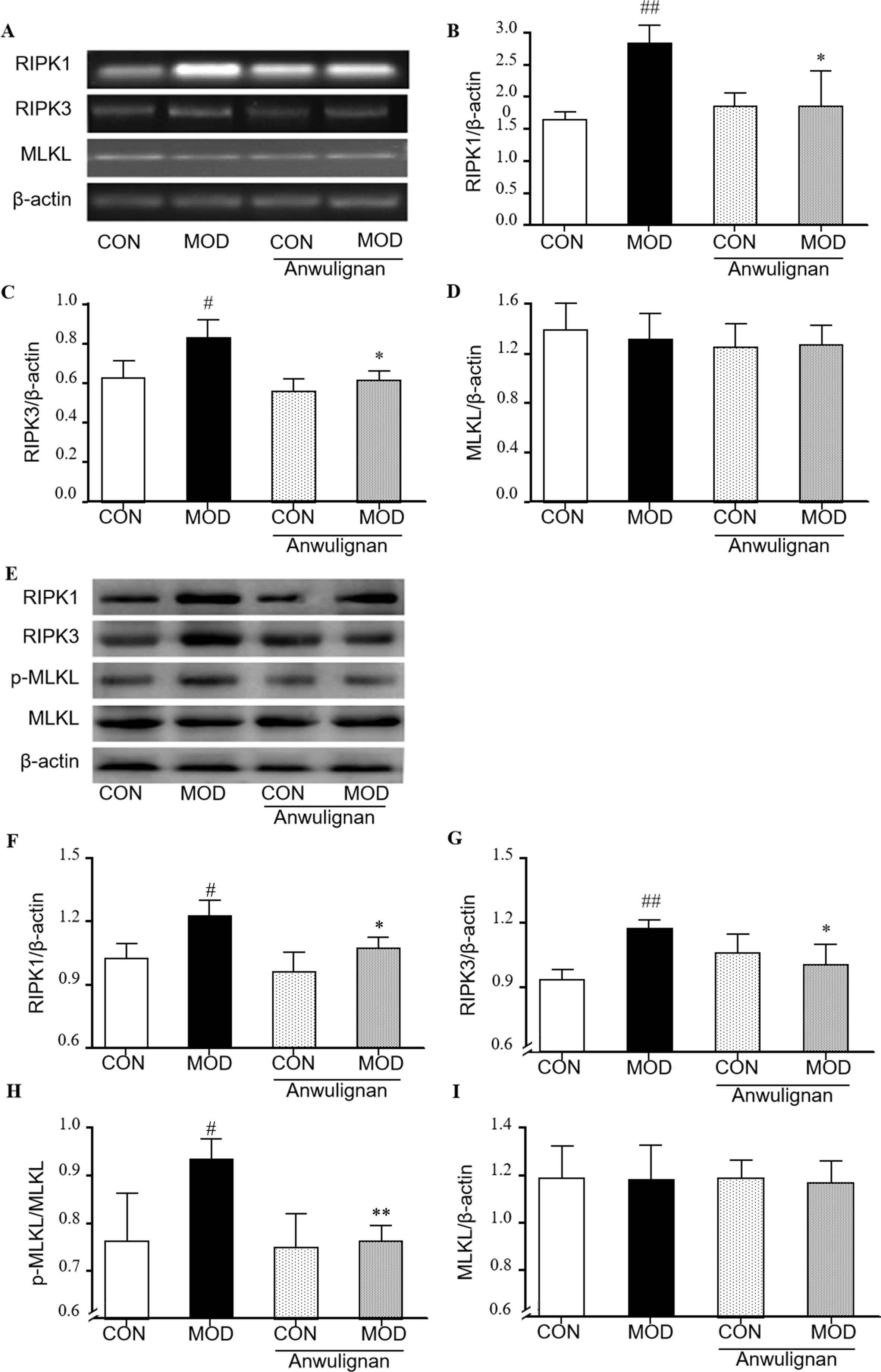
Effects of anwulignan on RIPK1/RIPK3/MLKL pathway-related proteins in liver tissue of mice. (A) Image of RT-PCR detection; (B) PIPK1 mRNA expression level detected by RT-PCR; (C) PIPK3 mRNA expression level detected by RT-PCR; (D) MLKL mRNA expression level detected by RT-PCR; (E) image of Western blot detection; (F) PIPK1 protein expression detected by Western blot; (G) PIPK3 protein expression detected by Western blot; (H) p-MLKL protein expression detected by Western blot; and (I) MLKL protein expression detected by Western blot. Compared with CON group, # P < 0.05, ## P < 0.01; compared with MOD group, *P < 0.05, **P < 0.01 (mean ± SD, n = 3). CON, control; mRNA, messenger ribonucleic acid; MOD, model; p-MLKL, phosphorylated mixed lineage kinase domain-like protein; RIPK, receptor-interacting serine/threonine-protein kinase; RT-PCR, reverse transcription polymerase chain reaction.
CCl4 is metabolized by cytochrome P450 2E1 in the endoplasmic reticulum of hepatocytes to reactive oxygen species (ROS), resulting in membrane lipid peroxidation and oxidative stress injury, and finally hepatocyte necrosis and disfunction. 26 CCl4 creates an acute liver injury in a different way than D-galactose, which creates a chronic liver injury. Therefore, we examined the effects of anwulignan in several ways.
First, we measured liver index and serum ALT and AST activities, because they can directly reflect liver swelling and cell membrane damage. Anwulignan showed its protective effect by lowering their levels.
Second, we detected antioxidation-related enzymes and products, that is, SOD, GSH-Px, and MDA. SOD can catalyze free radicals into oxygen and hydrogen peroxide and then catalyzed by GSH-Px into nontoxic hydroxy compounds. Therefore, these 2 enzymes participate in the balance of free radical generation and elimination, and MDA is a product of lipid peroxidation. 17,18 The present results showed that anwulignan increased the activities of GSH-Px and SOD and reduced the content of MDA in the liver tissue, suggesting that anwulignan could eliminate more free radicals and reduce cell damage. Anwulignan reportedly increased the nuclear erythroid 2-related factor 2 antioxidant responsive element (NRF2-ARE) pathway-related protein expression level, 27,28 and NRF2 can regulate the expression levels of SOD and GSH-Px, and thus we can speculate that anwulignan increased the activities of GSH-Px and SOD through regulating the NRF2-ARE antioxidant pathway.
Third, CCl4 reportedly can induce the vascular endothelial cells and Kupffer cells releasing proinflammatory factors, such as IL-1β, IL-6, and TNF-α, etc. 19 -21 ; therefore, we measured these factors to evaluate the degree of inflammation by CCl4 and the anti-inflammatory effect by anwulignan. The results showed that anwulignan consistently decreased the expression levels of both mRNAs and proteins of IL-1β, IL-6, and TNF-α, suggesting that anwulignan may alleviate the liver injury in CCl4-treated mice partly through the inhibition of proinflammatory factors, such as IL-1β, IL-6, and TNF-α.
Finally, we performed a histopathological examination and necroptosis related to protein detection. In the model used in this study, CCl4-induced acute liver injury shows typical necrosis. This is manifested as mitochondrial dysfunction, the swelling and rupture of organelles and cells, the leakage of cell contents, and the inflammation of surrounding tissues. 21,29 Studies also showed that TNF-α can induce necroptosis, and during this process, receptor-interacting protein 1/3 (PIPK1/3) is activated, then MLKL is recruited, and after being phosphorylated by the necrosis complex, p-MLKL, is transferred to the cell membrane to activate the ion channels and promote cell necrosis. 30,31 Therefore, we detected the expression levels of the key regulatory factors of the necroptosis signaling pathway, RIPK1, RIPK3, and p-MLKL. Anwulignan decreased the expression levels of TNF-α, RIPK1, RIPK3, and p-MLKL in the liver tissue, suggesting that it alleviates the histopathological changes partly through its inhibition of necroptosis-related factors.
Conclusion
Anwulignan alleviated the CCl4-induced acute liver injury in mice, which may be related to its antioxidation, anti-inflammation, and inhibition of necroptosis effects in liver cells.
Experimental
Experimental Animals
Male Institute of Cancer Research (ICR) mice, weighing 20 ± 2 g, were provided by China Changchun Yisi Experimental Animal Research Center (Certificate of Quality No: SCXK (JI)-2016‐0003). The mice were housed in separate cages with a 12-hour light-dark cycle and free access to standard laboratory feed and water. All animal experiments were approved by the Ethical Committee of Beihua University. All procedures were conducted in accordance with the guidelines for the care and use of laboratory animals (China).
Animal Administration and Experimental Protocol
Anwulignan (Sichuan Vicke Biotechnology Co., Ltd .), Auto-Scaled Chromatogram (Supplementary Material 1). Forty male ICR mice were randomly and evenly divided into 4 groups, that is, CON group (sodium carboxymethylcellulose + oil), MOD group (sodium carboxymethylcellulose + CCl4), anwulignan-CON group (6 mg/kg anwulignan + oil), and anwulignan-MOD group (6 mg/kg anwulignan + CCl4). Sodium carboxymethylcellulose or anwulignan was administered successively for 14 days, 10 mL/kg, once a day by gavage. One hour after the last administration, 20 mg/kg CCl4 (10 mL/kg) was given to mice in the MOD and anwulignan-MOD groups intraperitoneally to make the acute liver injury model, and an equal volume of oil solution was given to mice in the CON and anwulignan-CON groups. Ten hours after the modeling, blood samples were taken by removing the eyeballs of mice anesthetized with ether. The blood samples were centrifuged at 4000 rpm for 10 minutes to obtain the serum, which was stored at −80 °C. After blood sample collection, the mice were sacrificed, the livers removed for calculation of the liver index, histopathological observation, immunohistochemical staining, measurement of SOD and GSH-Px activities, the content of MDA, detection of RT-PCR, and Western blot.
Calculation of Liver Index
At the end of the last administration, the weight of each mouse and its liver were measured, respectively. The liver index (%) = the liver weight/the body weight × 100%.
Determination of Serum AST and ALT Activities
Serum ALT and AST activities were measured according to the instructions provided for the corresponding kit.
Histopathological Observation of Liver Tissue
The left lobe of the liver was cut out and fixed in formalin solution for 72 hours. The fixed liver was soaked overnight with 0.01 mol/L phosphate buffer solution, dehydrated with ethanol, paraffin-embedded, and sectioned into 5 µm paraffin sections. After dewaxing and ethanol rehydration, the sections were stained with HE for 20 seconds, then dehydrated with 75% ethanol and 75% ethanol with xylene, and finally, the sections were sealed for observation.
Determination of SOD and GSH-Px Activities and MDA Content in Liver Tissue
The liver tissue samples were made into a 10% homogenate, which was used for the measurement of SOD and GSH-Px activities, and MDA content, according to the instruction of the corresponding kit.
ELISA Detection
The serum TNF-α content was determined according to the instructions for the TNF-α ELISA kit.
Immunohistochemical Detection
The TNF-α expression level in the liver tissue was detected by immunohistochemical staining. The liver tissue was routinely dewaxed and rehydrated by gradient concentrations of ethanol. The antigen was prepared with sodium citrate antigen repair solution. After blocking, the first antibody, anti-TNF-α, (1:100 diluted) was added to the sample and incubated at 4 °C overnight, and then the second antibody, anti-TNF-α, was added and incubated at room temperature (1:250 diluted) for 30 minutes. After 3,3′-diaminobenzidine staining, the slices were restained with hematoxylin, and the sections were mounted with neutral resin. The sections were examined under an optical microscope, and the accumulated optical density values were measured.
RT-PCR Detection
The total RNA in the liver tissue was extracted using an RNA extraction kit, the complementary deoxyribonucleic acid was synthesized according to the instructions of the reverse transcription reaction kit, and RT-PCR amplification was carried out according to the instructions of the kit. The sequence numbers of each gene were found in Genebank, and their primers were designed by using primer software 6 and synthesized by Beijing Dingguo Changsheng Biotechnology Co., Ltd. β-actin was used as the reference gene. All the primers are shown in Table 1. The PCR amplification conditions were as follows: predenaturation at 94 °C for 5 minutes, followed by denaturation at 94 °C for 30 seconds, annealing for 30 seconds (annealing temperature: 55 °C for IL-6, IL-β, TNF-α, and β-actin, 56 °C for RIPK1 and RIPK3, and 63.5 °C for MLKL), and extending at 72 °C for 30 seconds, with 30 cycles; then kept at 72 °C for 5 minutes and stored at 4 °C. Agarose gel (1.5%) was prepared for the electrophoresis at 100 V and 70 mA for 15 minutes. The images were scanned and analyzed by an image analyzer.
Primer Sequence and Product Length.

Abbreviations: IL, interleukin; p-MLKL, phosphorylated mixed lineage kinase domain-like protein; RIPK, receptor-interacting serine/threonine-protein kinase; TNF-α, tumor necrosis factor-alpha.
Western Blot Detection
Liver tissue samples of 3 mice randomly selected from each group were added to lysis buffer and homogenized; the homogenate was incubated on ice for 1 hour, centrifuged at 12 000 rpm at 4 °C for 10 minutes, and then its supernatant was collected. The protein concentration in the supernatant was determined by the bicinchoninic acid assay method, and the target proteins were separated by 10% sodium dodecyl sulfate-polyacrylamide gel electrophoresis and transferred onto polyvinylidene fluoride membranes for 2 hours. Then, Tris-buffered saline and Tween 20 (TBST) buffer containing 5% skim milk powder was used for blocking for 1 hour. After incubation at room temperature, the blocking buffer was discarded, and the first antibodies, that is, anti-TNF-α, RIPK1, RIPK3, MLKL, and p-MLKL (1:1000) were added to the membranes at 4 °C overnight. The membranes were rinsed 3 times with TBST buffer, 10 minutes each time, and then incubated at room temperature for 1 hour with the corresponding second antibody (1:5000). The membranes were rinsed 3 times with TBST buffer, 10 minutes each time. Finally, the enhanced chemiluminescent developer was added onto the membranes, and the target protein bands were observed and analyzed by a chemiluminescence instrument.
Statistical Treatment
SPSS 19.0 software was used for statistical analysis of the experimental data; one-way analysis of variance was used for the comparison among groups, and the data were expressed as mean ± SD. A P-value less than 0.05 was defined as a significant difference in statistics.
Supplemental Material
Supplementary Material 1 - Supplemental material for Anwulignan Alleviates Carbon Tetrachloride-Induced Acute Liver Injury in Mice
Supplemental material, Supplementary Material 1, for Anwulignan Alleviates Carbon Tetrachloride-Induced Acute Liver Injury in Mice by Zhang Yuchong, Zhang Xinyun, Wang Yao, Lin Huijiao, Yu Chunyan, Jing Shu, Zhuang Wenyue, Wang Chunmei, Sun Jinghui, Li He and Chen Jianguang in Natural Product Communications
Supplemental Material
Supplementary Material 2 - Supplemental material for Anwulignan Alleviates Carbon Tetrachloride-Induced Acute Liver Injury in Mice
Supplemental material, Supplementary Material 2, for Anwulignan Alleviates Carbon Tetrachloride-Induced Acute Liver Injury in Mice by Zhang Yuchong, Zhang Xinyun, Wang Yao, Lin Huijiao, Yu Chunyan, Jing Shu, Zhuang Wenyue, Wang Chunmei, Sun Jinghui, Li He and Chen Jianguang in Natural Product Communications
Footnotes
Statement of Human and Animal Rights
All animal experiments were approved by the Ethical Committee of Beihua University. All procedures were conducted in accordance with the guidelines for the care and use of laboratory animals (China).
Declaration of Conflicting Interests
The author(s) declared no potential conflicts of interest with respect to the research, authorship, and/or publication of this article.
Funding
The author(s) disclosed receipt of the following financial support for the research, authorship, and/or publication of this article: this project was financially supported by the Department of Science and Technology of Jilin Province (20200201521JC, 20200404053YY, 20200404022YY), Jilin Development and Reform Commission (2020C033-2), Jilin Province Traditional Chinese Medicine Administration (2020121), the Education Department of Jilin Province (JJKH20200076KJ), and Jilin City Science and Technology Innovation Development Plan Project (20190601177).
Statement of Informed Consent
There are no human subjects in this article and informed consent is not applicable.
References
Supplementary Material
Please find the following supplemental material available below.
For Open Access articles published under a Creative Commons License, all supplemental material carries the same license as the article it is associated with.
For non-Open Access articles published, all supplemental material carries a non-exclusive license, and permission requests for re-use of supplemental material or any part of supplemental material shall be sent directly to the copyright owner as specified in the copyright notice associated with the article.