
Abstract
Bladder cancer (BCa), ranked as the 10th most common malignancy worldwide, presents significant challenges due to its high recurrence rates and clinical heterogeneity. The current standard diagnostic methods, cystoscopy and urine cytology, demonstrate limited sensitivity, particularly for low-grade tumors, highlighting the urgent need for minimally invasive biomarkers. Extracellular vesicles (EVs), membrane-bound nanoparticles that carry extracellular RNA (exRNA), have emerged as critical players in BCa diagnostics and therapeutics. Recent advancements show that EVs-exRNA facilitates early detection, dynamic monitoring of disease progression, and prognosis prediction. EVs-exRNA, as promising biomarkers, have made substantial progress in BCa research. In this review, we examine the role of EVs-exRNA in BCa, investigate its diagnostic and prognostic value and discuss the challenges and future directions for its clinical application, aiming to provide new insights for future research in this field.
Introduction
Overview of bladder cancer
Bladder cancer (BCa) is the 10th most common cancer worldwide and the 13th leading cause of cancer-related deaths, 1 arising from the urothelium, which lines the bladder wall. The bladder wall consists of five to seven layers of epithelial cells, including the urothelium, underlying lamina propria, detrusor muscle, and perivesical fat. 2 Urothelial carcinoma, accounting for approximately 95% of BCa cases, is characterized by a high degree of heterogeneity in clinical staging, pathological types, and prognosis. 3 At initial diagnosis, approximately 70%–75% of patients are classified as having non-muscle-invasive bladder cancer (NMIBC), 20%–25% have muscle-invasive bladder cancer (MIBC), and approximately 5% have metastatic disease. The 5-year survival rate for BCa is closely related to the stage of the disease at diagnosis. For instance, patients diagnosed with early-stage carcinoma in situ (Tis) can have a 5-year survival rate of up to 95.8%, while the survival rate drops to just 4.6% once the cancer has metastasized. 4 These statistics underscore the critical importance of accurate and timely diagnosis in improving the prognosis for BCa patients.
Traditional diagnostic methods and limitations of BCa
At present, the diagnosis of BCa is based on two principal methods: urine cytology examination and cystoscopy. 5 Urine cytology is an extremely accurate method for diagnosing high-grade lesions, with a sensitivity of approximately 80%–90% and a specificity of 98%–100%. 6 However, its efficacy is diminished for low-grade tumors due to the lesser disruption of intercellular adhesion and the poor cell shedding capabilities,7,8 resulting in a sensitivity that ranges from 4% to 31%, along with a high false-positive rate.8–11 Cystoscopy is regarded as the gold standard for the diagnosis of BCa. In comparison to traditional cystoscopy, advanced fluorescence cystoscopy offers enhanced detection of smaller lesions and carcinoma in situ.9,10 However, cystoscopy is an invasive procedure that can result in a number of complications, including difficulty urinating (50%), hematuria (19%), and urinary tract infections (3%). 11
Given the limitations of traditional diagnostic methods, the development of non-invasive diagnostic tools for BCa has attracted considerable attention. Among these, bladder tumor antigen (BTA) and nuclear matrix protein 22 (NMP22) have emerged as promising biomarkers. BTA is a glycoprotein present in BCa cells and has been proposed in recent years as an early diagnostic biomarker for BCa. Studies have demonstrated that BTA exhibits high sensitivity and specificity, particularly showing excellent detection capability for low-grade bladder tumors. On the other hand, NMP22, a nuclear matrix protein, has also been extensively studied for its role in BCa diagnosis. NMP22 testing shows high sensitivity, especially in the early stages of BCa, and effectively enhances diagnostic rates. 12 In addition to BTA and NMP22, other non-invasive biomarkers have also been proposed and applied to BCa detection. For instance, urinary DNA methylation biomarkers are considered an important complement to BCa diagnosis, with particularly high sensitivity for detecting both primary and recurrent BCa. 13 While these methods have shown remarkable performance in the early screening of BCa, they are also associated with certain limitations, such as a high false-positive rate, especially in the presence of other pathological conditions like urinary tract infections, which may lead to misdiagnosis. 14 In light of the invasive nature of cystoscopy and the associated risks, as well as the insufficient sensitivity of urine cytology, there is an urgent need for extensive research to identify new and effective biomarkers.7,15–17 This would facilitate enhanced screening, diagnosis, prognostic assessment, and follow-up for at-risk populations.
Advances in the treatment of BCa
The standard treatment for NMIBC is transurethral resection of the bladder, supplemented postoperatively with intravesical adjuvant therapy.18,19 This can take the form of small molecule chemotherapy drugs or Bacillus Calmette–Guérin (BCG) instillation therapy.8,15,17 Nevertheless, even after complete endoscopic resection, the recurrence rate of NMIBC remains high, at 50%–70%, with 10%–30% of cases progressing to MIBC.18,19 Postoperative intravesical adjuvant therapy, which typically involves frequent and prolonged BCG instillations, is associated with reduced patient adherence, substantial financial burden, and a notably increased incidence of adverse events. Furthermore, the necessity for routine follow-up involving repeated cystoscopic and cytological evaluations introduces significant physical and psychological strain, collectively contributing to a marked decline in patients’ overall quality of life.
In recent years, novel therapies for NMIBC have shown promising results. A meta-analysis comparing intravesical gemcitabine plus docetaxel with standard BCG therapy revealed slightly lower clinical recurrence rates in the gemcitabine plus docetaxel group, along with fewer severe side effects, indicating its potential as a safer alternative.
20
Radical cystectomy represents the primary treatment method for MIBC. The combination of preoperative therapy with cisplatin-based neoadjuvant chemotherapy has been demonstrated to enhance long-term survival outcomes. In addition to chemotherapy, immunotherapy—especially the use of immune checkpoint inhibitors (ICIs)—has revolutionized the treatment of BCa. ICIs work by blocking the signaling pathways that suppress T-cell activity, thus inhibiting tumor growth. By preventing T-cell exhaustion, they enhance the immune system’s ability to recognize and attack cancer cells more effectively, significantly boosting the overall immune response against the tumor.
21
The most commonly targeted checkpoint proteins are programmed cell death-1 (PD-1) and cytotoxic T-lymphocyte-associated protein 4
EVs have emerged as a novel cell-free strategy for the treatment of many diseases including cancer. Due to their natural ability to mediate cell-to-cell communication and their high physicochemical stability and biocompatibility, EVs are considered excellent delivery vehicles for various therapeutic agents. EVs have been used as delivery vehicles for therapeutic RNAs such as mRNAs, siRNAs, and miRNAs. 24 The Cytosine Deaminase-Uracil Phosphoribosyltransferase mRNA/protein complex is loaded into EVs and has tumor cell-killing activity, suggesting a great significance of EVs in cancer therapy. 25 The delivery of miRNAs (e.g., miR-206, 26a, and124a) by mesenchymal stem cells-derived EVs has been described in various types of tumors, including osteosarcoma, hepatocellular carcinoma, Non-Small Cell Lung Cancer, breast cancer, and glioma, and has shown promising anti-tumor effects.26–30 Similarly, engineered EVs have also shown great potential in the field of BCa. For example, Brancolini and Vago’s study found that EVs can carry chemotherapy drugs, such as epirubicin, mitomycin, methotrexate, and mitoxantrone. Furthermore, EVs loaded with epirubicin exhibited stronger cytotoxicity against BCa cells compared to the free drug, resulting in a better therapeutic effect for BCa treatment. 31 Lin et al. constructed engineered integrin α6 (ITGA6) EVs to inhibit the lymphatic pre-metastatic niche, effectively suppressing lymphatic metastasis and prolonging survival in preclinical models. This engineered EV-based strategy shows promise in combating lymphatic metastasis in BCa, offering new therapeutic prospects for patients with advanced metastatic BCa. 32
Research progress of EVs and EVs-exRNA in BCa
Extracellular RNA (exRNA) was discovered decades ago, 33 and in recent years, studies have confirmed that exRNA can bind to shielding vectors such as lipoproteins and protein complexes, or be encapsulated in EVs and thus resist ribonuclease (RNase) degradation. EVs are heterogeneous aggregates of lipid bilayer-enclosed particles that are actively synthesized and secreted by cells into the extracellular environment. All types of cells are capable of secreting EVs, which can load and release a range of molecular cargoes in a non-selective or selective manner.34–36 These cargoes include RNA, DNA, proteins, and lipids.34,37,38 In comparison to normal cells, cancer cells have been observed to release a greater number of EVs.34,39 EVs are generally considered to include various subtypes, such as exosomes (30–150 nm), microvesicles (100–1000 nm), apoptotic bodies (1000–5000 nm), 40 and micron-sized particles referred to as “tumor bodies” (1–10 μm). 41 EVs possess a bilayer membrane structure and physical characteristics that include small size, stability, and low immunogenicity, which enables them to resist degradation by ribonucleases and to stably exist in bodily fluids such as serum, plasma, cerebrospinal fluid, saliva, and urine. Consequently, EVs have become ideal carriers for transporting various exRNAs, including mRNA, miRNA, ribosomal RNA, transfer RNA (tRNA), long non-coding RNA (lncRNA), piwi-interacting RNA, circular RNA (circRNA), small nuclear RNA, and small nucleolar RNA as well as mitochondrial RNA42,43 (Figure 1).

EVs consist of several different types of vesicles, including exosomes, microvesicles, apoptotic bodies, and large oncosomes. EVs contain a variety of proteins, DNA, and RNA species, including miRNA, lncRNA, mRNA, piRNA, tRNA, circRNA, and others.
These RNA molecules, especially for small RNAs, are involved in intercellular communication and play a significant role in a number of physiological and pathological processes, including cell proliferation, migration, angiogenesis, immune suppression, epithelial-mesenchymal transition (EMT), invasion, and metastasis.41–46 In addition, these exRNAs show marked differential expression levels from the earliest stages of tumorigenesis and display tissue specificity, potentially conveying molecular signatures associated with specific phenotypes.35,36,47,48 This enables them to reflect specific changes occurring in tumors.
In the context of liquid biopsy, exRNAs are commonly referred to as circulating free RNA (cfRNA), which encompasses RNA molecules released from both tumor and normal cells. Specifically, circulating tumor RNA refers to the subset of cfRNA that is derived from tumor cells.49,50 These RNAs can provide crucial information about cancer, including genetic mutations and the biological characteristics of the tumor. cfRNA has been demonstrated to be a promising source of tumor-associated biomarkers in a range of biological fluids and cancer types.51–53 For example, the urinary level of UBE2C cfRNA has been proposed as a means of distinguishing BCa from healthy controls (HCs). 54 Additionally, vascular endothelial growth factor (VEGF) mRNA fragments have been detected in the urine samples of renal cell carcinoma patients, and their potential as diagnostic molecular markers has been studied. 55 Furthermore, compared to tumor biopsies, the detection of exRNAs is more cost-effective, facilitating the widespread adoption of early screening and diagnosis.
In conclusion, exRNA, as a crucial element of liquid biopsy, exhibits considerable potential and prospects for early tumor diagnosis. In the field of BCa research, the unique physiological structure of the bladder has resulted in urine becoming a rich source of EVs. The presence of EVs in urine enables the transfer of a range of molecular signals, offering new avenues for advancing our comprehension of BCa diagnosis, disease progression, prognosis assessment, and the evaluation of treatment options.
The role of EVs-exRNA in BCa
The application of EVs-exRNA for BCa diagnosis
Due to the specificity of the physiological structure, the clinical diagnosis of BCa, especially the early and precise diagnosis, has put forward new requirements. Recent studies have demonstrated that exRNA transported in EVs is emerging as a key factor in the diagnosis of BCa. 56 For example, Lin et al. conducted a high-throughput sequencing analysis of urine samples from BCa patients and healthy individuals, identifying differentially expressed miR-93-5p and miR-516a-5p. The researchers validated these miRNAs in an independent cohort through real-time fluorescent quantitative reverse transcription polymerase chain reaction (RT-qPCR) and constructed a receiver operating characteristic curve to assess their diagnostic performance, thereby demonstrating their potential as biomarkers for BCa. 57 El-Shal et al. employed RT-qPCR to detect the expression of miR-96-5p and miR-183-5p in urine exosomes, identifying a significant elevation in these miRNAs in the urine of BCa patients relative to the control group. The method demonstrated high sensitivity and specificity in distinguishing BCa from non-BCa patients (with sensitivity and specificity reaching 88.2% and 87.8% when used in combination). These findings provide new scientific evidence for the development of non-invasive diagnostic tools. 58
Research on EV-derived lncRNAs has also primarily focused on evaluating their potential as pre-selection diagnostic candidates. Zhang et al. observed that the expression levels of lncRNA PCAT-1, UBCa1, and SNHG16 were elevated in BCa patients relative to HCs. The panel of these three lncRNAs demonstrated a sensitivity of 85% and specificity of 78% for the detection of BCa, exhibiting superior performance compared to urine cytology and further enhancing the accuracy of BCa diagnostics. 59 Another study by Zhan et al. isolated EVs and assessed the levels of eight lncRNAs that have been shown to play a role in tumorigenesis. This led to the creation of a diagnostic panel comprising three lncRNAs (MALAT1, PCAT-1, and SPRY4-IT1) for BCa diagnosis, which was then validated in a separate cohort. 60 Furthermore, the panel demonstrated greater efficacy than urine cytology (area under the curve (AUC) of 0.619) in identifying potential novel biomarkers. Notably, lncRNA PCAT-1 and another mediator of DNA damage repair, ANRIL, were also elevated in the EVs derived from the urine of early-stage (T1 and T2) BCa patients compared to HCs. This suggests that detecting ANRIL and PCAT-1 levels may facilitate earlier diagnosis of BCa. 61
In addition to miRNAs and lncRNAs, tRNA fragments (tRFs) and circRNAs have also been identified as potential biomarkers for BCa. In a study conducted by Strømme et al., small RNA next-generation sequencing was performed on EVs derived from urine and serum, as well as from serum supernatant. This identified a series of tRFs that exhibited differential expression between BCa patients and healthy individuals, including tRF-18-MBQ4NKDJ, tRF-20-40KK5Y93, and tRF-31-PER8YP9LON4VD. 62
Since EVs-exRNA play such a crucial role in the development and progression of BCa, multiple clinical trials have been initiated to focus on the potential clinical applications of EVs-exRNA. Specifically, the miR Sentinel™ BCa test evaluated the clinical performance of urine-derived exosomes as a diagnostic biomarker for BCa in patients with hematuria (NCT04155359), whereas another clinical trial was dedicated to exploring the potential of EV-derived lncRNA as a new target and preoperative diagnosis for lymph node metastasis of BCa (NCT05270174). These studies aim to harness the unique properties of these molecular entities to improve the accuracy and efficiency of BCa detection, potentially leading to earlier diagnosis and better patient outcomes.
These findings provide new foundations for the development of non-invasive diagnostic strategies. The ability of a biomarker to distinguish between two classes of samples at different thresholds is measured by AUC. The higher the AUC value, the stronger the discriminative ability. This makes it suitable for the preliminary evaluation of the overall performance of a biomarker. Therefore, we have selected the top 10 EVs-exRNA based on AUC. These findings provide new foundations for the development of non-invasive diagnostic strategies (Table 1).
Top 10 EVs-exRNA biomarkers in BCa: diagnostic performance.

AUC, area under the curve; BCa, bladder cancer; EMT, epithelial-mesenchymal transition; EVs, extracellular vesicles; exRNA, extracellular RNA; HC, healthy control; MIBC, muscle-invasive bladder cancer; NMIBC, non-muscle-invasive bladder cancer; N/A, not available.
Regulatory role of EVs-exRNA in BCa development
EVs-exRNA promotes BCa proliferation, migration, and invasion
In pathological conditions, abnormal cell proliferation is a key mechanism leading to disease progression, encompassing abnormal cell division and cell differentiation. 68 This principle is also applicable to BCa. It has been demonstrated that exRNAs encapsulated in EVs are not only remarkable biomarkers for distinguishing tumors from normal populations but also play an important function in the proliferative and metastatic capacity of cancer cells. For example, Lin et al. observed a notable elevation in the levels of miR-93-5p and miR-516a-5p in urine exosomes derived from BCa patients. Additionally, miR-93-5p exhibited a more pronounced increase in muscle-invasive BCa specimens compared to those of non-muscle invasive forms. Further investigation revealed that BTG2 (B-cell translocation gene 2) is a key target of miR-93-5p, which suppresses BTG2 expression and enhances BCa cell proliferation, invasion, and migration, thereby, contributing to disease progression and metastasis. 57 Similarly, Hou et al. 69 showed that miRNA-217 was significantly upregulated in BCa tissues and accelerated the proliferation and migration ability of BCa cells by suppressing the levels of the tumor suppressor KMT2D.
Conversely, some miRNAs have been observed to be downregulated in tumors and may function to inhibit cancer progression. For example, miR-139-5p and miR-133b were identified to be downregulated in the serum exosomes of BCa patients. Jia et al. 70 demonstrated that the overexpression of miR-139-5p inhibited tumor cell proliferation through the targeting of polycomb repressor complex 1. Similarly, Cai et al. reported that overexpression of miR-133b resulted in a reduction in cell proliferation and the induction of apoptosis in BCa cells, leading to a notable decrease in tumor volume and weight in xenograft models. This suggested an oncogenic function of miR-133b. 71
Another category of EVs-RNAs, lncRNAs, has been the subject of extensive investigation with regard to their role in modulating BCa cell proliferation. For example, it has been demonstrated that exosomal LINC00355 derived from cancer-associated fibroblasts (CAFs) promotes the proliferation and invasion of BCa cells via the miR-15a-5p/HMGA2 axis. Modulation of LINC00355 expression in CAF-derived exosomes may represent a potential therapeutic strategy for targeting BCa pathogenesis. 72 Yang et al. 73 observed that LINC01133, a lncRNA with lower expression in BCa cell exosomes, inhibits BCa cell viability, and proliferation, by regulating the Wnt signaling pathway, thereby restraining BCa progression.
Apoptosis plays a pivotal role in maintaining tissue homeostasis and regulating tumor progression. Several studies have underscored the role of EVs-exRNA in regulating apoptosis in BCa cells. For example, DUSP1 (dual specificity phosphatase 1) was demonstrated to impede cancer progression via the SAPK/JNK signaling pathway. 74 The exosomal miR-133b was found to promote apoptosis in 5637 and T24 cells by targeting DUSP1, resulting in a notable reduction in tumor burden in xenograft mouse models. 73 Exosomal miR-9-3p derived from bone marrow mesenchymal stem cells was found to regulate apoptosis in the BCa cells by targeting endothelial cell-specific molecule 1. The expression levels of proliferation-associated factors (Ki67 and PCNA) and invasion-associated factors (matrix metalloproteinase-2 (MMP-2) and matrix metalloproteinase-9 (MMP-9)) were reduced after Exo-miR-9-3p treatment, resulting in decreased viability, migration, and invasion of BCa cells, while promoting apoptosis. 75 It has also been demonstrated that miR-29c induces apoptosis in BCa BIU-87 cells by downregulating the anti-apoptotic genes B-cell lymphoma 2 and myeloid cell leukemia-1. 76
In addition to microRNAs, exosomal lncRNAs have also been demonstrated to participate in the regulation of apoptosis in BCa. PTENP1, a lncRNA that functions as a tumor suppressor in a number of cancers, was found to be significantly downregulated in both BCa tissues and plasma exosomes of BCa patients (p < 0.05). Zheng et al. 77 discovered that PTENP1 is transferred to BCa cells via exosomes secreted by normal cells, where it suppresses BCa malignancy by promoting apoptosis and reducing invasion and migration behaviors.
The regulation of apoptosis by EVs-exRNA offers significant insights into the development of novel targeted therapies. The targeting of specific exRNA molecules or associated pathways may induce cancer cell apoptosis and inhibit BCa tumor growth, thereby improving therapeutic outcomes. Nevertheless, the function of other EVs-exRNA in BCa cell apoptosis remains poorly understood, and further investigation is necessary. Moreover, despite substantial evidence indicating that EVs-exRNA plays a crucial role in regulating BCa progression, the precise mechanisms remain unclear. An in-depth investigation of the mechanisms of exRNAs regulating the proliferation, migration, and invasion of BCa cells is of great significance in clinical applications, such as providing a theoretical basis for prognostic assessment and enabling the development of personalized and precise treatment plans for patients by analyzing the specific exRNA expression patterns in the tumors.
Involvement of EVs-exRNA in EMT in BCa
ExRNAs derived from EVs exert a profound influence on tumor cell phenotypes, including proliferation and migration, through intercellular communication. Additionally, they regulate a multitude of cellular functions by interacting with key molecules in various signaling pathways, such as EMT. EMT is closely associated with tumor progression and characterized by the loss of apical-basal polarity, loss of adhesion, and the acquisition of mesenchymal-like migratory properties. 78 This process is of critical importance for cancer cells, including those of BCa, to detach from the epithelium and invade surrounding tissues. Furthermore, it is regarded as a principal instigator of metastasis and resistance to treatment.
Generally, a single exRNA does not directly induce EMT but instead acts by interacting with upstream or downstream protein or RNA targets to modulate the expression of key genes, thereby indirectly influencing the EMT process. For example, microRNAs frequently inhibit pivotal EMT regulators through their gene-silencing mechanisms. The marked reduction in miR-203 levels in BCa cells results in the removal of its inhibition on Twist1, thereby disrupting the equilibrium of EMT and promoting the metastatic behavior of BCa cells. 79 Similarly, the inhibition of SphK1 by miR-613 represents a critical factor in the prevention of BCa EMT and metastasis, accompanied by an increase in E-cadherin and a decrease in Snail, vimentin, and N-cadherin. 80 Other microRNAs, including miR-22, 81 miR-186, 82 and miR-200c, 83 have also been demonstrated to regulate EMT progression in BCa.
Extensive research has also focused on the role of lncRNAs in regulating BCa migration. Xue et al. 66 elucidated that BCa cells under hypoxia can secrete large amounts of exosomes encapsulating lncRNA-UCA1, which can be internalized by BCa cells both in vivo and ex vivo, contributing to the reduction of the Ki67, waviness proteins, and MMP-9 in the cancer cells, and significantly increasing the expression level of E-cadherin, which ultimately promotes tumor growth and progression. Berrondo et al. demonstrated that HOX transcript antisense RNA (HOTAIR) was present at elevated levels in the urine exosomes of patients with high-grade muscle-invasive BCa. HOTAIR knockdown suppressed the expression of epithelial-related genes, including ZEB1 and TWIST1, thereby inhibiting EMT and reducing migration and invasion in BCa cell lines. 84 The targeted inhibition of HOTAIR may represent a potential therapeutic strategy to impede muscle invasion in BCa patients with high HOTAIR expression. The role of exosomal circRNAs in BCa regulation is less well studied, but the importance of circRNAs in BCa progression cannot be denied. Chen et al. found increased expression levels of circPRMT5 in serum and urinary exosomes of patients diagnosed with uroepithelial carcinoma and verified that circPRMT5 promotes, through uptake of microRNA-30c, the EMT process in uroepithelial cancer cells.
The regulation of EMT by exRNA in BCa contributes to a deeper understanding of the molecular interactions at both genetic and epigenetic levels, which are key regulators of cancer metastasis. In light of the high rates of metastasis and mortality associated with BCa, future research should prioritize the elucidation of the detailed molecular networks through which exRNAs regulate EMT. For example, the combination of multi-omics techniques with advanced experimental models represents a crucial direction for future research. The integration of the mechanisms of various exRNAs may facilitate the development of effective therapeutic strategies for BCa, including the creation of exRNA-based targeted therapies or the utilization of exRNAs as biomarkers for prognosis. These approaches may prove beneficial in enhancing the survival and quality of life of BCa patients.
EVs-exRNA in lymphangiogenesis of BCa
BCa is typified by a high propensity for metastasis and recurrence, which are pivotal determinants of patient survival. The most common sites for metastasis in BCa patients are the lymph nodes and lungs. Nevertheless, the precise molecular mechanisms underlying metastasis remain unclear. Typically, tumor cells express or secrete signaling molecules that promote their invasion through blood or lymphatic vessels, as evidenced by the EMT. Some circulating tumor cells necessitate the formation of pre-metastatic niches at distant sites to facilitate colonization. For example, tumor cells can secrete cytokines such as vascular endothelial growth factor C (VEGF-C) and vascular endothelial growth factor D (VEGF-D), which act on lymphatic endothelial cells (LECs) to induce the proliferation and growth of new lymphatic vessels.85,86 Additionally, tumor cells may alter the phenotype of LEC through the release of exosomes containing nerve growth factor receptors. This process promotes LEC proliferation, enhances lymphatic branching, and increases tumor cell adhesion, ultimately facilitating metastasis. Tumor-associated lymphangiogenesis is predominantly driven by VEGF-C, but approximately 20% of BCa cases with lymph node metastasis exhibit low VEGF-C expression, indicating the existence of VEGF-C-independent mechanisms for lymphangiogenesis and lymphatic metastasis in BCa.87,88
The latest research suggests that EVs-exRNA plays a crucial role in lymphangiogenesis and metastasis. For instance, Chen et al. demonstrated that the lncRNA lymph node metastasis-associated transcript 2 (LNMAT2) can stimulate the formation of tubes and the movement of cells in human lymphatic endothelial cells (HLECs), which in turn enhances both the development of tumors and the spread of cancer to lymph nodes in BCa. From a mechanistic perspective, exosomal LNMAT2 secreted by BCa cells interacts with heterogeneous ribonucleoprotein A2B1 (hnRNPA2B1), which upregulates PROX1 (LEC marker), thereby driving lymphangiogenesis and promoting lymphatic metastasis. These findings highlight a VEGF-C-independent pathway for promoting lymphatic metastasis in BCa and suggest that LNMAT2 may serve as a novel therapeutic target. 89 Concurrently, Zheng et al. found that lncRNA BCYRN1 is markedly elevated in BCa patient urine-derived exosomes and stimulates tube formation and migration in HLECs both in vitro and in vivo. The specific inhibitor SAR131675 was found to significantly suppress lymphatic metastasis induced by BCYRN1. In clinical practice, elevated exosomal BCYRN1 levels are associated with decreased survival rates in BCa patients, suggesting that BCYRN1 may serve as a prognostic marker and a promising therapeutic target. 90
Other EVs-exRNA species, including miRNAs and circRNAs, in promoting BCa lymphangiogenesis and metastasis have also been widely reported. Zeng et al. 91 suggested that cancer-derived exosomes are the main drivers of cancer-induced pre-metastatic ectopic niche formation and that miR-25-3p in exosomes regulates the expression of VEGFR2, ZO-1, occludin, and Claudin5 in endothelial cells by targeting KLF2 and KLF4, thereby promoting vascular permeability and angiogenesis. Lin et al. identified a novel EVs subpopulation derived from BCa that delivers circRNA-LIPAR to LECs, inducing E-selectin (SELE)-marked lymphatic remodeling in pre-metastatic niches and promoting lymph node metastasis. Mechanically, circRNA-LIPAR interacts with ITGA6 and the switch II domain of RAB5A (member RAS oncogene family), thereby maintaining RAB5A in its GTP-bound active state. 32 These findings offer new insights into BCa lymphatic metastasis and propose an engineered EVs-based therapeutic strategy that may prove an effective means of inhibiting lymphatic metastasis in BCa. However, the mechanisms by which exosomal circRNA facilitates lymphangiogenesis remain to be fully elucidated. Further studies should be carried out to gain a deeper understanding of the interactions of these EVs-exRNA with classical lymphatic metastasis-associated molecules, which will lead to more valuable therapeutic strategies for the clinical management of patients with BCa. The mechanisms of key EVs-exRNA species in BCa progression are depicted in Figure 2.

EVs-exRNA is involved in the processes of proliferation, migration, invasion, EMT, and lymphangiogenesis in BCa.
The role of EVs-exRNA in the prognosis of BCa
The identification of tumor prognostic biomarkers is of critical importance for the guidance of treatment decisions, the evaluation of prognosis, and the monitoring of recurrence in the context of cancer therapy. As previously stated, EVs-exRNA offers the benefits of stability and convenient accessibility, making it an emerging tool for predicting survival outcomes in BCa patients, including cancer-specific mortality, overall survival (OS), progression-free survival, and disease-free survival.
A number of studies have demonstrated that EVs-exRNA molecules serve not only as molecular markers for early diagnosis but also as valuable predictive biomarkers for BCa progression and patient survival. For example, in a study by Zhan et al., a BCa detection panel comprising urinary exosomal lncRNAs revealed that elevated levels of PCAT-1 and MALAT1 were significantly correlated with poorer recurrence-free survival (RFS) in NMIBC (p < 0.001 and p = 0.002, respectively). Furthermore, multivariate Cox regression analysis indicated that overexpression of exosomal PCAT-1 was an independent prognostic factor for RFS in NMIBC (p = 0.018). These findings underscore the substantial clinical value of lncRNAs in BCa prognosis. 60 In addition to urinary lncRNAs, serum lncRNAs have also been demonstrated to possess significant potential as prognostic markers. In a study by Zhang et al., 11 candidate serum lncRNAs were analyzed in both BCa patients and HCs, resulting in the identification of three upregulated lncRNAs (PCAT-1, UBCa1, and SNHG16) in BCa patients. The Kaplan–Meier analysis demonstrated that elevated UBCa1 expression was associated with diminished RFS in NMIBC (p = 0.01), and the Cox analysis substantiated its independent correlation with tumor recurrence (p = 0.018). 59 Furthermore, serum exosomal lncRNA H19 was found to be significantly elevated in BCa patients and was correlated with shorter survival, indicating its potential as a non-invasive prognostic biomarker for BCa. 67
In a large-scale study, Wang et al. employed matched urinary and tissue samples from BCa patients and HCs to validate miR-214 as a potential prognostic biomarker for BCa. The researchers observed that lower miR-214 expression in BCa patients was associated with significantly worse RFS and OS compared to patients with higher miR-214 levels. Additionally, urinary miR-214 levels were identified as independent prognostic predictors for both RFS and OS in MIBC. 92 In a separate investigation, researchers employed next-generation sequencing on both preoperative and postoperative urinary and serum samples from BCa patients, thereby identifying two miRNAs, miR-451a and miR-486-5p, as prospective biomarkers for RFS in T1 BCa. These miRNAs were markedly elevated in preoperative T1 BCa samples, underscoring their potential for predicting RFS. Furthermore, low expression of serum exosomal miR-185-5p and miR-106a-5p, and high expression of miR-10b-5p, were found to be associated with poorer survival outcomes in BCa patients. 93
Notwithstanding the advances made in the identification of lncRNA and miRNA biomarkers, research on other EVs-exRNA molecules for BCa prognosis remains scarce, and there are currently no clinically available diagnostic or prognostic kits based on these markers. This suggests that the full clinical potential of EVs-exRNA in cancer progression and prognosis has yet to be realized. ICIs work by blocking the immune evasion signals between tumor cells and immune cells, thereby enhancing the immune system’s ability to target and eliminate tumors. These inhibitors have become a crucial therapeutic option for advanced BCa. However, not all BCa patients respond to immunotherapy. This lack of response may be related to the role of EVs-exRNA in modulating immune cell infiltration and activity within the tumor microenvironment, thereby aiding tumor immune evasion or inducing immune resistance, 94 which in turn impacts patient prognosis. For instance, a study by Yang et al. 95 found that circTRPS1 in BCa exosomes promotes CD8+ T cell exhaustion by modulating glutamine metabolism (GLS1 pathway), affecting the response to immunotherapy. Similarly, Luo et al. 96 demonstrated that exosomal LINC00355 derived from CAFs promotes resistance to cisplatin in BCa cells by regulating the miR-34b-5p/ABCB1 axis, thereby influencing treatment outcomes and altering patient prognosis. In the context of immunotherapy, the role of EVs-exRNA as predictive biomarkers is under investigation. Identifying specific EVs-exRNA expression profiles may help predict BCa patients’ responses to immunotherapy, guiding treatment decisions and monitoring disease progression to improve patient prognosis.
Based on this context, in this study, we systematically curated a comprehensive Table 2 synthesizing current evidence on diverse EVs-exRNA species across different pathological stages of BCa (Table 2). This table meticulously documents the origins and isolation methods of EVs-exRNA, along with their potential clinical applications in diagnosis, disease progression monitoring, therapeutic intervention, and prognostic evaluation. The construction of this table aims to provide clinicians with a clear reference framework, enabling them to more effectively utilize the latest advancements in EVs-exRNA research when participating in diagnostic and therapeutic decision-making for BCa patients, thereby offering more evidence-based recommendations.
Examples of EVs-exRNA in different pathological stages of BCa.
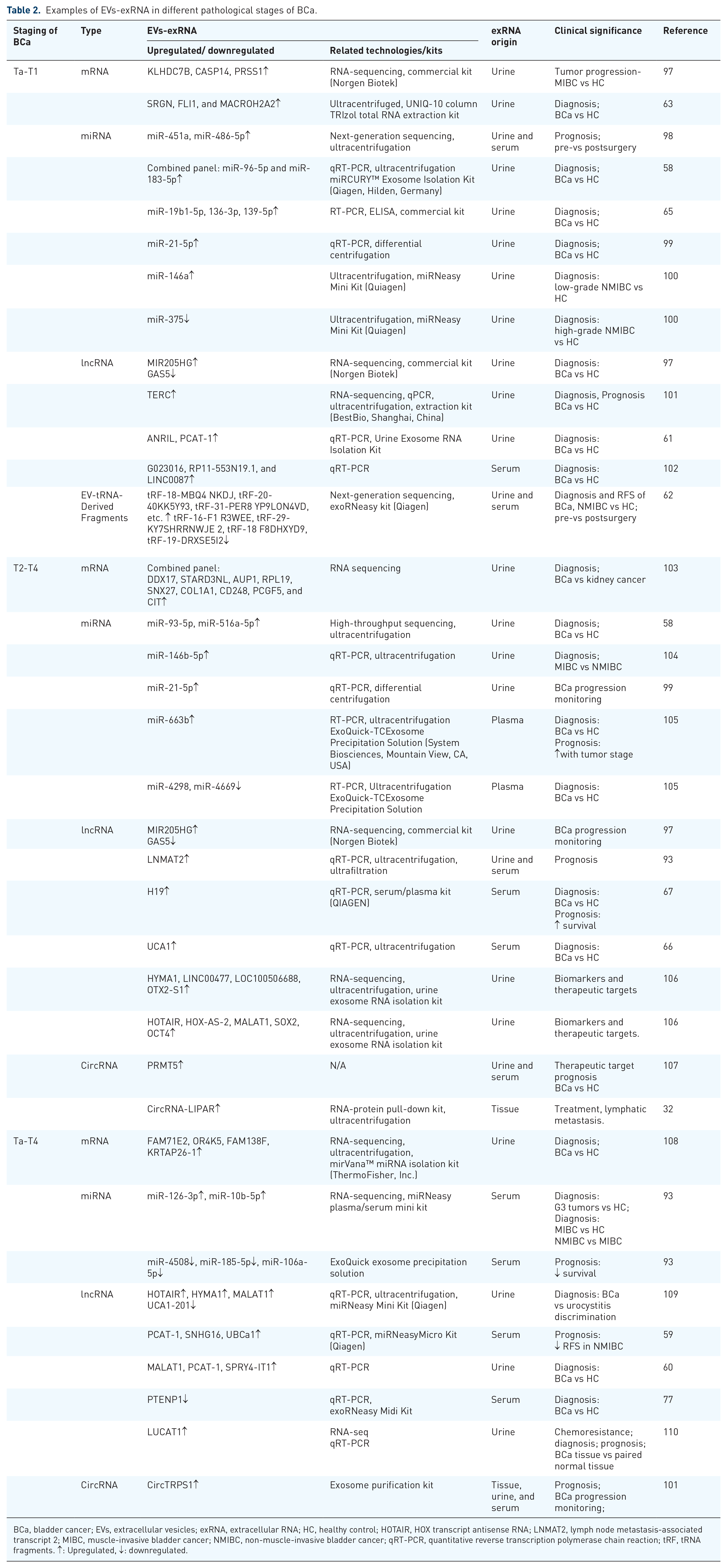
BCa, bladder cancer; EVs, extracellular vesicles; exRNA, extracellular RNA; HC, healthy control; HOTAIR, HOX transcript antisense RNA; LNMAT2, lymph node metastasis-associated transcript 2; MIBC, muscle-invasive bladder cancer; NMIBC, non-muscle-invasive bladder cancer; qRT-PCR, quantitative reverse transcription polymerase chain reaction; tRF, tRNA fragments. ↑: Upregulated, ↓: downregulated.
Future prospect
EVs-exRNA, particularly miRNAs and lncRNAs, have demonstrated remarkable potential in the diagnosis, monitoring, and prognosis of BCa. Studies reveal that these RNA molecules are differentially expressed in BCa tissues, urine, and blood, with significant enrichment in urine and plasma, making them ideal biomarkers for non-invasive liquid biopsies. Compared with traditional tissue biopsy, EVs-exRNA-based liquid biopsy offers advantages such as minimal invasiveness, high reproducibility, and the ability for real-time monitoring. To improve the diagnostic and prognostic accuracy of BCa, combining multiple EVs-exRNA, such as SRGN, FLI1, and MACROH2A2, can improve diagnostic accuracy. 63 Moreover, combining EVs-exRNA with other biomarkers, such as proteins, metabolites, and circulating tumor DNA, has become a research focus. For example, miRNA-21 combined with NMP22 or BTA markedly improves diagnostic performance.111,112 This multi-biomarker approach not only provides more comprehensive tumor metabolic and gene expression information but also compensates for the shortcomings of single biomarker detection, making it more effective in identifying BCa patients.
Beyond their diagnostic potential, EVs-exRNA derived from specific exosome subpopulations are being increasingly recognized for their roles in modulating the tumor microenvironment. For instance, CAF-derived exosomes tRNA molecules such as miR-21 and circRNA circ_0067557 to promote cancer stem cell (CSC) phenotypes and chemoresistance.113,114 Immune cell-derived exosomes (e.g., from tumor-associated macrophages) regulate immune evasion through RNAs like miR-155 and miR-222. 115 Additionally, exosomes from CSCs and endothelial cells play crucial roles in tumor heterogeneity, angiogenesis, and metastasis. 116 These subpopulation-specific EVs-exRNA not only serve as potential biomarkers for BCa subtyping and personalized therapy but also offer novel insights into tumor biology. Nevertheless, despite the growing body of research, significant variability and inconsistency remain across studies, limiting the clinical utility of EVs-exRNA. For instance, while Wang et al. 117 found that the miR-200 family is downregulated in patients with BCa suggesting its potential role in the pathogenesis of BCa. While Long et al. 118 observed a significantly higher expression of miR-200c in the urine BCa fraction compared to HCs. miR-21, though frequently identified as a cancer biomarker, is also highly expressed in many other physiological and pathological conditions, reducing its specificity. 119 These discrepancies may result from differences in tumor stages, patient populations, sample sizes, or quantification methodologies. Therefore, large-scale, prospective, and multicenter clinical studies with standardized protocols are urgently needed to validate the diagnostic and prognostic value of EVs-exRNA. 111
From a technical perspective, current limitations in EV isolation and RNA detection technologies further hinder clinical translation. Common EV isolation methods include ultracentrifugation, precipitation, ultrafiltration, immunoaffinity capture, and size exclusion chromatography.120–122 Each method has its advantages and disadvantages. For example, ultracentrifugation can achieve relatively pure EVs, but it is time-consuming, complex to operate, and prone to batch-to-batch variation; whereas precipitation is simple to perform but results in lower purity, which may affect downstream applications.123,124 To address these limitations, novel technologies are emerging. For example, the EV-Osmoprocessor uses osmotic-driven filtration to efficiently isolate EVs with improved yield and structural integrity. 125 On the detection front, single-particle imaging and fluorescence in situ hybridization (SPIRFISH). SPIRFISH combines single-particle interferometric reflectance imaging with single-molecule fluorescence in situ hybridization, enabling high-throughput RNA analysis. SPIRFISH can be used to detect specific RNAs within EVs. This may have major utility for EV therapeutics, which are increasingly focused on EV-mediated RNA delivery. SPIRFISH should enable single-particle analysis of a broad class of RNA-containing nanoparticles. 126 Similarly, Zhang et al. reported an approach for profiling single-EV multi-miRNA signatures by combining total internal reflection fluorescence imaging with a deep learning algorithm for the first time. This innovative technique allows for the precise characterization of EV miRNAs at the single-vesicle level, overcoming the challenges posed by EV heterogeneity. 127 Multi-type RNA single-vesicle analysis, as an emerging high-throughput technique, enables simultaneous detection of multiple RNA species within individual EVs, providing a novel tool for deciphering EVs-exRNA heterogeneity and their functional roles in tumor biology. This technology offers superior resolution and sensitivity in BCa diagnosis, capable of identifying low-abundance RNA biomarkers and revealing molecular signatures associated with tumor heterogeneity and treatment response. Furthermore, multi-type RNA single-vesicle analysis can be employed for dynamic monitoring of EVs-exRNA variations, offering potential for real-time diagnosis and therapeutic surveillance in BCa. 128 These strategies provide novel opportunities to explore more specific EVs-exRNA biomarkers for BCa diagnosis, progression monitoring, treatment, and prognostic evaluation.
Despite these advances, the translation of EV-based technologies into clinical practice is further complicated by regulatory and manufacturing hurdles. EV-based therapeutics must comply with rigorous standards regarding cell sourcing, production, quality control, and safety testing. However, global regulatory frameworks remain fragmented and inconsistent, posing significant challenges to standardization. In this regard, the development of harmonized international guidelines—led by organizations like the International Society for Extracellular Vesicles—is urgently needed. Moreover, industrial-scale production of EVs must ensure batch-to-batch consistency, scalability, and cost-effectiveness to meet clinical demands. Currently, many EV-based workflows remain expensive and technically demanding compared to FDA-approved diagnostic platforms, limiting their broader implementation. 129
Therefore, future research should prioritize the following directions: First, developing high-purity and high-specificity exosome isolation techniques to accurately identify distinct exosome subpopulations and their RNA cargo. Second, employing single-cell sequencing and multi-type RNA single-vesicle analysis to elucidate the precise mechanisms of specific exosome subsets within the tumor microenvironment. By unraveling EVs-exRNA heterogeneity and its functional roles in tumor biology and integrating multi-omics approaches with multi-type RNA single-vesicle analysis technologies, we may revolutionize the current paradigm for BCa management. Building on these insights, the therapeutic engineering of EVs presents a particularly promising direction. Engineered EVs can be designed to carry therapeutic RNA molecules, small-molecule drugs, or immune modulators specifically targeted to tumor cells or the tumor microenvironment. Such modifications enhance the delivery efficiency, biological stability, and tumor specificity of EVs, potentially overcoming the limitations of conventional therapies. Future studies should focus on optimizing loading methods, improving targeting strategies, and ensuring the safety and scalability of engineered EVs for clinical application (Figure 3).
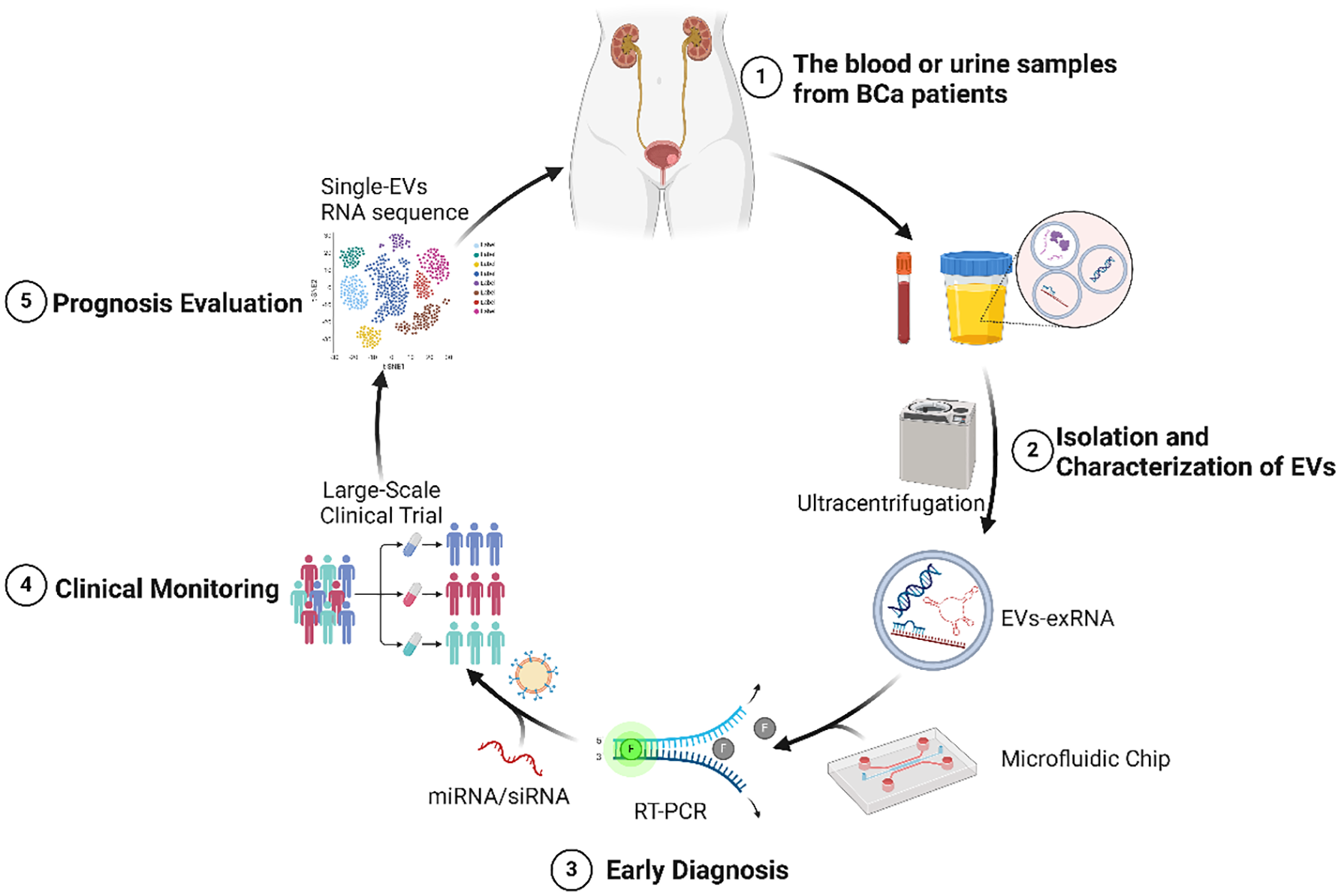
The future prospect of EVs-exRNA in BCa.