
Abstract
Background:
Post-stroke cognitive impairment (PSCI) is commonest clinical disorder in which peripheral cholinergic activity is important. Oleuropein (OLP) is polyphenol is present in olive oil. Here we evaluated the effect of OLP in cognitive dysfunction rats in post cerebral stroke model.
Methods:
The post cerebral stroke cognitive dysfunction PSD rat model was created by occlusion of transient middle cerebral artery. The rats were divided into 6 groups named, Sham + Vehicle, Sham + OLP (50 mg/kg), PSD rats + Vehicle, PSD rats + OLP (20, 50 or 100 mg/kg). The spatial learning was assessed by Morris water maze (MWM). The expression of choline acetyltransferase (ChAT), acetylcholine (ACH), extent of histone acetylation and phosphorylation of cAMP response element-binding protein (CREB) were evaluated by Western blot assay and immunofluorescence staining.
Results:
Treatment of OLP at various doses showed higher number of spontaneous and rewarded alterations and lesser percentage bias compared to vehicle treated PSD rats. OLP resulted in decreased levels of ChAT and ACH, whereas the degree of histone acetylation and phosphorylation of CREB improved in dose dependent pattern.
Conclusion:
treatment of OLP improved PSCI via increasing the phosphorylation of CREB and histone acetylation, thus attenuating cholinergic activity.
Introduction
Post-stroke cognitive impairment (PSCI) comprises any impairment which happens after stroke irrespective of causes such as degenerative or vascular disorder. 1 Studies suggest that increased occurrence of PSCI is due to accelerated aging and decline in mortality post stroke. Presently, PSCI has been identified to be the second most cause after Alzheimer’s contributing for dementia. A study recently have suggested that as compared to normal patients without PSCI the patients with PSCI have higher chances of death and such subjects also have significantly impaired living style, 2 hence it becomes utmost important for researchers to find out a therapy for treating PSCI which would not only increase the life expectancy but will also improve the living quality.
The central cholinergic system contributes majorly for normal cognitive function, the area of brain specifically in hippocampus, forebrain and cortex are responsible for cognition. 3,4 A Study recently have reported that homeostasis of histone acetylation at chromatin level is very crucial for cognitive functioning. 5 Also, a study demonstrated that acetylation of histone governed the levels of cognition associated proteins. 6 A report also evidenced that in CNS of animal model of Alzheimer’s disorder the degree of histone acetylation decreased significantly, treating them with a histone deacetylase inhibitor (HDACI) modulated cognitive impairment. 7 In a study, HDACI (trichostatin A) which encourages acetylation of histone was blocked in experimental animals having CREB mutation clearly suggested that the histone acetylation activity of HDACI is associated to CREB. 8 In addition to this, it has been found that the activation of CREB is regulated by its phosphorylation. 9
The Mediterranean diet has been discovered to play a key protective role against disorders of neurons, cardiovascular and cancer. 10 -12 Literatures have come up and have suggested that the inclusion of extra virgin olive oil in diet exerts beneficial effect in Mediterranean population. 13,14 Extra virgin olive oil contains vitamin E, vitamin K, saturated fats in low amount but contains high amount of monounsaturated fatty acids and small amounts of bioactive compounds such as polyphenols responsible for its useful activity. 15 OLP is the glycated derivative of the major secoiridoid precursor 3,4-dihydroxyphenylethanol-elenolic acid (3,4-DHPEA-EA) present in olive oil responsible for bitter taste in its leaves. OLP has been reported to exhibit neuroprotective, hepatoprotective and cardioprotective effects. 16,17 In addition to this OLP has been found to show potential effect against major malignancies such as colon, lung and breast cancer. 18,19 Very interestingly a study suggested that OLP may cross the blood brain barrier and reduce the edema of brain in rats. 20 All these findings made it logical to study the effect of OLP in post stroke dementia condition. Here we evaluated the protective effect of OLP in PSCI animal model and also the involved mechanism.
Material and Methods
Post MCAO PSCI Rat Model
The rat model of PSCI was created for which Sprague dawley rats weighing between 210-230 g aging between 6-7 weeks. The animals were housed in polypropylene cages at 25ºC and exposed to 12 h dark-light cycle; the rats were pellet diet and water ad libitum. The animals were provided by Jinan Pengyue Experimental Animal Breeding Co., Ltd (Jinan, China). The experiments were approved by the institutional animal ethical committee of Basic Medical School of Jining Medical University China. The animal experiments were in accordance to Regulations for the Administration of Affairs Concerning Experimental Animals laid by Ministry of science and technology, China. The PSCI rat model was created by middle cerebral artery occlusion (MCAO) as described earlier with slight changes. 21 Briefly, the rats were anesthetized (1% sodium pentobarbital 40 mg/kg). The rats were operated and right carotid bifurcation was exposed, the middle cerebral artery was inserted with a piece of rounded tip filament, after 45 minutes of MCAO the filament was removed and blood reperfusion was carried. For monitoring the cerebral blood flow (CBF) laser Doppler flowmetry was used. The sham rats underwent same operational procedure devoid of MCAO. After 2 weeks, all the rats were submitted to MWM test. The rats showing increased searching distance and escape latency compared to normal rats were confirmed to be PSCI.
Screening and Treatment Groups
The rats after MCAO were submitted to MWM test and PSCI rats were screened out. The rats were divided into 6 groups as Sham operated + Vehicle, Sham operated + OLP (50 mg/kg), PSD rats + Vehicle, PSD rats + OLP (20, 50 and 100 mg/kg), each group had about 12 Morris water maze screened rats. As the vehicle for solublizing OLP was water (deionized water), the vehicle treated group received water through intra-gastric route for next 28 days. The rats receiving OLP (Sigma Aldrich USA) were given the defined doses once in a day for next 28 days via gavage route.
Spatial Learning by Morris Water Maze Test
The rats were evaluated for spatial learning using MWM test as per the procedure described earlier with slight modifications. 22 Briefly, the rats were evaluated for their ability for site navigation in the MWM, 2 criteria’s i.e the distance traveled and escape latency for searching the submerged platform were monitored and were considered for learning and memory functions. The rats were trained for first 3 days by an experienced pharmacologist in the MWM tank, on the fourth day the searching distance traveled and escape latency by the rats to reach the platform were observed and recorded.
Brain Tissue Preparation
As described earlier, Hippocampus, basal forebrain and cortex in the brain are mainly responsible for cognitive functioning, hence for the further tissue studies the occluded side of these sites was selected. After treatment regimen of 28 days and spatial memory evaluation 6 rats from each group were anesthetized using sodium pentobarbital (45 mg/kg) and were decapitated. The rats were pre-fixed to formaldehyde which was infused through heart, after 5 minutes of infusion the brains were removed. The areas of special interest i.e hippocampus, cortex and forebrain were identified with the help of experienced pharmacologist of our university. The brain tissues of 3 rats were fixed in formaldehyde (4%) for 1 day and then fixed in paraffin for obtaining sections of 10 µm using a rotary microtome. The sections were processed for immunofluorescence staining and specifically the Hippocampus, basal forebrain and cortex were studied. The 3 other brain tissues were removed and freezed in liquid nitrogen for analyzing the activity of ACH, protein expression by western blot and qRT-PCR analysis.
Acetyl Choline (ACH) Assay
The levels of ACH were determined using ACH determination kit (Abcam, USA) following the supplied instructions. The content of ACH were determined by formula ACH (g/mg) = [Optical density of sample solution − Optical density of Blank] / [Optical density of Standard solution- Optical density of blank] x 400 x dilution factor.
Immunofluorescence Staining
The isolated brain tissues were submitted for immunofluorescence staining as reported earlier. 23 Briefly, the brain tissue sections were washed with Tris-buffered saline (pH 7.4) and were then maintained in donkey serum (5%) and Triton X-100 (0.25%) in Tris-buffered saline at 25ºC for 60 minutes. For examining the presence of ChAT and AcH3 positive cells, the sections were incubated with goat anti-ChAT antibody (1:500) and rabbit anti-Ac-H3 antibody (1:500) for 12 hours at 4º C. After incubation with antibodies, the sections were washed with Tri-buffered saline 3 times and again maintained with donkey anti-rabbit and anti-goat secondary antibodies (1:500) respectively for 2 hours at 25ºC in dark conditions. The resultant tissue sections were viewed under fluorescence microscopy. The red colored cells were ChAT positive and cells showing green staining were Ac-H3 positive. Cells positively stained for ChAT and Ac-H3 were recorded and expressed as cells/mm 2 .
qRT-PCR Studies
The RNA was isolated using Trizol RNA isolating kit following the supplied instructions. RNA was reverse transcripted into cDNA with the help of AMV reverse transcriptase. The PCR conditions for detecting ChAT were 94ºC for 5 minutes followed by 35 cycles of 94ºC for 50 seconds, 60ºC for 55 seconds, 72ºC for 50 seconds. The qRT-PCR was performed on a Applied Biosystems Real-Time System.
Western Blot Analysis
Total proteins were isolated from the brain tissues from rats specifically from Hippocampus, basal forebrain and cortex in the brain as per the process described earlier. 24 The proteins were loaded on PVDF membranes and the membranes were incubated with primary antibodies against Ac-H3 (1:500), H3 (1:500), ChAT (1:500), H4 (1:500), Ac-H4 (1:500), p-CERB (1:500), CERB (1:500) all the antibodies were procured from Bioworld USA and SantaCruz Biotech USA. The membranes were then exposed to Horseradish peroxidase-conjugated anti-goat and anti-rabbit secondary antibodies and then visualized with the help of enhanced chemiluminescence and then exposed to film. Densitometric analysis was performed for intensity of blots against the loading control GAPDH.
Statistical Analysis
All the results are presented as mean±SD, the data was processed by Graphpad Prism pro software version 6. The variations between the experimental groups were established by performing 1-way analysis of variance (ANOVA) followed by the post hoc test. The value of P < 0.05 was regarded significant.
Results
OLP Ameliorated Spatial Learning Ability and Memory in Experimental PSCI Rats
For assessing the spatial learning and memory we used Morris water maze test. We observed that the rats which were confirmed as PSCI showed significantly increased the searching distance and escape latency compared to sham rats (Figure 1).When treated with various doses of OLP we observed a decrease in both escape latency and searching distance at all doses, but the dose of OLP 50 and 100 mg/kg showed a significant decrease compared to PSCI vehicle treated rats. The results clearly suggested that OLP improved the memory and cognition with increasing dose. The outcomes of OLP treatment on sham operated rats exhibited no any significant decrease in both searching distance and escape latency against the sham operated rats.
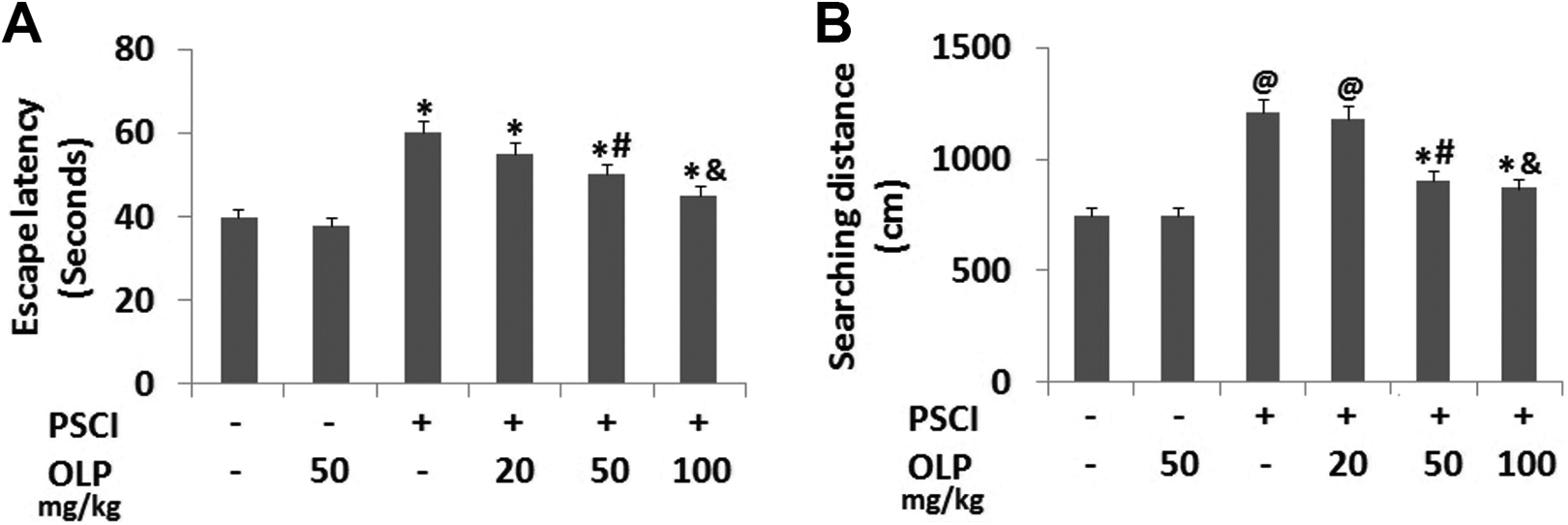
Effect of OLP treatment on spatial memory (learning and memory) of post-stroke cognitive impairment (PSCI) rats. *P < 0.05 compared to PSCI(-)/OLP(-) sham operated rats. @P < 0.01 compared to PSCI(-)/OLP(-); #P < 0.05 compared to PSCI(+)/OLP(-); &P < 0.01 compared PSCI(+)/OLP(-).
OLP Attenuated the Cholinergic Activity in PSCI Rats
ACH is one of the very important marker in the study of cholinergic activity of brain, hence the ACH levels in the brain cholinergic circuits was analyzed. The results suggested that the rats with PSCI exhibited decreased levels of ACH in Hippocampus, basal forebrain and cortex of the brain tissue compared to sham operated rats (Figure 2A), especially in the cortex region of the brain. It was noted that after receiving treatment of OLP specifically at 50 mg/kg and 100 mg/kg showed a significant increase in all the 3 regions of brain i.e hippocampus, basal forebrain and cortex compared PSCI rats treated with vehicle. It was observed that in the cortex and hippocampus region the levels of ACH in rats treated with 100 mg/kg dose of OLP showed a significant increase. However, the levels of ACH in Sham rats treated with OLP showed a non-significant increase in levels of ACH compared to sham operated vehicle treated group.

OLP attenuated the cholinergic activity in PSCI rats.
After levels of ACH, ChAT is another important marker for evaluating the functioning of cholinergic system. The outcomes of qRT-PCR suggested that the mRNA levels of ChAT levels in the hippocampus, basal forebrain and cortex in the brain of the PSCI rats were significantly decreased compared to the sham operated rats. After treating the rats with varied doses of OLP, the ChAT mRNA levels in the hippocampus, basal forebrain and cortex in the brain increased gradually with dose, especially with 50 mg/kg and 100 mg/kg there was a significant increase compared to PSCI vehicle treated rats Figure 2B). However, it was noticed that the mRNA ChAT levels in the hippocampus, basal forebrain and cortex in the brain of Sham+OLP treated rats showed non-significant increase in ChAT mRNA levels against the sham operated rats (Figure 2B).
The results of western blot analysis were parallel to the outcomes of qRT-PCR. The protein levels of ChAT in the hippocampus, basal forebrain and cortex in the brain of PSCI rats were analyzed and showed that levels were decreased significantly compared sham operated rats. The expression levels of ChAT protein improved in rats treated with OLP in dose dependent manner with rats treated with 50 and 100 mg/kg of OLP showed a significant increase (Figure 2C-2F).
Treatment of OLP Improved the Histone Acetylation in the Brain Tissues of PSCI Rats
As an indicator of histone acetylation homeostasis in CNS, the expression of Ac-H3 and Ac-H4 in cholinergic system was evaluated. The results suggested that the levels of both Ac-H3 and Ac-H4 in the hippocampus, basal forebrain and cortex regions of the brain of PSCI rats were suppressed significantly compared to that of sham operated rats, however the levels of Ac-H4 were more significantly suppressed (Figure 3). After the PSCI rats were treated with various doses of OLP, the levels of Ac-H3 increased with all the OLP treatments, however the treatment of 50 mg/kg and 100 mg/kg showed significant increase in Ac-H3 levels specifically in the cortex and hippocampus region of brain compared to sham operated vehicle treated rats. The expression levels of Ac-H3 in sham operated OLP treated rats demonstrated no significant increase compared to sham operated rats (Figure 3A-D). However, the expression of Ac-H4 showed no any alterations after any of the treatments dose of OLP treated PSCI rats (Figure 3A-C and E).

OLP improves histone acetylation in the brain tissue of PSCI rats. A-C: Western blot analysis for expression of Ac-H3 and H3, Ac-H4 and H4 in cerebral cortex, hippocampus and basal forebrain region of PSCI rats. D: Quantitative results for expression of Ac-H3, H3, Ac-H4 and H4 in cerebral cortex, hippocampus and basal forebrain region of PSCI rats after receiving defined treatments. *P < 0.05 compared to PSCI (-)/OLP(-) Sham operated; @P < 0.01 compared to PSCI(-)/OLP(-); #P < 0.05 compared to PSCI(+)/OLP(-); &P < 0.01 compared to PSCI (+)/OLP(-).
Further the results of expression of ChAT and Ac-H3 levels were confirmed by performing the immunofluorescence staining of brain tissue sections for the presence of ChAT and AC-H3 positive cells. We performed the immunofluorescence staining specifically for the cortex, hippocampus and basal forebrain regions of the brain. The results of immunofluorescence staining (Figure 4) were parallel to that of results of western blot analysis for expression of ChAT and Ac-H3 levels.

Effect OLP treatment on alteration of the ChAT positive and Ac-H3 positive cells in cortex, hippocampus and basal forebrain region of PSCI rats.
OLP Ameliorated the Suppression of the p-CREB in Brain of PSCI Rats
Further in our study we also studied the levels of p-CREB in the cortex, basal forebrain and hippocampus regions of the brain of rats submitted to PSCI. The results suggested that the levels of p-CREB in the cortex, basal forebrain and hippocampus regions of the PSCI rat brain were decreased significantly compared to sham operated rats. After the PSCI rats received various dose of OLP, the expression of p-CERB increased in all the treatment groups, with dose of OLP 50 mg/kg and 100 mg kg specifically in cortex and hippocampus region showed significant increase compared to those in vehicle treated PSCI rats (Figure 5A-D). The levels of p-CERB levels in sham operated OLP treated rats showed no significant increase compared to sham operated rats.
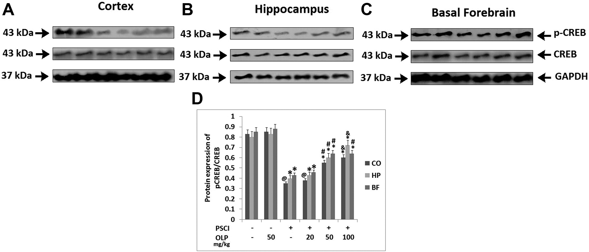
Treatment of OLP improved the inhibition of p-CREB protein in the brain tissue of PSCI rats. A-C: Western blot analysis for protein expression of p-CREB and CREB in the cortex, hippocampus and basal forebrain region of PSCI rats. Quantitative results of protein expression of p-CREB and CREB in the cortex, hippocampus and basal forebrain region of PSCI rats. *P < 0.05 compared to PSCI (-)/OLP(-) Sham operated; @P < 0.01 compared to PSCI(-)/OLP(-); #P < 0.05 compared to PSCI(+)/OLP(-); &P < 0.01 compared to PSCI (+)/OLP(-).
Discussion
Homeostasis of cholinergic system is very important for maintaining the cognitive function, 25 the cholinergic neurons are reported to be centralized in cortex, hippocampus, brain stem and basal forebrain region of the brain. 26 Among the regions of brain, the hippocampus-forebrain region and cortex forebrain region are very vital in the learning, memory, attention, stimulation and awakening. 27,28 In the present study, a PSCI model was created by submitting the rats to MCAO surgery, mimicking the real stoke condition in patients. Though MCAO is mainly found to alter functioning of basal ganglia, inadequacy of cholinergic neurons were found in basal forebrain, cortex and hippocampus, which may be due to the intervention in the synaptic, link in cholinergic circuits.
OLP is reported to exert neuroprotective effect against Alzheimer’s and Parkinson disease by forming a non-covalent complex with Aβ peptide. 29 However in a study earlier OLP had been screened for its effect on post-traumatic stress disorder which resulted in its beneficial effect on the cognitive deficits by altering the brain derived neurotrophic factors in hippocampus region. 30 The findings of the present study were in agreement to this study in which treatment of OLP resulted in improvement of cognitive function post stroke. OLP has been reported to exert neuroprotective effect against cognitive dysfunction induced by colchicines in the hippocampus region of brain in rats. This study suggested involvement of OLP affecting Acetylcholine activity in specific region of brain. 31 However a clear study confirming the involved mechanism for OLP is lacking and is poorly understood. In the present study, we demonstrated that OLP significantly ameliorated cognition and promoted the levels of ChAT and ACH in the hippocampus, cortex and basal forebrain region of brain of PSCI rats
A study had confirmed about acetylation of histone is involved in cognitive function. 32 Also a study emerged suggesting over expressed Ac-H3 levels in hippocampus region resulted in improvement of learning and memory in experimental rats. 33 In our study, the acetylation levels of histone H3 in PSCI rats decreased significantly in important zones of brain such as basal forebrain, cortex and hippocampus region which are responsible for all the major cholinergic activity, this decline in Ac-H3 levels was positively associated with decline in cognitive activity of PSCI rats. However our study found that suppressed levels of Ac-H3 could be inverted by OLP at 50 and 100 mg/kg in PSCI rats. It is a first study of its type in which PSCI rats when screened were reported for alteration in levels of histone acetylation after treating them with OLP.
It has been evidenced that 2 enzymes named and histone deacetylases (HDACs) and histone acetyltransferases (HATs) are involved in maintaining equilibrium of histone acetylation homeostasis. HATs is responsible for histone acetylation and promotion of transcription of gene, whereas the HDAC works in opposite manner by halting gene transcription. Studies earlier have suggested that acetylation of histone could initiate transcription of ChAT mRNA, increase the synthesis of ChAT protein and upregulate the expression of ChAT as a result of DNA methylation. 34 The outcomes of present study showed that the protein as well as mRNA levels of ChAT in PSCI rats were in agreement with the levels of AcH3 after receiving treatment of OLP indicating that engagement of histone acetylation in transcription of ChAT played a important part in therapeutic role of OLP in Post stroke cognitive impaired condition. The results of the present study also showed that, p-CERB was responsible for significant decrease in cholinergic activity in PSCI rats which was attenuated by treatment of OLP. CREB is protein from leucine zipper family, phosphorylation of CREB (p-CREB) directs the CREB interacting protein i.e CBP to form a complex of CREB-CBP which further combines to CREs i.e cAMP response elements activating the process of histone acetylation and encourage transcription of ChAT gene. 35,36 In the present study, the alteration of p-CREB levels conferred with Ac-H3 hence it is believed that OLP promoted histone acetylation via the CREB activity. Further studies are required using agents which could suppress the levels of p-CREB and modulate histone acetylation and study its effect on PSCI.
Conclusion
The present study is first of its kind which evidenced that, MCAO resulted in cognitive impairment which was accompanied with suppressed ChAT and ACH levels in specific regions of brain such as hippocampus, basal forebrain and cortex which are regions of cholinergic activity. It was also confirmed that homeostasis of histone acetylation in these brain regions was suppressed in PSCI rat model, in addition to this the levels of p-CERB in these brain areas decreased accordingly. We also found that OLP ameliorated the cognitive function in rats confirmed for PSCI, the treatment also increased the expression of ChAT and ACH in the hippocampus, cortex regions and basal forebrain of the brain. The findings suggested that OLP corrected and reversed the phosphorylation of CERB.
Footnotes
Authors’ Note
All the authors contributed in preparing the manuscript. BY and YG Planned the work, XL, RX, YG performed the animal studies. HY, RT, ZQ, GL and JL performed all the rest of experiments. All the authors contributed directly or indirectly in all the parts of study working as team. All the authors prepared the manuscript and reviewed it. All the data is presented in paper. The supporting study data for work is under ethics laws of university and is hence not presented here. Here by we declare that our institutes are aware of the work and declare consent for publication of the manuscript. The experiments were approved by the Institutional Animal Ethical Committee of Basic Medical School of Jining Medical University China.
Acknowledgments
The authors are thankful to Basic Medical School of Jining Medical University China. The authors are also grateful to the funding agencies The National Natural Science Foundation of China, The Undergraduate Innovation and Entrepreneurship Training Program of Shandong, The Undergraduate Innovation and Entrepreneurship Training Program of Jining Medical University and NSFC cultivation project of Jining Medical University for supporting the work.
Declaration of Conflicting Interests
The author(s) declared no potential conflicts of interest with respect to the research, authorship, and/or publication of this article.
Funding
The author(s) disclosed receipt of the following financial support for the research, authorship, and/or publication of this article: This study was supported by The National Natural Science Foundation of China (NO.81703490), The Undergraduate Innovation and Entrepreneurship Training Program of Shandong (No. S201910443008), The Undergraduate Innovation and Entrepreneurship Training Program of Jining Medical University (No. cx2019026), NSFC cultivation project of Jining Medical University (NO.JYP2018KJ06).