
Abstract
Wistar rats were divided into six groups, which were given La(NO3)3 at 20.0, 10.0, 2.0, 0.2, and 0.1 mg/kg, and the control group, which was given physiological saline, respectively, for six months. Pathological changes of liver were observed via light microscopy and transmission electron microscopy. Glutamic-oxalacetic transaminase, glutamic-pyruvic transitanase, gamma-glutamyl transferase, and alkline phosphatase activities in the serum were measured. Superoxide dismutase (SOD), glutathione peroxidase (GSH-Px), and malondialdehyde of liver were determined. The metabolic accumulation of lanthanum in rat liver was investigated via inductively coupled plasma mass spectrometry. Results showed no abnormal biochemical changes. In the group of 20.0 mg/kg La(NO3)3, there were loss of weight, decrease of glycogen in the hepatocytes, denser matrix of the mitochondria, and deformation of the nuclei of some hepatocytes with different degrees and infiltration of inflammatory cells in the portal area. The higher was the dose, the higher was the number of bodies contain high electronic dense gravel-like granules, and secondary lysosomes with dense bodies were observed. In the group fed 0.1 mg/kg La(NO3)3, intracellular glycogen showed an increasing tendency, particularly increased animal growth and increased activities of SOD and GSH-Px. The content of La in the liver increased regularly with increase in close and time of administration. The results further proved that low-dose La(NO3)3 produced some specific biologic effects. This study illustrated the influence of La(NO3)3 on rat liver at cellular and subcellular levels and it provides an experimental basis for the purpose of setting a reasonable standard for safely utilizing rare earth elements.
INTRODUCTION
Rare earth elements (REEs) widely exist in nature. China has the richest mineral resources of REEs in the world. Owing to their unique electronic structure and many kinds of fine physiochemical properties, REEs are widely used. There have been rapidly advancing applications of REEs in agriculture, forestry, stock raising, and medicine since REEs were discovered to accelerate animal and plant growth. Some biological effects of REEs, such as the absorbance, distribution, and deposition in various visceral organs of animals, aroused people's extensive attention to the effects of REEs on health. Lanthanum (La) is a light REE, and liver is the chief organ of accumulation of light REEs. In this study, we observed the changes of hepatic fine structure and blood biochemistry in rats after they were fed different doses of La(NO3)3. The results provide a theoretical basis for the purpose of setting down a reasonable standard of REEs for safe application.
MATERIALS AND METHODS
Animals
Wistar rats weighing about 80 g (180 total; male: 84.3 ± 20.3 g, female: 80.9 ± 16.7 g) were purchased from the Division of Experimental Animals of Jilin University and kept under routine laboratory conditions. Rats were randomly divided into 6 groups, 15 female and 15 male in each group.
Administration of La(NO3)3
La(NO3)3 was provided by the China Rare Earth Center for Agriculture Development (purity >99.9%). Animals in experimental groups were given La(NO3)3 via gavage at 20.0, 10.0, 2.0, 0.2, and 0.1 mg/kg and the control group was given physiological saline, respectively, at about 9:00 a.m., once daily, six times a week for six months.
Measurement of the Ratio of Liver to Body Weight
The animals were sacrificed after six months; the liver was removed and weighed immediately and accurately to calculate the ratio of liver to body weight.
Assay of Liver Function
After ether anesthesia, blood from the rat eyeball was centrifuged to prepare serum. The levels of glutamic-oxalacetic transaminase (GOT), glutamic-pyruvic transitanase (GPT), gamma-glutamyl transferase (γ-GT), and alkline phosphatase (ALP) in the serum were measured with biochemical autoanalyzer (Hitachi, 7250).
Examination of Histology and Histochemistry
Animals were sacrificed by decapitation. The right lateral lobe of the liver was excised and fixed with Carnoy solution and embedded in paraffin. The specimens were stained via HE and PAS method for observing the general structure and glycogen change of the liver.
Transmission Electron Microscope (TEM)
The blocks of fresh liver were removed and fixed by immersion for 2 h in a solution of 2% polyformaldehyde and 2.5% glutaraldehyde in 0.1 mol/L phosphate buffer (pH 7.4), postfixed in osmium tetroxide for 1.5 h, dehydrated through a series of graded alcohols, and embedded in Epon 812 mixture. Ultrathin sections were double stained with uranium acetate and lead citrate and examined with a JEM-1200EX electron microscope.
Assay for Superoxide Dismutase (SOD), Glutathione Peroxidase (GSH-Px), and Malondialdehyde (MDA)
SOD activity was assayed by the inhibition of the photochemical reduction of nitroblue tetrazolium. GSH-Px was determined in the homogenates by using DTNB. MDA was measured as the amount of thiobarbituric acid reactive substances determined by the thiobarbituric acid reaction. Each absorbance was measured at wavelengths of 420, 340, and 532 nm via spectral photometer.
Assay for La Content
Liver was thawed and homogenized with Tris-HCl buffer, and centrifuged for 30 min at 10,000 rpm. Sediment was collected and digested with concentrated HNO3. After digestion was completed, 2.0 mL solution was 17 dissolved with 2 mL 1 % HNO3, added to 1.0 mL 200 μg/L In(NO3)3 that was used as an interior label, and brought to volume with 1% HNO3. La content was measured via inductively coupled plasma mass spectrometry (ICP-MS).
Statistical Analysis
Student's t-test was used to evaluate the statistical significance of the data.
RESULTS
Body Weight and Ratio of Liver to Body Weight
The study showed that the male rats put on weight slowly in 20.0 mg/kg La(NO3)3 in contrast with the control (P < 0.002), but gained weight quickly with decrease of La(NO3)3 dose. The body weight put on by male rats of the 0.1 mg/kg group was markedly, compared with the control (P < 0.001). The results of ratios of liver to body weight of animals in each group are shown in Table 1. From Table 1, it is suggested that the ratio of liver to body weight of rats in the males of the 20.0 mg/kg group was far higher than those in the control group.
Ratio of liver to body weight of rats (%)

Mean ± standard division, n = 13;
P < 0.05,
P < 0.0001 are compared to control.
Analytical results of serum of female rats after different doses La(NO3)3 administration for six months

Mean ± standard division, n = 13.
Effects of REEs on Hepatocyte Function
The results of serum biochemical measurement showed that the content of ALP in the males increased obviously for 20.0, 2.0, 0.2, and 0.1 mg/kg (Table 3).
Analytical results of serum of male rats after being administrated with different doses La(NO3)3 for six months

Mean ± standard division, n = 13;
P < 0.05,
P < 0.01,
P < 0.001 are compared to control.
Light Microscopic Finding
The structure of the hepatic lobule and portal area was normal in the control. There were turbulent arrangements of hepatocyte cord and infiltration of inflammatory cells in the portal area in the 20.0 mg/kg La(NO3)3 group (Fig. 1). Hepatocytes were normal predominantly in lower dose groups compared with the control. Glycogen granules in hepatocytes were purplish red, granular, and well-distributed in the control group. Glycogen decreased at different degrees and even diminished at the 20.0 mg/kg La(NO3)3 dose (Fig. 2). There was an upward tendency of hepatin in the 0.1 mg/kg La(NO3)3 group.

HE staining for rat liver. There were a few lipid droplets in some hepatocytes and inflammatory cells in the portal area after 20.0 mg/kg La(NO3)3 administration for six months.

PAS staining for rat liver to observe content of glycogen. Glycogen granules in hepatocytes were purplish red and granular. Glycogen decreased in different degrees and even diminished at the dose of 20.0 mg/kg La(NO3)3 compared with the control group.
TEM Finding
Ultrastructure of the hepatocytes in the control group are normal. The nuclei of hepatocytes have an irregular outline in different degrees for the 20.0 mg/kg La(NO3)3 group, glycogen granules are reduced, and a number of lipid droplets emerged in some hepatocytes. The hepalocytes possessed many lysosomes that contained particles with high electronic density and dense bodies which had no membrane. They gathered mostly around bile canaliculi and occasionally in the perinuclear cytoplasm. Hepatocytes contained many high electronic densities and lysosomes containing high electronic densities in the 20.0 mg/kg La(NO3)3 group (Fig. 3). Although lysosomes with high electronic densities were also found in >10.0 mg/kg groups, the same distribution as that in the 20.0 mg/kg group, amount of deposit particles and electronic density decreased along with reduction of administered.

Ultrastructurally, in 20.0 mg/kg La(NO3)3 group, we observed that density of matrix mitochondria enhanced and glycogen granules reduced. In hepatocytes, there were many lysosomes which contained particles with highly electronic density and dense bodies which had not enclosed membrane. They distributed mostly around bile canaliculi.
SOD, GSH-Px, and MDA in Liver
The activities of SOD and GSH-Px increased markedly in the groups of 0.1 and 0.2 mg/kg La(NO3)3, but the concentration of MDA decreased obviously in the 0.2 mg/kg La(NO3)3 group compared with the control (Tables 4 and 5).
Activities of SOD, GSH-Px, and MDA concentrations in liver of female rats

Mean ± standard division, n = 5;
P < 0.05,
P < 0.01,
P < 0.001 are compared to control.
Activities of SOD, GSH-Px, and MDA concentrations in liver of male rats
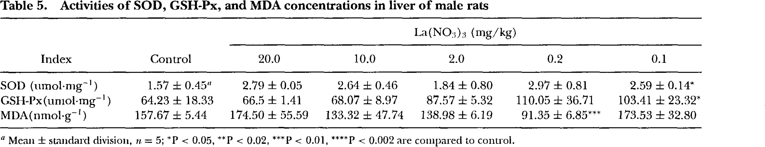
Mean ± standard division, n = 5;
P < 0.05,
P < 0.02,
P < 0.01,
P < 0.002 are compared to control.
Amount of La in Liver
Table 6 shows La content in water-miscible ingredients in Wistar rat liver. La content in every studied group was much more than that in the control. At 20.0 mg/kg La(NO3)3, it was 100 times as much as drat in the control.
La content in water-miscible ingredients in Wistar rat liver (ng/g protein)

Mean n = 5.
DISCUSSION
High-dose REEs can bring about acute morphological changes in the liver. The increase of hepatin at 0.1 mg/kg made a remarkable contrast with the control group, which might be related to increased insulin content in serum by administration of low-dose REEs (Zhou et al., 1996). Low-dose REEs were given to rats by way of oral administration, and intraperitoneal and intravenous injection every other day, respectively, at 0.05 mg/kg LaCl3, 0.05 mg/kg YbCl3 for 12 iterations (Nie et al., 1997). Nie et al. observed hepatin increase in hepatocytes and drought the change in heparin content might be connected with administration procedures, dose, and change of some hormone content, because experiments clearly demonstrated increased GH and insulin in serum after administration of 0.05 mg/kg LaCl3 for one month. In the 20.0 mg/kgLa(NO3)3 group, increase of body weight was far slower than that of the control group, infiltration of inflammatory cells were found in the portal area, and the lightly deformed nuclei of hepatocytes and higher density of matrix mitochondria and reduction of glycogen granules and a number of lipid droplets were found in some hepatocytes. These showed in 20.0 mg/kg La(NO3)3 injured rat liver. Some people gave mice PrCl3 via intravenous and intraperitoneal injection alternatively every other day (intravenous injection 40 mg/kg, intraperitoneal injection 60 mg/kg, for two days, respectively, total dose of 200 mg/kg, 24 h after the last injection), and by light microscopy discovered that the hepatocytes were in acidophilic or vacuolar degeneration. Ultrastructually, these cells presented nuclear deformation, enlargement of endoplasmic reticuli, lack of hepatin, and lysosomes containing high electronic density particles, which were similar to what we found (Zou et al., 1993). In this paper, we found by TEM and ICP-MS that particles in hepatocytes and the accumulation of lanthanum in liver increased by degrees with increase of dose. Many particles were around bile canaliculi. The ICP-MS is an efficient multi-element analytical method that enables the detection of metals at (ultra) trace levels. These results demonstrated that La was deposited in hepatocytes and might be excreted through bile canaliculi.
The content of GOT, GPT, and γ-GT in serum in experimental groups was not in sharp contrast to that of control group. This showed that REEs did not significantly affect the liver function of rats. Conversely, the ALP level of the males in the groups of 20.0, 2.0, 0.2, and 0.1 mg/kg obviously increased, but the level for the females was not clearly different in comparison with the control. Liver is not unique organ in that it generates ALP. The mechanism of ALP increase may be more complex. This needs to be further studied.
Antioxidant enzymes constitute an important defense system to clear up detrimental free radicals, such as SOD and GSH-Px. MDA is a kind of oxidative stress indicator and can damage biological molecules. In low-dose La(NO3)3 groups, animals grew faster, and the activities of SOD and GSH-Px increased markedly, but the MDA level decreased obviously compared with that of the control. The finding of the present study was that low-dose La(NO3)3 could promote the antioxidant defense system and prevent peroxidation. Similar observations and suggestions implicating free radicals in experiments about rare earths were made by some other researchers as well. For example, a study showed that rare earths could increase activities of SOD, GSH-Px, and catalase, reduce lipid peroxidation level, and inhibit free radical production (Zeng et al., 1999). Wu et al. (1994) found that rare earths could inhibit free radicals produced by extrinsic compounds, and Re(NO3)3 or LaCl3, CeCl3 could significantly restrain · OH radicals induced by chrysotile at 2–20 mg (Wu et al., 1994). It was discovered that 12 mmol/L Ce3+ and Ce4+ could clear the amount of superoxide radical produced by irradiating flavin (J. Wang et al., 1997). Some researchers gave mice the carcinogens ethyl carbamate and dimethylhydrazine by hypodermic injection, let the mice drink water containing 0.25–0.025% ReCl3 for 120–210 d, and found notably increased SOD activities and lowered lipid peroxidation levels in mice drinking ReCl3 compared to the control (Liu et al., 1998).
The results suggested that La(NO3)3 has different effects on animals concerning dose and sex. Low-dose La(NO3)3 promotes growth of the animal, has antioxidation properties, and protects animal hepatocytes. On the contrary, higher dose La(NO3)3-–20.0 mg/kg—damages liver to different degrees.