
Abstract
This study investigated the quantitative polyphenolic constituents and gastroprotective effects of methanol extract of Vernonia amygdalina leaf (MEVA) against aspirin-induced gastric ulcer in rats. Ulceration was induced by 3 days’ oral administration of aspirin (150 mg/kg body weight). Wistar rats were pretreated with cimetidine (reference drug) at a dose of 100 mg/kg body weight and MEVA at 200, 300, and 400 mg/kg body weight once daily for 28 days prior to ulcer induction. At the end of the experiment, gastric secretions, antioxidant status, and histopathological alteration were evaluated. We observed that the significantly increased ulcer index, gastric volume, free and total acidity, malondialdehyde level, and pepsin activity were effectively reduced following treatment with 200 and 300 mg/kg MEVA. The extract also markedly attenuated the reduced activity of superoxide dismutase and reduced glutathione level as well as pH and mucin content in the ulcerated rats. Administration of the extract also significantly attenuates necrosis of the stomach tissue of the ulcerated rats. The results suggested that the MEVA leaf, preferably at 200 and 300 mg/kg body weight, ameliorated aspirin-induced gastric ulceration via antioxidative and H2 receptor antagonist.
Introduction
Peptic ulcer is one of the most serious gastrointestinal diseases in the world. Till date, it is one of the leading causes of morbidity and mortality in Nigeria and many developing countries in the world. 1 These diseases are more common in low-income people and most especially in elderly people. The incidence of peptic ulcer is increased due to stress, smoking, alcohol, Helicobacter pylori, and ingestion of nonsteroidal anti-inflammatory drugs (NSAIDs). 2 –5 It has been suggested that reactive oxygen species (ROS) are the harmful species known to cause the gastric ulcer development. 6,7 To scavenge ROS, gastric cells have several enzymatic and nonenzymatic antioxidants, including superoxide dismutase (SOD), catalase, glutathione peroxidase, and endogenous glutathione (GSH), but excessive generation of ROS enhances lipid peroxidation and depletes these antioxidants enzymes and/or causes erosion of mucus content that protects the gastric mucosa epithelial layer. 8 –10
Controlling the formation of ROS and secretion of gastric acid is essential for the treatment of these pathologies. In this regard, medicinal plants containing a wide variety of antioxidant compounds are some of the most attractive sources of new drugs and have been shown to produce promising results in the treatment of gastric ulcers. 11 –13
Vernonia amygdalina (VA; Asteraceae) leaf is commonly known as bitter leaves and is used in vegetable soups in some parts of Africa (most especially, Nigeria). This plant has been found to possess some important phytochemical elements and has been used to treat various illnesses in traditional medicine. The efficacy of VA extract against malarial 14 and bacterial and viral infections 15 had been reported. Moreover, the antifertility 16 and antidiabetic 17 –21 effects of VA had been reported. However, its protective effects against drug-induced gastric ulcer are scanty. Hence, this study is designed to study the antiulcer effect of methanol extract of Vernonia amygdalina (del.) (MEVA) leaves on experimental aspirin-induced gastric acid secretion and gastric injury in rats.
Materials and Methods
Drugs and Chemicals
Aspirin, 300 mg tablets, was purchased from SKG-Pharma Ltd (Ikeja, Lagos); cimetidine, 200 mg tablet, was purchased from Jiangsu Baosheng Longcheng Pharm. Co, Ltd (Jiangsu, China); ketamine hydrochloride (50 mg/10 mL) injection was purchased from Popular Pharmaceuticals Ltd (Bangladesh); and propylene glycol was procured from Biovision (Milpitas, California). Methanol and carboxyl methylcellulose (CMC) were analytical-graded chemicals purchased from Sigma (St Louis, Illinois).
Plant Extraction
Vernonia amygdalina leaves were procured from a garden in Ile-Ife, Nigeria, and certified by a taxonomist (Mr Omole) at the herbarium of the Department of Botany, Obafemi Awolowo University. The extraction method of MEVA leaves was carried out as described by Oyedeji et al. 22 Fresh VA leaves were air-dried and pulverized using laboratory blender (DIK-2910; Daiki Rika Kogyo Co Ltd, Tokyo, Japan). The pulverized specimen of VA (1.7 kg) was soaked in 70% methanol and shaken for 72 hours using an electric shaker. The mixture was filtered with Whatman no. 1 filtered paper. The filtered extract was evaporated under reduced pressure using rotary evaporator at a temperature of 40°C. The resulting concentrate was freeze-dried using a lyophilizer (Ilshin Lab Co Ltd, Seoul, Republic of Korea) to yield final product called MEVA.
The sample obtained as a product of freeze-drying was weighed to calculate for the percentage yield of the plant extract.


Stock Solutions of MEVA
Methanol extract of Vernonia amygdalina leaves was prepared at graded doses of 200, 300, and 400 mg. Two grams of MEVA was dissolved in 20 mL of propylene glycol to obtain a sample preparation (stock solution) for 200 mg/kg of MEVA. From the stock solution, the rats received 0.2 mL/100 g/d of the extract orally. Stock solutions for 300 and 400 mg/kg of MEVA were prepared by dissolving 3 and 4 g of MEVA in 20 mL of propylene glycol, respectively. The control rats received 0.2 mL/100 g/d of propylene glycol. This was the stock solution for the pharmacological tests.
Other doses used were prepared and administered either orally or intraperitoneally in acute toxicity phase of the study. For each other dose used, we calculated the volume as follows:

where D is dose used (g/kg body weight [bw]), P is body weight (kg), C is concentration of the extract (g/mL), and V is volume of extract (mL).
Animals
Adult Wistar rats of both sexes weighing 150 to 200 g were used for this study. They were purchased from the Animal Holding of the College of Health Sciences, Obafemi Awolowo University (Ile-Ife). The rats were housed in the laboratory plastic cages for 2 weeks under normal laboratory conditions at a room temperature of about 32°C and photoperiodicity of 12-hour light/12-hour dark before the commencement of the study. They were allowed to have access to rat chow and water ad libitum. The animal experimental procedures were conducted in accordance with the National Institutes of Health Guide for the Care and Use of Laboratory Animals (NIH Publications No. 8023, revised 1978) and approved by Obafemi Awolowo University Research Animals Ethics Committee.
Acute Toxicity Studies (LD50) of MEVA
The acute toxicity of MEVA was determined by the procedure outlined by Lorke. 23 Rats of either sex were divided into 2 phases. In the first phase of the study, 9 rats were divided into 3 groups of 3 rats each and they were treated with MEVA by gavage at the doses of 10, 100, and 1000 mg/kg. In the second phase, 8 rats were divided into 4 groups of 2 rats each and they were treated with MEVA by gavage at the doses of 850, 1700, 3400, and 6800 mg/kg. The general behavior of the animals was observed continuously for 1 hour after treatment, then intermittently for 4 hours, and finally hourly for the next 24 hours. The LD50 was determined using the formula:

where a = least dose that killed a rat and b = highest dose that did not kill any rat.
Ulcer Induction
Gastric ulceration was induced in the animals by administered oral dose of aspirin (150 mg/kg bw) for 3 days. Twenty-four hours after the 3 days, the animals were deprived of food but had free access to water for manifestation of ulcer induction. Various degrees of ulceration have manifested after 3 days of aspirin administration.
Experimental Design
A total of 30 rats were divided into 6 groups (5 animals per group). Group 1 received an oral dose (2 mL/kg/d) of propylene glycol for 28 consecutive days. Group 2 received 150 mg/kg/d of aspirin suspended in 3 mL of 1% CMC in water for 3 days during which the rats were fasted for induction of ulcer. Group 3 rats pretreated with 150 mg/kg bw aspirin suspended in 3 mL of 1% CMC in water for 3 days, followed by cimetidine at 100 mg/kg bw suspended in 3 mL of 1% CMC in water orally for 28 consecutive days. Groups 4, 5, and 6 rats pretreated with 150 mg/kg bw of aspirin suspended in 3 mL of 1% CMC in water for 3 days and thereafter received 200, 300, and 400 mg/kg bw of MEVA, respectively, for 28 consecutive days. Twenty-four hours after administration of aspirin, all the animals were killed under ketamine anesthesia. The stomach of the rats was removed and opened on the greater curvature and gastric contents were collected into plain tubes and centrifuged for measurement of gastric secretion parameters, such as gastric volume, total and free acidity, pepsin activity, mucin, and pH levels. Gastric mucosal lesions were examined for evaluation of the degree of ulceration, which was expressed as ulcer score, ulcer index (UI), and percentage inhibition (PI). Part of the stomach of each rat was carefully excised, weighed, and fixed in 10% formal saline for histopathological studies, while the other stomach tissues were processed for assay of lipid peroxidation and antioxidant enzymes.
Determination of Total Phenolics
The total phenol contents in the plant extracts were determined by the method of Singleton and Rossi 24 and as described by Gulcin et al. 25 Briefly, an aliquot of the extract (1 mL) was mixed with 4 mL of Folin-Ciocalteu phenol reagent which already being diluted with distilled water (1:10 vol/vol). The resulting mixture was vortexed. After 5 minutes of standing, 3 mL of 7% (wt/wt) sodium carbonate (Na2CO3) solution was added and thereafter incubated for 90 minutes at room temperature for color development. Using an ultraviolet (UV)–Vis spectrophotometer (Beckman, DU 7400, California, United State), the absorbance was read at a wavelength of 765 nm against a negative control containing 1 mL of distilled water. Extracts were evaluated at a final concentration of 1 mg/mL. Total phenolic content was expressed as mg/g gallic acid equivalent using the equation obtained from a calibration curve of gallic acid.
Determination of Total flavonoids
Total flavonoid content of the leaf extract was determined using aluminum chloride colorimetric assay method according to Zhilen et al 26 and as described by Miliauskas et al. 27 Briefly, 0.8 mL of distilled water and 0.4 mL of 5% sodium nitrate solution were added to 0.4 mL of the extract samples. After 5 minutes, 0.4 mL of 10% aluminum chloride and 0.8 mL of sodium hydroxide solutions were added to the resulting mixture. Against blank, the absorbance, at a wavelength of 420 nm, was read using a UV–Vis spectrophotometer (Beckman, DU 7400). The development of yellow color was taken as indication of the presence of flavonoids. Extract samples were evaluated at a final concentration of 1 mg/mL. Total flavonoid content was calculated as quercetin equivalent (mg/g) using the equation obtained from the calibration curve. Quercetin with varying concentrations 0.1, 0.2, 0.3, 0.4, and 0.5 mg/mL was used as standard in comparison to the extract sample.
Measurement of Body Weight Change and Food Consumption
Weekly body weight of the rats for all groups was measured with the aid of a digital weighing balance (Hanson, Zhejiang, China) to assess weekly weight gain or weight loss, while the food intake was measured and calculated with the aid of the metabolic cages and digital weighing balance (Hanson).
Measurement of Gastric Acid Secretion
Gastric acidity was performed as described earlier by Gehan et al. 28 Twenty-four hours after the induction of gastric ulcer, the rats were killed under ketamine hydrochloride; the abdomen was opened to remove the stomach. The stomach was opened along the greater curvature and gastric content was drained into a centrifuge tube. Five milliliters of distilled water was added and the resultant solution was centrifuged at 3000 rpm for 10 minutes. The pH of gastric juice was determined using a pH meter (PHS-25 PH meter; Microfield, England), while the procedures of Shay et al 29 and Corne et al 30 were used to determine specific pepsin activity and mucin concentration, respectively.
The free and total acid content of the gastric juice was determined by titrating gastric juice with 0.01 N NaOH, using Topfer reagent and phenolphthalein as indicator, and was expressed as MEq/L/h. 31,32 A burette was set up in the laboratory, 0.01 N NaOH was prepared and poured into the burette. Fifty milliliters of distilled water was added to the gastric content (aliquot) inside the plain bottles. Twenty-five milliliters of gastric juice was pipetted into a beaker and 3 drops of Topfer reagent added to make up for the free acid. The NaOH inside the burette was titrated against the acidic solution in the beaker and observed until yellow coloration was obtained. The volume of the alkali used, which corresponds to the free acidity, was noted.
The procedure above was repeated using phenolphthalein as an indicator. In this experiment, the color change was from colorless to red; the total volume of alkali added was recorded for total acidity and used for the determination of concentration of gastric acid as stated below.
The following formulas were used to determine the concentration of the acid:


where C
A = concentration of acid used, V
A = volume of acid used, C
B = concentration of base used, V
B = volume of base used, N
A = mole ratio of acid used, and N
B = mole ratio of base used. The unit mol/dL was converted to MEq/L.
Measurement of Gastric Ulcer Scoring
Twenty-four hours after the induction of gastric ulcer, the stomach was opened along greater curvature and washed with normal saline. Gastric mucosal lesions were expressed in terms of UI according to Peskar et al, 33 depending on the calculation of a lesion index using of a 0 to 3 scoring system based on the severity of each lesion. The severity factor was defined according to the length of the lesions: severity factor 0 = no lesions, 1 = lesions <1 mm length, 2 = lesions 2 to 4 mm length, and 3 = lesions >4 mm length.
The lesions/ulcer score for each rat was calculated as the number of lesions in the rat multiplied by their respective severity factor.
The mean UI was calculated by the method of Raji et al. 34
Thus,

The PI of a given drug was calculated by the equation of Hano et al. 35


Biochemical Assays
The stomach samples of the rats were homogenized in 50 mM Tris–HCl buffer (pH 7.4) containing 1.15% potassium chloride, and the homogenate was centrifuged at 10 000 rpm for 15 minutes at −4°C. The supernatant was collected for the estimation of SOD and was assayed by the method described by Misra and Fridovich, 36 reduced GSH was determined using the method described by Beutler et al, 37 while lipid peroxidation was measured as malondialdehyde (MDA) according to the method described by Ohkawa et al. 38
Histological Analysis
The stomach tissue biopsies of the rats were fixed in 10% formalin, dehydrated in graded alcohol, cleared in xylene, and embedded in paraffin wax. The tissues were then cut into 2- to 3-μm-thick sections by a microtome, fixed on the slides, and stained with hematoxylin–eosin. The slides were examined under a light microscope (Olympus CH; Olympus, Tokyo, Japan), and photomicrographs were taken with a Leica DM 750 camera at ×100 magnification.
Statistical Analysis
The results obtained were expressed as mean ± standard error of the mean. Data were analyzed using 1-way analysis of variance followed by post hoc test using Student-Newman-Keuls test, and P value less than .05 was considered statistically significant. The statistical analysis was performed with the aid of GraphPad Prism 5.03.
Results
Acute Oral Toxicity Test (LD50) of MEVA
Administration of MEVA up to 3400 mg/kg orally did not produce any sign of toxicity in rats. There was no significant change in daily body weight or organ weight during the next 4 weeks (results not shown). In addition, no symptoms of diarrhea or abnormal behavior were observed during this period. None of the rats died. However, oral administration of MEVA at 6400 mg/kg caused 100% mortality in rats. The oral LD50 of MEVA was determined to be ≥4808.33 mg/kg bw in adult Wistar rats (Table 1).
Acute Oral Toxicity Test (LD50) of MEVA.a

Abbreviations: MEVA, methanol extract of Vernonia amygdalina; LD50, lethal dose.
a LD50 of MEVA =
Total Phenolic and Flavonoid Contents of MEVA
Quantitative phytochemical analysis of MEVA revealed the presence of total phenols and flavonoids (Table 2).
Total Phenolic and flavonoid Contents of Methanol Extract of Vernonia amygdalina Leaf.a

a Values were expressed per gram of plant extract and are means of triplicate (n = 3) determination (standard deviation).
Body Weight Change and Food Consumption
Group 2 had a significantly lower (P < .05) body weight than the control group. There was a significantly higher (P < .05) body weight change in group 3 (cimetidine) and group 4 (200 mg/kg bw) animals treated with MEVA when compared with the control. However, group 5 (300 mg/kg bw) had no significant difference (P > .05) in body weight change when compared with the control. Group 6 (400 mg/kg bw) had a significantly lower (P < .05) body weight change than the control and other experimental groups (Figure 1).

Effect of MEVA leaves on percentage body weight of rats. Each value represents mean ± SEM (n = 5). * indicates significantly different from control (P < .05), α indicates significantly different from ulcer control group (aspirin alone; P < .05), β indicates significantly different from 100 mg/kg CMD (P < .05), # indicates significantly different from 200 mg/kg MEVA (P < .05), and ω indicates significantly different from 300 mg/kg MEVA (P < .05). CMD indicates cimetidine; MEVA, methanol extract of Vernonia amygdalina; SEM, standard error of the mean.
The food intake of rats treated with aspirin for 3 days was significantly lower (P < .05) when compared with the control and groups treated with cimetidine and MEVA. The food intake of groups 3 to 5 (cimetidine, 200 mg/kg and MEVA, 300 mg/kg) was significantly higher than the control. However, group 6 showed a significantly lower food intake than the control. Thus, treatment with MEVA prevents the deleterious effect of aspirin on food intake after 3 days of induction of ulcer (Figure 2).

Effect of MEVA leaves on food intake of rats. Each value represents mean ± SEM (n = 5). * indicates significantly different from control (P < .05), α indicates significantly different from ulcer control group (aspirin alone; P < .05), β indicates significantly different from 100 mg/kg CMD (P < .05), # indicates significantly different from 200 mg/kg MEVA (P < .05), and ω indicates significantly different from 300 mg/kg MEVA (P < .05). CMD indicates cimetidine; MEVA, methanol extract of Vernonia amygdalina; SEM, standard error of the mean.
Effects on Gastric Volume, pH, and Gastric Acid Secretion
The gastric volume and free and total acidity of ulcer control group (group 2) were significantly higher (P < .05) than the normal control. However, the gastric pH of ulcer control group was significantly lower (P < .05) than the control group.
There was a significantly lower (P < .05) gastric volume in cimetidine- (group 3) and MEVA-treated groups (groups 4 and 5) than ulcer control group (group 2). However, the gastric pH of MEVA-treated groups and cimetidine (groups 4, 5, and 3 respectively) was significantly higher (P < .05) than ulcer control group (group 2; Table 3). The free and total acidity of the rats treated with MEVA and cimetidine was significantly lower (P < .05) than ulcer control group. Thus, MEVA prevented or reduced the increase in gastric acid secretion induced by aspirin treatment (Table 3). However, groups 4 and 5 offered better protection against increase in gastric acid secretion caused by aspirin than group 6 and they were compared well with the standard drug (cimetidine) used (Table 3).
Effect of Methanol Extract of Vernonia amygdalina (MEVA) Leaves on Aspirin-Induced Gastric Ulcer in Rats.a

Abbreviations: ASPN, aspirin; CMD, cimetidine.
a Each value represents mean ± standard error of the mean (SEM; n = 5).
b Significantly different from control (P < .05).
c Significantly different from toxic group (3 days ASPN; P < .05).
d Significantly different from 100 mg/kg CMD (P < .05).
e Significantly different from 200 mg/kg MEVA (P < .05).
f Significantly different from 300 mg/kg MEVA (P < .05).
Effect on Pepsin Activity and Mucin Content
Aspirin treatment significantly (P < .05) increased specific pepsin activity as well as significantly caused reduction (P < .05) in mucin content of gastric juice of ulcerated rats when compared with the normal control (Table 4). The MEVA leaf significantly attenuated (P < .05) the observed changes in these parameters. Thus, treatment with 200 and 300 mg/kg of MEVA revealed more potent efficacy in the modulation of both pepsin and mucin contents of gastric juice of ulcerated rats (Table 4).
Effect of Methanol Extract of Vernonia amygdalina (MEVA) Leaves on Pepsin and Mucin Content of Rats Induced With Gastric Ulcer.a
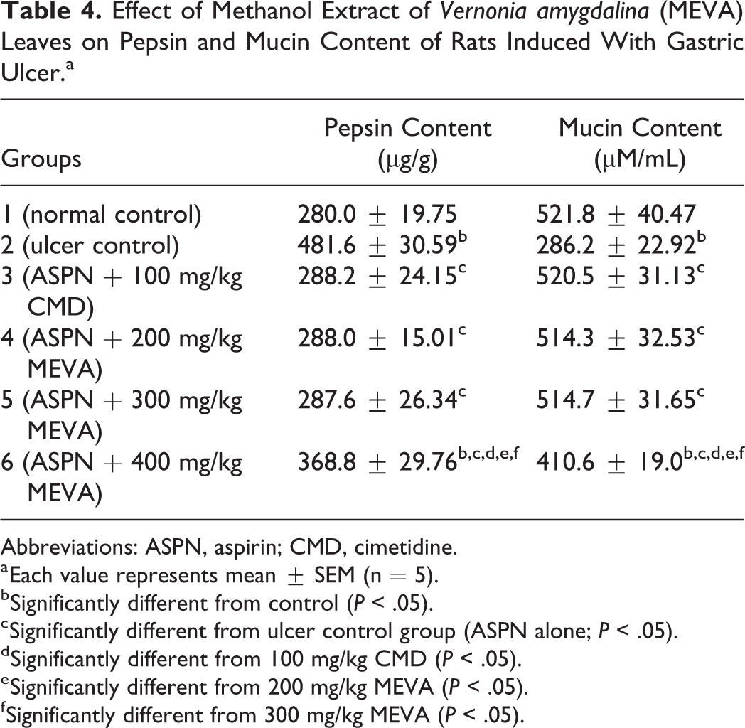
Abbreviations: ASPN, aspirin; CMD, cimetidine.
a Each value represents mean ± SEM (n = 5).
b Significantly different from control (P < .05).
c Significantly different from ulcer control group (ASPN alone; P < .05).
d Significantly different from 100 mg/kg CMD (P < .05).
e Significantly different from 200 mg/kg MEVA (P < .05).
f Significantly different from 300 mg/kg MEVA (P < .05).
Effects on Gastric Ulcer Scoring
As shown in Table 3, MEVA-treated groups had a significantly lower (P < .05) ulcer scores in the treated rats than the ulcer control (Table 3). The percentage of ulcer inhibition of the extract-treated rats at 200 and 300 mg/kg was similar to that of the standard drug (cimetidine) used in this study. Thus, the increase in ulcer score and index was prevented by MEVA in this study (Table 3).
Assessment of Oxidative Stress Indicators
Figures 1 to 3 revealed the effects of MEVA on the SOD activity, GSH, and MDA levels of gastric mucosal of aspirin-ulcerated rats. A significant reduction (P < .05) was observed in the activity of SOD in the aspirin-induced animals (Figure 3). Similarly, GSH level was also reduced in the ulcer control group (Figure 4). However, MDA level was significantly increased (P < .05) in the ulcerated animals (Figure 5). Treatment with the MEVA resulted in significant improvement (P < .05) in these parameters and the observable effects compared favorably well with both normal control and standard drug (cimetidine) used in this study.

Effect of MEVA leaves on SOD of rats. Each value represents mean ± SEM (n = 5). * indicates significantly different from control (P < .05), α indicates significantly different from ulcer control group (aspirin alone; P < .05), and # indicates significantly different from 200 mg/kg MEVA (P < .05). MEVA indicates methanol extract of Vernonia amygdalina; SOD, superoxide dismutase; SEM, standard error of the mean.

Effect of MEVA leaves on GSH of rats. Each value represents mean ± SEM (n = 5).* indicates significantly different from control (P < .05), α indicates significantly different from ulcer control group (aspirin alone; P < .05), and # indicates significantly different from 200 mg/kg MEVA (P < .05). GSH indicates glutathione; MEVA, methanol extract of Vernonia amygdalina; SEM, standard error of the mean.

Effect of MEVA leaves on MDA of rats. Each value represents mean ± SEM (n = 5). * indicates significantly different from control (P < .05), α indicates significantly different from ulcer control group (aspirin alone; P < .05), β indicates significantly different from 100 mg/kg CMD (P < .05), # indicates significantly different from 200 mg/kg MEVA (P < .05), and ω indicates significantly different from 300 mg/kg MEVA (P < .05). MDA indicates malondialdehyde; MEVA, methanol extract of Vernonia amygdalina; CMD, cimetidine; SEM, standard error of the mean.
Histological Observations
The representative photomicrographs of stomach sections of control and treated rats are shown in Figure 6. The histology of the stomach sections of control rats was structurally normal having the normal epithelial architecture and laminal, submucosa, and muscularis propria. In contrast, the histology of aspirin-ulcerated group revealed severe epithelial erosion and necrotic and distorted glands accompanied by marked cellular infiltration by mononuclear cells and degenerative changes in forestomach and fundic regions of the stomach. The morphological characteristics of the stomach of rats treated with aspirin plus MEVA or cimetidine showed significant prevention of aspirin-induced ulcer and were comparable to those in the control group.

Photomicrograph of stomach of CN, ulcer control, ASPN + CMD, ASPN + 200 mg/kg MEVA, ASPN + 300 mg/kg MEVA, and ASPN + 400 mg/kg MEVA. Control shows intact epithelium, laminal propria, submucosa, and muscularis propria. Ulcer control showed degenerative changes in forestomach and fundic regions of the stomach. There is also marked ulceration with loss of cellular constituents (brown and yellow arrow) in forestomach. Yellow arrow indicates development of subepithelial space, and brown arrowhead indicates distortion of mucosal architecture in the ulcerated rats. There was intact architecture of the stomach tissue in ASPN + CMD, ASPN + 200 mg/kg MEVA, and ASPN + 300 mg/kg MEVA. But ASPN + 200 mg/kg MEVA and ASPN + 300 mg/kg MEVA show sign of hemorrhage (black arrow). There is sign of sloughing of submucosa and mucosa layer (brown arrow) in ASPN + 400 mg/kg; ×100 magnification H&E. ASPN indicates aspirin; CMD, cimetidine; CN, control; MEVA, methanol extract of Vernonia amygdalina; SEM, standard error of the mean.
Discussion
Quantitative phytochemical screening of the MEVA leaves revealed the presence of flavonoids and phenolic contents (Table 2). The large amount of total flavonoid and phenolic contents detected in MEVA can be attributed to the antioxidant potential formerly described for this species. 39 –41
In this study, aspirin treatment decrease food intake and body weight in rats. The significant decrease in food intake and body weight in the aspirin-ulcerated group indicates that the general metabolic functions of the animals are deranged and demonstrates the induction of ulcer by aspirin as reported previously. 42 Warner et al 43 and Reuter et al 44 reported that NSAIDs, such as aspirin, decreased nutrient digestion and absorption, although they have a direct effect on the gastric and intestinal mucosal cells. However, the direct effect of NSAIDs on gastric and intestinal mucosal cells is as a result of inhibitory effects of NSAIDs on constitutive cyclooxygenase in the gastrointestinal cells. An increase in food consumption and body weight was observed in the MEVA-treated groups compared to the aspirin-ulcerated group. The gain in body weight that was observed in MEVA-treated groups could be attributed to the increase in food intake or mobilization of endogenous prostaglandins in the gastric mucosa cells of the rats treated with MEVA.
The inhibitory actions of aspirin on prostaglandin synthesis coupled with free radical formation have been reported in previous studies to be critical biochemical events in the pathogenesis of gastric ulceration. 42,45,46 Prostaglandins normally protect the gastrointestinal mucosa from damage by maintaining blood flow and increasing mucosal secretion of mucous and bicarbonate. Its inhibition in gastric mucosa by NSAIDs, such as aspirin, caused elevation in gastric acid secretion and reduced mucosal blood flow, mucus, and bicarbonate secretion. 47,48 It is therefore seemed that MEVA probably prevented the inhibitory action of aspirin on prostaglandin synthesis in the gastric mucosa cells.
The pH shows the level of gastric acidity and secretion volume. The low value of gastric pH is an indication of decreased hydrogen ion concentration in gastric juice. This has been linked to pathogenesis of ulcer and gastric damage in experimental animals. 49 Aspirin treatment in this study caused decrease in pH value and increase in gastric secretion volume in the ulcer control group. Also, aspirin treatment significantly increase UI, ulcer scores, and gastric volume following oral administration of aspirin in the ulcerated rats, and this may be attributed to either free radicals’ formation or inhibition of prostaglandin synthesis. Decreased prostaglandin level has been attributed to impaired gastroprotection and increased gastric acid secretion, which are important events in the etiology of mucosal ulceration. This agrees with the reports of Bech et al, 50 Debnath et al, 46 and Wang et al, 42 where NSAIDs such as aspirin were reported to have caused alterations in gastric secretions of rats. However, the results of this study showed that MEVA caused an increase in gastric pH and reduction in free and total gastric acid concentration in rats which is similar to that produced by cimetidine. This suggests that MEVA has inhibitory effect on gastric acid secretion and its inhibitory action might mimic cimetidine effect on gastric acid secretion. It has been established that inhibition of histamine release as a result of blockage of H2 receptors, inhibit intracellular adenylate cyclase, Na+-K+ ATPase, and proton pump of parietal cells, thereby reducing the gastric acid secretion. 51,52
Researches have confirmed that mucus secretion, wound retraction, and reepithelialization are involved in ulcer healing process after gastric mucosa injury. 53,54 Besides providing significant buffering capacity for the neutralization of luminal acid, mucus also provides protection against both endogenous aggressors and exogenous gastrotoxic agents such as aspirin, thereby enhancing the rate of local healing process. 55 In this study, the increased pepsin activity coupled with decrease in mucin secretion in the aspirin-ulcerated rats indicated altered hydrophobicity and reduced protective ability of the mucosal membrane against hemorrhagic erosion, thus resulting in tissue damage. Indeed, the histology of aspirin-ulcerated group revealed severe epithelial erosion of gastric mucosa and necrotic and distorted glands accompanied by marked cellular infiltration by mononuclear cells (Figure 6). This inferred the decreased ability of the gastric mucosa to withstand the offensive onslaught of aspirin. Besides antioxidant action that protects the mucus layer and arrests ulcer progression, drugs that increase the synthesis and secretion of gastric mucus would accelerate gastric ulcer healing. Pretreatment with the MEVA, however, facilitated ulcer healing process, which is associated with decreased pepsin activity and elevated mucin content in the gastric mucosa. This in turn facilitates speedy wound healing of the ulcerated areas of the mucosa epithelial layer and protects the gastrointestinal membrane, thus preventing aspirin gastric mucosa injury in the ulcerated rats. 54 This is suggestive of improved mucus secretory potential of the extracts and indicative of their significant role in ulcer healing process. Healing of mucosa epithelial cells was prominently displayed by the extracts at 200 and 300 mg/kg bw dose, depicting a better ulcer healing capacity and compared favorably well with the reference drug used in this study.
The sternness of oxidative damage depends on the degree of disturbances in normal redox state within the cells. Endogenous antioxidants such as SOD and GSH can curb the effects of ROS, but they quickly become overwhelmed by enormous quantities of ROS. 56 Aspirin has previously been reported to decrease antioxidant enzyme activity in rat stomach, thereby inducing gastric ulceration. 57 This is associated with overpowering of the cellular antioxidant defense systems by influence of free radicals damaging that subsequently results in gastric mucosa oxidative injury. In this study, aspirin treatment resulted in depletion of stomach antioxidant status as evident in the significant decrease in the activity of SOD as well as in the GSH level. The progressive elevation of the MDA level observed in stomach of aspirin-ulcerated rats indicates a state of stress and induction of lipid peroxidation as a result of depletion in the gastric mucosa antioxidant system. 45 However, the significantly reduced concentration of MDA coupled with marked increase in the activity of SOD and GSH level in MEVA-treated rats is an obvious indication of antiperoxidative and antioxidative potentials of the extract.
Overall, protection presented by the MEVA leaves against aspirin-induced gastric ulceration may be attributed to its phytochemicals constituent, such as polyphenolic compounds and other bioactive principles of the plant. These include the ability to scavenge free radicals and regulate mucosal membrane permeability, thereby countering the effect of aspirin on gastric acid secretion. This is in agreement with the works of Hussain et al, 11 Mota et al, 58 and Wang et al, 42 where gastroprotective potentials of plant extracts against aspirin-ulcerated rats were associated with their polyphenolic compounds and other various bioactive principles. Also, since cimetidine is a H2 receptor antagonist, the effect produced by the extract might mimic its mechanism of action via modulating cells in the mucosal lining of the stomach against excessive acid secretion. 59 –61
Conclusion
The protection offer by the MEVA leaves against aspirin-induced gastric injury is indicative of its outstanding gastroprotective and antioxidative potentials in rats. In addition, the presence of flavonoids content in MEVA may certainly contribute to the antiulcerogenic activity described in this study. However, higher dose of MEVA (400 mg/kg) seems not to have antiulcerogenic effect on aspirin-induced gastric ulcer.
Footnotes
Declaration of Conflicting Interests
The author(s) declared no potential conflicts of interest with respect to the research, authorship, and/or publication of this article.
Funding
The author(s) received no financial support for the research, authorship, and/or publication of this article.