
Abstract
Introduction
Breast cancer is the most common disease and the second leading cause of death for women worldwide. 1 In China, the incidence of breast cancer has rapidly increased. 2 About 30% of early-stage breast cancer develop metastasis and about 5% of patients are diagnosed with advanced distant metastasis. 3 Improvement of treatment does not significantly change the prognosis of metastatic breast cancer. The 5-year survival rate of metastatic breast cancer patients is only 23%, which is much lower than 84% to 99% in early-stage without distant metastases. 4
Chemotherapy plays an important role in the treatment paradigms for metastatic breast cancers. However, chemotherapy cannot eliminate the cancer cells. The resistant cells will cause cancer recurrence or metastasis, which impedes the success of chemotherapy. Hence, it is necessary to identify effective biomarkers for patients with drug-resistant risk, especially for those with metastatic breast cancers. Therefore, searching for highly sensitive, specific, non-invasive predictive biomarkers in peripheral blood of metastatic breast cancer patients is very urgent and practical.
The microRNAs (miRNAs) are a class of small noncoding RNA molecules (19-25 nucleotides), which play an important role in the regulation of proliferation, differentiation, apoptosis, and migration of cancer cells. 5,6 The panel of miRNA expression is quite different between cancers and normal tissues. 7 Previous studies showed that circulating miRNAs in peripheral blood may derive from cancer cells and reflect the pathological characteristics of primary tumors. 8,9 The plasma or serum miRNAs are quite stable and suitable for biomarker screening. 10 –12 The origins of miRNAs in plasma are currently unclear. The blood cells and tumor cells may contribute to the miRNAs in peripheral blood. And tumor cells release amount of miRNAs by active 13 or passive 14 ways. Furthermore, tumor-derived exosomes and microvesicles in the peripheral blood contain many miRNAs. 15 Some miRNAs have been reported as the promising features for predicting chemotherapy response in early stage of breast cancer patients. 16,17 We hypothesized that miRNAs in plasma are promising noninvasive biomarkers in metastatic breast cancer.
In our study, 14 miRNAs, which were reported to regulate metastasis formation, epithelial mesenchymal transition, and stem cell characteristics in the previous studies, 18 –23 were chosen from literatures as the candidate biomarkers. Due to the small amount of microRNAs in the limited volume plasma, we used a serum-direct Multiplex RT-PCR (SdM-qRT-PCR) to quantify the plasma levels of the miRNAs in metastatic breast cancer patients. 18 Here, we for the first time evaluated the potential of the microRNAs to predict chemotherapy resistance in metastatic breast cancer patients; miR-200a and miR-210 have been identified as new predictors.
Materials and Methods
Study Cohort
All the plasma were taken from the specimen bank of Beijing Cancer Hospital. We selected 40 metastatic breast cancer patients in the training stage and 103 metastatic breast cancer patients in the validation stage who were treated between June 2009 and June 2014. All the patients received chemotherapy with single-agent docetaxel or combination with docetaxel, who had response evaluation results. Eight age-matched healthy women with no history of cancer and in good health condition were recruited as controls at the same period with breast cancer patients. This study was approved by the ethics committee of Beijing Cancer Hospital (Beijing, China; 2016-KT45), and written informed consent was obtained from all the patients. This study strictly conformed to the principles outlined in the Declaration of Helsinki.
Before starting the treatment, plasma was obtained using the following procedures: 4 mL of peripheral blood from patients was collected in heparin-containing tubes and incubated at room temperature within 1 hour and then centrifuged at 2000 rpm for 15 minutes at room temperature. The supernatant was transferred to a microfuge tube and stored at −80°C for further use. Specimens that showed hemolysis were excluded for further miRNA detection.
Patient Treatment and Assessment
All the patients received single-agent docetaxel or combination with docetaxel every 21 days for 4 to 6 cycles. None of the Her2-positive patients received trastuzumab in this study because of economic reasons.
Demographic and clinic pathological details of patients were obtained from the medical records of the Department of Breast Oncology. The estrogen receptor (ER), progesterone receptor (PgR), and Her2 status were the result of the primary tumor. Estrogen receptor and PgR status were considered positive when 10% or more tumor cells exhibited nuclear staining for the receptors. Her2 positivity was defined as either as score of 3+ by Immunohistochemistry (IHC) or positivity by fluorescence in situ hybridization (FISH).
Treatment response was assessed by the RECIST criteria. 19 Complete response (CR), partial response (PR), and stable disease (SD) were considered as sensitive(S) group. Progressive disease (PD) was considered as resistant(R) group.
Quantitative RT-PCR of Plasma miRNAs
Serum-direct Multiplex qRT-PCR (SdM-qRT-PCR) was performed following our previous study. 18 Briefly, reverse transcription was performed using the plasma samples without RNA extraction and the Prime Script™ RT reagent Kit (Takara Bio Inc, Kyoto, Japan). The specific miRNAs RT primers were synthesized by Ribo Bio (Guangzhou, China). The 20-μL reverse transcription reaction mixture was incubated at 42°C for 30 minutes and 85°C for 15 seconds. Then, cDNA was obtained. The total of 2 μL cDNA was used per 25 μL qPCR reaction, including the SYBR Green I dye and miRNA-specific detection primers (Ribobio, Guangzhou, China). The quantitative real-time PCR reaction was performed at 95°C for 2 minutes in 40 cycles at 95°C for 15 seconds and 60°C for 60 seconds. To obtain reproducible results, plasma from each patient was split into 3 aliquots and performed the whole detection assay.
The relative expression of each miRNA was calculated from the following equation: relative expression = 2−ΔCt, where Ct is the threshold cycle for a sample and ΔCt = mean CtmiRNA – mean CtmiR-191.
Statistical Analysis
The statistical analyses were performed using the SPSS software package, version 19.0. The differences of miRNAs expression levels between R group and S group were identified using an unpaired t test. Data were presented as mean (standard deviation). In the validation stage, the χ2 or 2-tailed Fishers exact test was used to identify potential associations of plasma miRNAs and clinic pathological risk factors with PD. And the factors that might were associated with PD were analyzed by using the univariate and multivariate logistic regression analyses. P ≤ .05 in all cases was considered statistically significant.
Results
Characteristics of Patients and Study Design
In total, 143 breast cancer patients were included in this study. All the patients received chemotherapy with single-agent docetaxel or combination with docetaxel. In the training stage, 20 patients achieved PR or SD, defined as sensitive (S) group and 20 patients achieved PD, defined as resistant (R) group. Eight age-matched healthy women were recruited as controls. The median age was 53 (35-70) years old. In the validation stage, 34 patients with PR, 52 patients with SD and 17 patients with PD were studied.
The clinical characteristics of all the patients are presented in Table 1.
Clinicopathological Characteristics of the Patients With Metastatic Breast Cancer.

Abbreviations: CBR, clinical benefit rate; ECOG, Eastern Cooperative Oncology Group; ER, estrogen receptor; HR, hormone receptor; HER2, epithelial growth factor 2; IDC, invasive ductal carcinoma; ILC, invasive lobular carcinoma; ORR, overall response rate; PD, progression disease; PR, progesterone receptor; PR, partial response; SD, stable disease.
Selection of Candidate miRNAs
Previous studies indicated that the role of microRNAs in cancer was associated with the progression of breast cancer. We found 14 microRNAs: miR-150, miR-145, miR-155, miR-21, miR-200a, miR-200b, miR-200c, miR-210, miR-203, miR-221, miR-375, miR-451, miR-34a, and miR-122, which were particularly associated with prognosis, drug resistance, and stem cell characteristics in the previous reports. 20 –25 MiR-16 and miR-191 were taken as reference controls as previously indicated. 10,26 Thus, we selected the 16 miRNAs as the candidates to perform the integrative analyses in the plasma of metastatic breast cancer patients.
The endogenous control for detection serum miRNAs was determined as described in our previous study. 18 By using GeNorm and NormFinder, miR-191 was chosen as the most stably expressed miRNA internal control (Supplementary data, Figure S1).
Plasma microRNA Candidates for Breast Cancer Prognosis in the Training Set
The expression level of 14 plasma miRNAs was measured by qPCR in the 40 breast cancer patients in training stage (Table 2). The difference between R group and S group was determined using an unpaired t test. We found 2 miRNAs (miR-200a (P < .001) and miR-210 (P < .001)) significantly increased and 1 miRNA (miR-451 (P<0.001)) significantly decreased in the R group (Figure 1A). In addition, the expression of the 3 miRNAs in the plasma of metastatic breast cancer patients was significantly higher than that in the healthy controls (Figure S2). Furthermore, the receiver operating characteristic (ROC) curve analysis showed that area under curve (AUC) was 0.847 (95% CI 0.717-0.978) for miR-200a, 0.825 (95% CI 0.695–0.955) for miR-210, and 0.855 (95% CI 0.731–0.979) for miR-451 (Figure 1B). These data indicated these 3 microRNAs could be the potential biomarkers to distinguish R group from S group of breast cancer patients.
Plasma Levels of 14 microRNA Candidates in S Group and R Group in the Training Set.
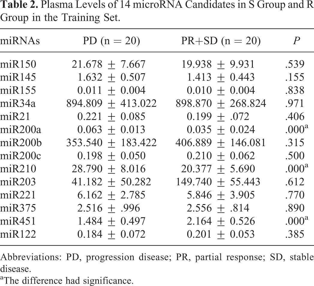
Abbreviations: PD, progression disease; PR, partial response; SD, stable disease.
aThe difference had significance.

Plasma levels and ROC analysis of miR-200a, miR-210, and miR-451 in the discovery set. A, The box plots showed the plasma levels of miR-200a, miR-210, and miR-451 in the discovery set composed of 20 resistant cases and 20 sensitive cases. B, ROC in the discovery set composed of 20 patients in R group and 20 in S group.
Validation of Plasma microRNAs for Breast Cancer Prognosis
To validate the 3 miRNAs identified in the training stage, we measured their plasma levels in an independent breast cancer population composing of 17 resistant cases (R) and 86 sensitive ones (S). We found that the relative levels of miR-200a and miR-210 were significantly higher in the R group than those in the S group. The relative level of miR-451 expression was much lower in the R group than that in the S group (Figure 2A). Next, the ROC curve analysis showed that the cut-off point of the 3 miRNAs in the training set was used directly for the validation set and the combined set. The results of AUC analysis of miR-200a, miR-210 in the validation set had the same trends with those in the training set. Unfortunately, the specificity of miR-451 was quite low (Table S1, Figure 2B). Moreover, we analyzed the combined data of training and validation stages including 37 resistant cases and 106 sensitive cases. Consistently, the 3 miRNAs showed a similar trend (Figure 3A and 3B). Hence, we focused on the results of miR-200a and miR-210 in the following analysis. Chemotherapy is the main treatment for triple negative breast cancer (TNBC). We further tested the predictive valued of miR-200a, miR-210 expression level in the TNBC. The relative levels of miR-200a and miR-210 were significantly higher in the 5 resistant cases than those in the 14 sensitive cases (P = .005, P = .007; Figure 3).

Plasma levels and ROC analysis of miR-200a, miR-210, and miR-451 in the validation set. A, The box plots show the plasma levels of miR-200a, miR-210, and miR-451 in the validation set composed of 17 resistant cases and 86 sensitive cases. B, ROC in the validation set composed of 17 patients in R group and 86 in S group.

Plasma levels and ROC analysis of miR-200a, miR-210, and miR-451 in the combined set. A, The box plots showed the plasma levels of miR-200a, miR-210, and miR-451 in the combined set composed of 37 resistant cases and 106 sensitive cases. B, ROC in the combination set composed of 37 patients in R group and 106 in S group.
Correlation Analysis of Plasma miR-200a and miR-210 Level With Clinicopatholgocial Characteristics of Breast Cancer
We assessed the association of plasma level of miR-200a and miR-210 with PD in the combined set and found miR-200a and miR-210 expression was correlated with the PD rate (P < .001, Table 3). Then, miR-200a/miR-210 with other clinicopatholgocial characteristics was further analyzed using univariate and multivariate logistic regression analysis to detect the association with chemotherapy response (Table 4). The levels of plasma miR-200a expression (odds ratio [OR] = 0.041 95% confidence interval [CI]: 0.010-0.169, P < .001) and miR-210 (OR = 0.062, 95% CI: 0.017-0.229, P < .001) were identified as independent factors for chemotherapeutic response.
Correlation of miRNA Expression and Chemotherapy Response in the Combined Set.

Abbreviations: CBR, clinical benefit rate; PD, progression disease; PR, partial response; SD, stable disease.
aExpression lower than the cut-off value.
bExpression equal to or higher than the cut-off value.
The Correlation of Clinicopathological Characteristics and miRNA Expression With Chemotherapy Response.

Abbreviations: ER, estrogen receptor; HR, hormone receptor; HER2, epithelial growth factor 2; PgR, progesterone receptor.
Further Analysis of the Relation of the miRNAs in the Plasma With Other Clinicopathological Characteristics
The expression of miR-200a was significantly associated with the stage in surgery, and miR-210 was associated with internal organ metastasis, as shown in Table 5. Then, we analyzed the expression levels of miRNAs and found that the patients with stage IV disease at diagnosis had higher expression levels of miR-200a (Figure 4A), and the patients with internal organ metastasis had higher expression levels of miR-210 (Figure 4B).The consistent results confirmed the association of miRNA levels and the clinical characteristics (Figure 5).
Expression of the Candidate miRNAs in the Plasma and Associations With Other Clinical Characteristics (P value).

Abbreviations: ECOG, Eastern Cooperative Oncology Group; ER, estrogen receptor; HR, hormone receptor; HER2, epithelial growth factor 2; IDC, invasive ductal carcinoma; ILC, invasive lobular carcinoma; PR, progesterone receptor.
aFisher exact test.

Plasma levels in the triple negative breast cancer (TNBC). A, The box plots showed the plasma levels of miR-200a of 5 resistant cases and 14 sensitive cases of TNBC. B, The box plots showed the plasma levels of miR-210 of 5 resistant cases and 14 sensitive cases of TNBC.

Plasma microRNA analysis in the subgroup of patients with breast cancer. A, The box plots showed the plasma levels of miR-200a according the stage in surgery. B, The box plots showed the plasma levels of miR-210 in the patients with or without internal organ metastasis.
Discussion
Chemotherapy response predictive markers are quite important in the clinical practice. Growing body of evidence showed miRNAs played important role in chemoresistance of breast cancer. Efforts have been made to evaluate circulating level of the microRNAs as biomarkers for prediction of prognosis or monitoring patient responses to therapy. Circulating level of miR-106b was found closely related to tumor size, metastasis, as well as shorter overall survival and progression-free survival. 27 Frères et al analyzed the plasma miRNA signature and showed high diagnostic or predictive accuracy with breast cancers. 13,14,28 Here, we for the first time showed miR-200a and miR-210 could predict metastatic breast cancer chemoresistance as new biomarkers. Compared with previous reports, we found that plasma miR200a and miR-210 has a good predictive performance for PD in the metastatic setting, which is a more complex group. All these studies may ultimately lead to better treatment options for breast cancer patients.
Our results suggested that high level of miR-200a and miR-210 in plasma of metastatic breast cancer patients was associated with chemotherapy resistance. MiR-200 family consists of 5 members: miR-200a/miR-200b/miR-429/ miR-200c/miR-141, which are classified into 2 categories according to their chromosomal locations at 1 and 12. MiR-200 family was reported to inhibit EMT and suppress the proliferation of stem cells. 15,29 –31 In breast cancer patients, San-Jian Yu et al 32 reported that the level of miR-200a in lymph node metastasis group increased more than 7-fold when compared with that in nonmetastasis patients. The data suggested that the high level of miR-200a was associated with metastatic behavior in breast cancer. Additionally, Madhavan et al 33 showed that the metastatic breast cancer patients with circulating tumor cells (CTC)-positive had higher level of miR200a than that in CTC-negative patients. Together with our findings of miR-200a in plasma, these consistent data indicated the prognostic value of the microRNA. We would further test the correlation of miR200a with the prognosis of the metastatic patients. In hypoxic microenvironment, miR-210 is a critical regulator for cell survival. 25,34 High level expression of miR-210 was detected in many cancers including breast cancer. 35 A meta-analysis from 511 breast cancer cases indicated that high level of miR-210 expression might predict poor survival in the patients. 36 Toyama et al 37 showed that the level of miR-210 in triple-negative breast cancers was significantly higher than that in estrogen receptor-positive/HER2-negative breast cancers. Moreover, high level of miR-210 was an independent factor for worse prognosis in breast cancer, especially in lymph node-negative triple-negative patients. 37,38 In this study, we found that miR-210 in plasma was associated with drug resistance, and the higher expression of miR-210 was associated with internal organ metastasis. Collectively, our results showed that high level of plasma miR-200a and miR-210 was associated with chemotherapy resistance in metastatic breast cancer patients. These results provided important evidence for the application of circulating microRNAs for the prediction of response to breast cancer treatment. Larger prospective, multi-institutional studies to validate the potential role of plasma miRNAs as chemotherapy predictive markers for metastatic breast cancer are expected in the future.
Supplemental Material
Supplementary_data - Plasma microRNAs Predict Chemoresistance in Patients With Metastatic Breast Cancer
Supplementary_data for Plasma microRNAs Predict Chemoresistance in Patients With Metastatic Breast Cancer by Bin Shao, Xiaoxia Wang, Lei Zhang, Deyu Li, Xiaoran Liu, Guohong Song, Huiqing Cao, Jun Zhu, and Huiping Li in Technology in Cancer Research & Treatment
Footnotes
Abbreviations
Authors’ Note
First author: Bin Shao, Department of Medical Oncology, Key laboratory of Carcinogenesis and Translational Research (Ministry of Education), Peking University Cancer Hospital & Institute, No. 52 Fucheng Road, Haidian district, Beijing Cancer Hospital, Beijing, P.R. China. Email:
Declaration of Conflicting Interests
The author(s) declared no potential conflicts of interest with respect to the research, authorship, and/or publication of this article.
Funding
The author(s) disclosed receipt of the following financial support for the research, authorship, and/or publication of this article: This study was supported by Grants from National Basic Research Program of the Chinese Ministry of Science and Technology (973 Grant no.2013CB531202) and National Natural Science Foundation of China (No.81502586).
Supplemental Material
Supplemental material for this article is available online.
References
Supplementary Material
Please find the following supplemental material available below.
For Open Access articles published under a Creative Commons License, all supplemental material carries the same license as the article it is associated with.
For non-Open Access articles published, all supplemental material carries a non-exclusive license, and permission requests for re-use of supplemental material or any part of supplemental material shall be sent directly to the copyright owner as specified in the copyright notice associated with the article.