
Abstract
Objectives
This study aimed to describe the epidemiology and clinical presentation of presumed hereditary or presumed breed-related ocular diseases in a population of cats in France.
Methods
Medical records from between September 2013 and August 2017 were reviewed to identify cats with at least one presumed hereditary or breed-related ocular disease. Cats with concurrent, or a history of, ocular or systemic infectious diseases were excluded. Signalment, history and clinical findings were recorded.
Results
Of the 1161 cats that presented to our institution during the study period, 129 were diagnosed with at least one presumed hereditary or presumed breed-related ocular disease (11.1%, 95% confidence interval [CI] 9.3–12.9). Five ocular abnormalities had a prevalence of >1%: entropion, corneal sequestration, persistent pupillary membrane, cataract and retinal dysplasia. The prevalence of entropion was 2.2% (95% CI 1.3–3.0), with Persians (P = 0.03), Maine Coons (P <0.01) and male cats (P <0.01) being over-represented. The prevalence of corneal sequestration was 2.4% (95% CI 1.5–3.3), with Persians (P <0.01) and Exotic Shorthairs (P = 0.02) being over-represented. Persistent pupillary membranes and cataracts had the same prevalence of 2.3% (95% CI 1.5–3.2), with no particular sex or breed significantly over-represented. Retinal dysplasia had a prevalence of 1.6% (95% CI 0.8–2.3) and Persian cats were over-represented (P = 0.04). Anterior segment dysgenesis had a low prevalence (0.9%, 95% CI 0.4–1.5), with all affected cats being domestic shorthairs and this breed therefore was over-represented (P = 0.04).
Conclusions and relevance
In a French population of cats, presumed hereditary or breed-related ocular diseases accounted for 11.1% of all ocular diseases. Cataracts, corneal sequestration, persistent pupillary membrane, entropion and retinal dysplasia were the most common conditions. Statistical breed over-representation was observed for entropion, corneal sequestration and retinal dysplasia. We recommend that more systematic screening of feline species is conducted.
Keywords
Introduction
Hereditary or presumed breed-related ocular diseases have been described in cats for over 50 years. Such diseases are less common in cats than in dogs.1,2 Pedigree analysis is required to prove inheritance, and should be followed by a candidate-gene approach or genome-wide analysis to determine the underlying mutations. 3 These analyses are only conducted for some diseases in feline ophthalmology, including rod–cone dysplasia, rod–cone degeneration, congenital glaucoma and progressive retinal degeneration in Bengals and Persians. Most of these diseases can be tested for with DNA-based tests.4–10 There is no current proof of inheritance for many diseases. Nevertheless, some breeds seem to be over-represented, or familial descriptions exist. The terms ‘presumed breed-related’ or ‘familial’ are preferred. 1 Presumed hereditary or breed-related ocular diseases are poorly described and considered rare in cats compared with dogs.1,2,11
To the best of our knowledge, there have been no studies on the epidemiology and clinical presentation of presumed hereditary or breed-related ocular diseases in a large population of cats of varying breeds. This study aimed to describe the prevalence and clinical features of presumed hereditary or breed-related ocular diseases in a French population of cats, including diseases for which inheritance is not suspected to this date in cats but has been proven for other species.
Materials and methods
Case selection
The clinical records of cats that presented at the ophthalmological unit of the National Veterinary School of Alfort, France, between September 2013 and August 2017 were reviewed. Cats were included if the ophthalmic examination revealed a single or multiple hereditary, presumed hereditary or presumed breed-related ocular diseases.
Cats with concurrent or a history of infectious ocular or systemic disease, cats for which the clinical features suggested an acquired ocular disease or cats with incomplete clinical records were excluded.
Data retrieved
Data collected and reviewed from medical records included signalment (age at diagnosis, breed, sex), medical history and clinical findings for each abnormality, including diagnosis, isolated or in combination with other ocular abnormalities, laterality, location and stage of development.
Ophthalmic examination
A complete ophthalmic examination was performed on each cat by a diplomate or resident of the European College of Veterinary Ophthalmologists. It included menace response, palpebral, dazzle and pupillary light reflexes, slit-lamp biomicroscopy (SL-15 or SL-17 portable slit-lamp; Kowa), indirect ophthalmoscopy (Heine Omega 200; Heine Optotechnik), rebound tonometry (TonoVet; Icare), Schirmer’s tear test type 1 (Dina Strip Schirmer-Plus; Gecis) and fluorescein staining (fluorescein 0.5% single-dose eyewash; TVM Laboratories). If required, pupillary dilation was performed using a topical 0.5% tropicamide solution (Mydriaticum; Théa).
Statistical analysis
Statistical analysis was performed using Microsoft Excel and BiostaTGV software (http://biostatgv.sentiweb.fr/). The prevalence of each ocular disease in the reference population and the 95% confidence interval (CI) of the estimates were calculated. To compare breed and sex proportions between cats in each ocular disease subpopulation and the reference population, a χ² test or Fisher’s exact test was used. The reference population consisted of all cats that presented to the Ophthalmology Unit of the National Veterinary School of Alfort during the study period. Odds ratios (ORs) were calculated when a variable was over-represented in our population compared with the reference population. Statistical analysis was not performed when the effect was ⩽ 2. A P value <0.05 was considered to be statistically significant.
Results
Of the 1161 cats that presented to our institution during the study period, 129 (11.1%, 95% CI 9.3–12.9) were diagnosed with a presumed hereditary or breed-related ocular disease.
Our population included 61 females (47.3%, 95% CI 38.7–55.9) and 68 males (52.7%, 95% CI 44.1–61.3), with no statistically significant difference (P = 0.8). At diagnosis, median age was 2 years (interquartile range 0.6–8.6, range 0.2–12). Twenty-one breeds were represented (Table 1). However, Persians (OR 2.9; P <0.01) and Exotic Shorthairs (ESHs; OR 9.3 [P <0.01]) were significantly over-represented.
Breeds and number of cats diagnosed with presumed-hereditary or breed-related ocular diseases that presented to the Ophthalmology Unit of the National Veterinary School of Alfort, France, between September 2013 and August 2017
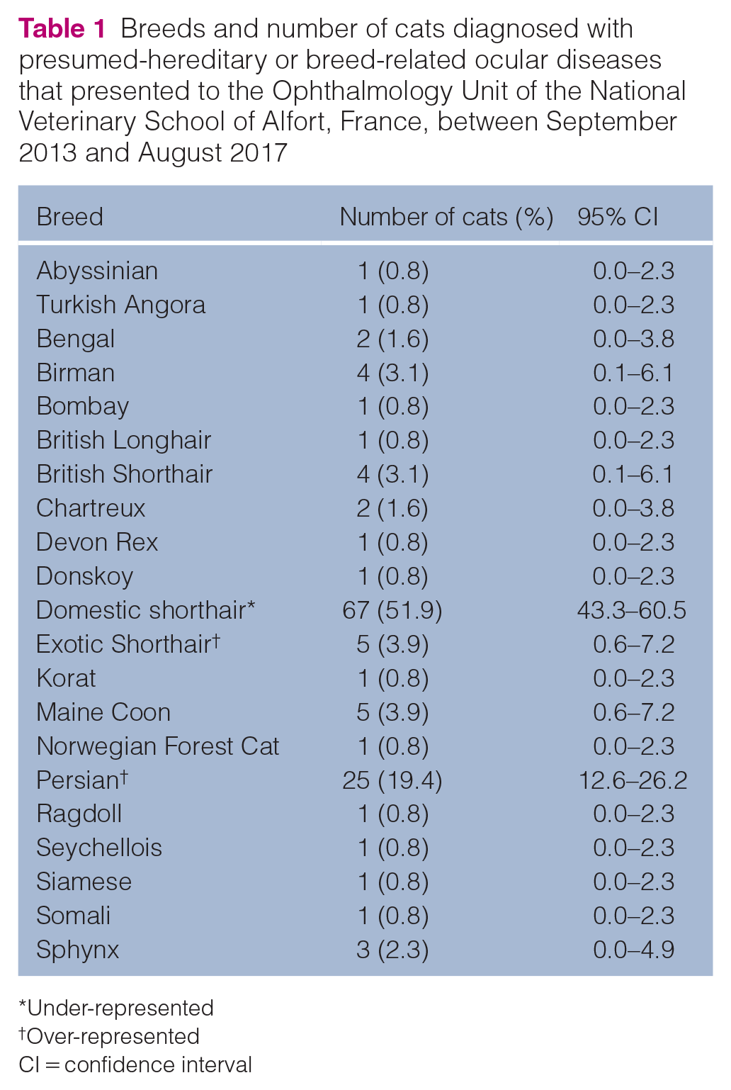
Under-represented
Over-represented
CI = confidence interval
Nineteen presumed hereditary or breed-related ocular diseases were observed: microphthalmia, strabismus, entropion, palpebral dermoid, distichia, palpebral coloboma, apocrine hydrocystoma, corneal sequestration, corneal dystrophy, persistent pupillary membrane (PPM), anterior segment dysgenesis (ASD), uveal cyst, cataract, primary lens luxation, microphakia, retinal dysplasia, progressive retinal degeneration, optic nerve coloboma and primary glaucoma (Tables 2 and 3). Thirty-eight cats had multiple ocular abnormalities (Table 4).
Number of cats of each breed affected with a presumed hereditary or breed-related ocular disease affecting the globe, the adnexas and the cornea

CI = confidence interval
Number of cats of each breed affected with a presumed hereditary or breed-related ocular disease affecting the intraocular structures
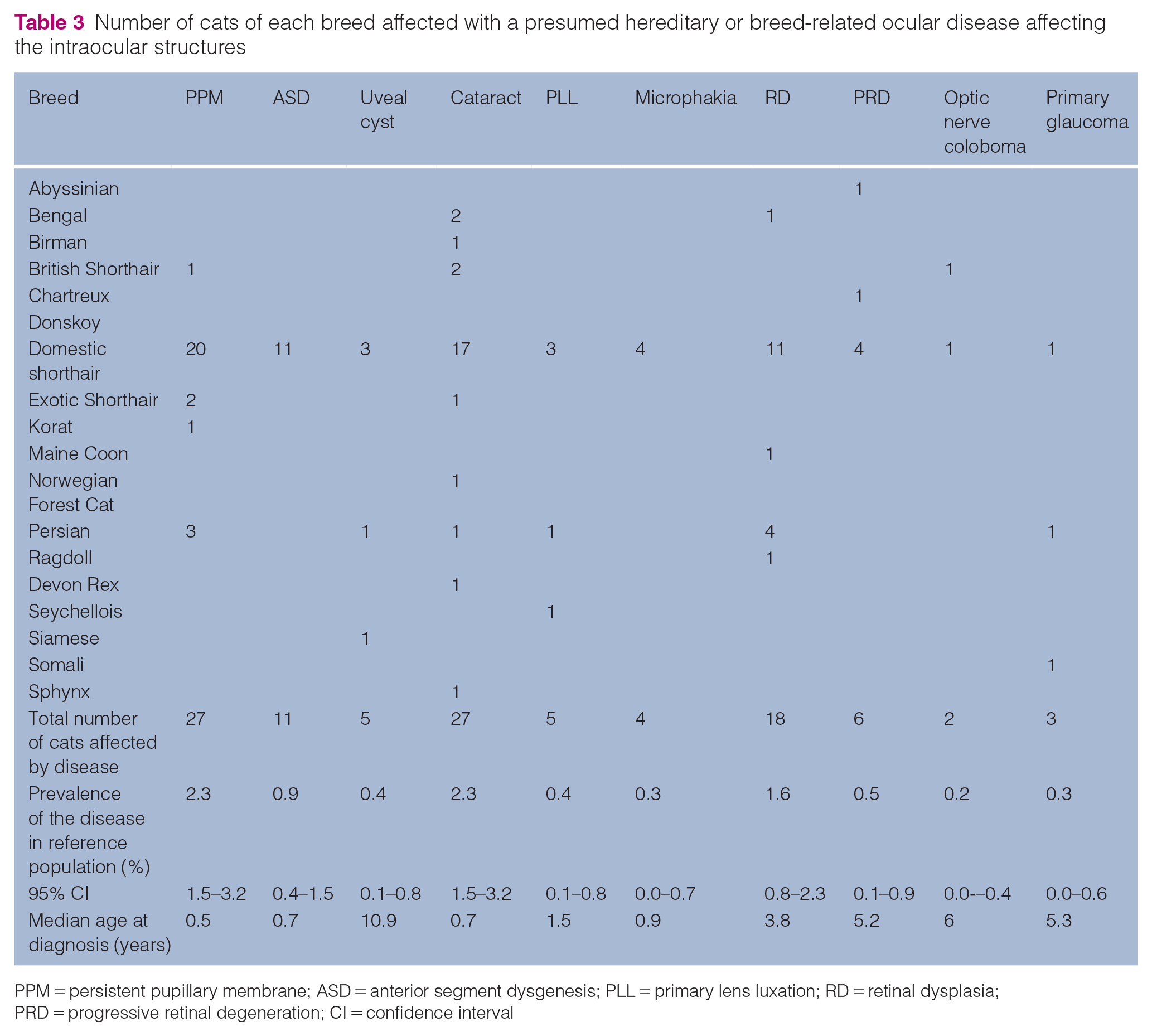
PPM = persistent pupillary membrane;
ASD = anterior segment dysgenesis; PLL = primary lens luxation; RD = retinal dysplasia; PRD = progressive retinal degeneration; CI = confidence interval
Number of presumed hereditary or breed-related ocular diseases per animal
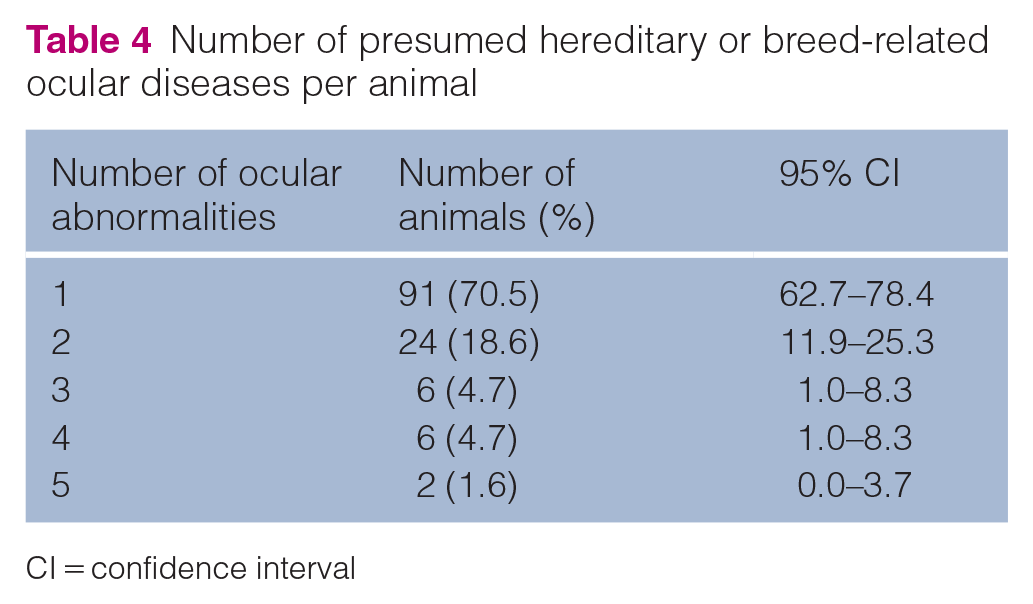
CI = confidence interval
Entropion
Presumed breed-related entropion (Figure 1) was identified in 25/129 cats (19.4%). The prevalence of this disease was 2.2% (95% CI 1.3–3.0).

Primary inferotemporal entropion with trichiasis and mucopurulent discharge in a 1-year-old male Maine Coon cat (affected bilaterally)
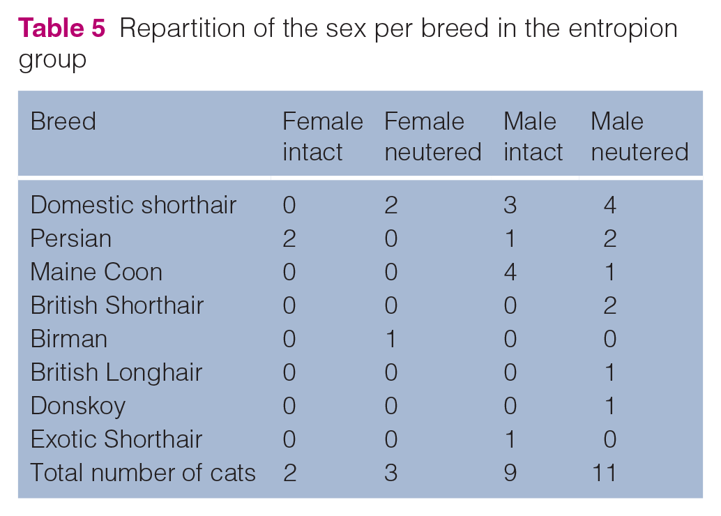
Eight breeds are represented in this subpopulation (Tables 2 and 5). Persians (OR 3.4; P = 0.03) and Maine Coons (OR 12.9; P <0.01) were over-represented. Although being the most frequent breed, domestic shorthair (DSH) cats were under-represented (OR 0.2; P <0.01). This subpopulation included five females (20.0%, 95% CI 13.1–26.9) and 20 males (80.0%, 95% CI 73.1–86.9). The repartitions of intact and neutered cats for each breed is shown in Table 5. Among all cats of this subpopulation, males were over-represented (OR 3.4; P <0.01). No sex was significantly over-represented among Persians and Maine Coons.
Repartition of the sex per breed in the entropion group
At the time of diagnosis, median age was 1.6 years (interquartile range [IQR] 1.0–4.7, range 0.2–15.0). For Persians, Maine Coons and DSHs, median age was 5.6 years (IQR 1.2–7.4, range 0.5–15.0), 1.0 year (IQR 0.8–1.6, range 0.7–4.4) and 1.7 years (IQR 1.1–5.3, range 0.2–7.6), respectively.
Twenty (80.0%) and five (20.0%) cats were bilaterally and unilaterally affected, respectively. Entropions were located temporally in the lower eyelid in 23/45 eyes (51.1%), medially in the lower eyelid in 20/45 eyes (44.4%) and temporally in the upper eyelid in 1/45 eyes (2.2%). Location was not recorded in one eye.
Among the cats affected with entropion located temporally in the lower eyelid, Maine Coons were over-represented (5/13; OR 31.9 [P <0.01]). In this subpopulation, there were 11 males (84.6%; five intact and six neutered) and two neutered females (15.4%). All the Maine Coons were male; four were intact and one was neutered. Males were over-represented (OR 4.7; P = 0.04).
Among the cats affected by entropion located medially in the lower eyelid, Persian was the most represented breed (5/10) and was over-represented (OR 12.9; P <0.01). No sex was over-represented (P = 0.35).
Ten cats (40%) presented with other ocular abnormalities. Corneal sequestration, cataract and persistent pupillary membrane were the most commonly associated with entropion.
Corneal sequestration
Corneal sequestration (Figure 2) was identified in 28/129 cats (21.7%), with a prevalence of 2.4% (95% CI 1.5–3.3). There were 10 females (35.7%; 95% CI 27.4–44.0) and 18 males (64.3%; 95% CI 56.1–72.6), and no sex was significantly over-represented (P = 0.28). Six breeds were represented (Table 2), with Persian being the most common and over-represented (OR 12.9; P <0.01). ESHs were also over-represented (OR 17.1; P = 0.02). Twenty-four (85.7%) and four cats (14.3%) were unilaterally and bilaterally affected, respectively.

Corneal sequestration in an 8-year-old female Persian cat
Median age at the time of diagnosis was 6.8 years (IQR 2.8–11.1, range 1.3–16.8). For Persians, median age was 6.7 years (IQR 3.6–11.1, range 1.3–16.8). The three ESHs were 1.3, 9.2 and 10.6 years old, respectively.
Six cats (21.4%) and one cat (3.6%) presented with one or two other concurrent ocular abnormalities: entropion (4/28 [14.3%]), PPM (2/28 [7.1%]) and retinal dysplasia (2/28 [7.1%]).
PPM
PPM, observed as iris-to-iris or iris-to-lens strands (Figure 3), was diagnosed in 27/129 cats (20.9%) from five breeds, representing a prevalence of 2.3% (95% CI 1.5–3.2; Table 3). No breed was statistically over-represented. They included 14 females (51.9%; 95% CI 43.2–60.5) and 13 males (48.1%; 95% CI 39.5–56.8), with no statistical difference (P = 0.55). Twelve (44.4%) and 15 cats (55.6%) were unilaterally and bilaterally affected, respectively. Median age at the time of diagnosis was 0.5 years (IQR 0.45–1.57; range 0.3–12.8). Twenty cats had other ocular abnormalities. Cataract was the most common ocular disease associated with PPM (12/27 [44.4%]), followed by eyelid colobomas (5/27 [18.5%]), entropion and retinal dysplasia (4/27 [14.8%]), microphakia (3/27 [11.1%]), distichia, microphthalmia, corneal sequestration, anterior segment dysgenesis, primary lens luxation (2/27 each [7.4%]), progressive retinal degeneration and optic nerve colobomas (1/27 [3.7%]).

Persistent pupillary membrane in a 2-month-old male domestic shorthair cat
ASD
ASD, observed as extensive and complete corneal opacity associated with a large adherence of the iris to the corneal endothelium, and a narrowed anterior chamber (Figure 4), was found in 11/129 cats (8.5%). The prevalence was 0.9% (95% CI 0.4–1.5). All cats were DSHs, and this group was over-represented (P = 0.04). Nine (81.8%) and two (18.2%) cats were affected unilaterally and bilaterally, respectively. Seven (63.6%; 95% CI 55.3–71.9) and four cats (36.4%; 95% CI 28.1–44.7) were female and male, respectively, with no statistically significant difference (P = 0.25). Median age at the time of diagnosis was 0.7 years (IQR 0.3–2.7; range 0.15–10.6). Five cats (45.5%) had other ocular diseases, including cataracts (2/11), microphakia (2/11) and microphthalmia (1/11).

Anterior segment dysgenesis in a 5-month-old male domestic shorthair cat: (a) front view; and (b) lateral view
Cataracts
Cataracts were found in 27/129 cats (20.9%), accounting for a prevalence of 2.3% (95% CI 1.5–3.2). Nine breeds were represented (Table 3), with none over-represented. Twelve (44.4%; 95% CI 35.9–53.0) and 15 (55.6%; 95% CI 47.0–64.1) cats were female and male, respectively, with no statistical difference (P = 0.87). Median age at the time of diagnosis was 0.7 years (IQR 0.4–2.2, range 0.15–18.3).
This subpopulation included 47 eyes. Twenty cats (74.1%) were bilaterally affected and five cats (18.5%) were unilaterally affected, and, for two cats, bilaterality could not be assessed owing to traumatic corneal opacity in one eye. Cataracts were incipient, immature, mature and hyper-mature in 18 (37.5%), 19 (39.6%), eight (16.7%) and two eyes (4.2%), respectively.
Opacities were located in both the nucleus and cortex in 21 eyes (43.8%), in the cortex in 11 eyes (22.9%) and the nucleus in 10 eyes (20.8%). The sites of opacities were not recorded in five eyes.
Among the cortical opacities, four were in the anterior cortex, three in the posterior cortex and the anteroposterior location was not recorded for two eyes.
Seventeen cats had at least one concurrent ocular disease. PPMs were most frequently found to be associated with cataracts (11/27), followed by retinal dysplasia (3/11), entropion (4/11), microphakia (3/11) and microphthalmia (3/11).
Retinal dysplasia
Retinal dysplasia, observed as multifocal retinal folds in the peripheral zone of the tapetal fundus, was diagnosed in 18 cats (13.9%) from five different breeds, accounting for a prevalence of 1.6% (95% CI 0.8–2.3; Table 3). Persians were over-represented (OR 3.7; P = 0.04). Four cats (22.2%) were affected unilaterally, and 12 (66.7%) were affected bilaterally. For two cats (11.1%), one eye was affected, but an assessment of retinal dysplasia by ophthalmoscopy was not possible for the second eye because of lens opacities or complete retinal detachment. Eight (44.4%; 95% CI 35.9–53.0) and 10 (55.6%; 95% CI 47.0–64.1) cats were female and male, respectively, with no statistical difference (P = 0.89). Median age at the time of diagnosis was 3.8 years (IQR 0.6–8.0, range 0.3–15.3). Eleven cats had at least one other ocular disease. PPM and cataracts were the most common ocular diseases associated with retinal dysplasia (4/18 for both).
Discussion
The prevalence of presumed hereditary or breed-related ocular disease in our feline population was 11.1% during the study period. Although these diseases are considered rare in cats,1,2,11 our results suggest that they are not uncommon findings. Six diseases were more commonly diagnosed: corneal sequestration (2.4%), entropion (2.2%), PPM (2.3%), cataract (2.3%), retinal dysplasia (1.6%) and ASD (0.9%).
Corneal sequestration had the highest prevalence in our study (2.4%). This corneal disease may be acquired, but genetic or breed-related origins have also been suggested.11,12 Our study excluded cats for which a primary cause was suspected or identified (previous corneal ulcers, feline herpesvirus-1 infection and entropion). Four cats had concurrent entropion that was not considered related to a corneal disease by a clinician, as it was distant from the location of the corneal sequestration. As we did not systematically explore infectious diseases with PCR, we cannot completely exclude a herpetic origin, but none of the selected cats had a history of viral infection. Our study showed an over-representation of brachycephalic cats (Persians and ESHs) among cats diagnosed with corneal sequestration, accounting for 60.7% of cats in this subpopulation. This result is consistent with previous studies, which have reported that 54.7–78.4% of cats with corneal sequestration are brachycephalic.12–14 The age at diagnosis varied widely (range 1.3–16.8 years), with no predilection, which is consistent with previous reports.12–14 Only one previous study suggested that young adult cats were more likely to be diagnosed with corneal sequestration, with 41% of cats being between 2 and 4 years old at the time of diagnosis. Nevertheless, this study reported a wide range of ages (range 0.4–17 years). 15 It has been suggested that corneal sequestration is more likely to occur in cats with chronic corneal irritation.1,16 Thus, the suspected brachycephalic predisposition to corneal sequestration could be linked to their facial features, leading to corneal irritation.1,16 Therefore, our results support the existence of primary corneal sequestration, but further studies are required to assess the genetic or conformational origin of corneal sequestration.
Primary entropion is an inward turning of the eyelid, leading to trichiasis, which develops at an early age and is due to facial conformation.2,17 Primary entropion is common in Persians and other brachycephalic breeds, and usually involves the medial aspect of the lower eyelid.2,11,18,19 Consistent with previous descriptions, our study showed an over-representation of Persians with primary lower lid medial entropion. We also found a statistical over-representation of Maine Coons with primary lower lid temporal entropion. A previous study described lower lid entropion in three male Maine Coons, with the authors suspecting a relationship between entropion and facial conformation, particularly their prominent jowls. 17 In our population of cats with primary entropion, male cats were statistically over-represented. In particular, all the Maine Coons were male. We could not prove statistical significance for sex predilection in this breed, possibly because of the lack of statistical power. Consistent with the study of Williams and Kim, 17 this trend suggests a predisposition for male Maine Coons, possibly due to conformational characteristics. Moreover, our study shows that primary entropion seems to develop later in Persians and DSHs than in Maine Coons.
PPM was the third most common ocular abnormality found in our study, with a prevalence of 2.3%. This ocular abnormality is considered rare in cats. 11 In a recent study conducted over a 7.5-year period with 13,977 cats, 20 presented with at least one ocular congenital abnormality, including two with PPMs. 20 Important differences in the recruitment protocol with our study make direct comparisons difficult, which could explain the high prevalence of PPMs in our study. Hereditary or familial PPM occurs in canine breeds, but there is no evidence yet of a breed or sex predisposition in cats.11,20,21 Nevertheless, familial predisposition for PPM was suspected in Persian and Birman cats. 22 Moreover, a possible familial form of PPM has been described in related DSHs affected by primary lens luxation. 23 Our study did not support a breed or sex predisposition in the feline species, as no breed or sex over-representation was shown. The median age at diagnosis was 6 months in our study, which is consistent with previous descriptions and the congenital nature of this anomaly.20,23,24
Presumed hereditary cataracts were observed in 27 cats of nine breeds, including DSH, Bengal, British Shorthair, Birman, ESH, Norwegian Forest Cat, Persian, Devon Rex and Sphynx. None of these breeds was over-represented. Previous studies have reported potential hereditary cataracts in all of these breeds, with recessive autosomal transmission suggested for some.1,25–30 In our study, 74.1% of the cats were bilaterally affected, which is similar to previous reports.20,25,28 Most of the presumed hereditary cataracts seem to have an early onset, as the median age at diagnosis was 8 months in our study, consistent with previous descriptions.20,25–27,29
Genetic retinal dysplasia is common in dogs,11,31 but there is a lack of data in this regard for the feline species. In our population, retinal dysplasia accounted for 1.6% of the prevalence, with Persians being over-represented. A form of suspected hereditary multifocal retinal dysplasia was previously suspected in related Somali cats in Sweden 2 and Norwegian Forest Cats in France, 22 supporting a possible genetic origin of retinal dysplasia in these breeds. Although this anomaly is congenital, the median age at diagnosis was 3.8 years, and 25% of the cats in our study were older than 8 years. As retinal dysplasia is mostly a non-progressive anomaly with a low impact on vision, a diagnosis was made fortuitously. Retinal dysplasia was found to be associated with other ocular diseases in 61.1% of cases, mainly those with PPMs and cataracts, and we suspect that retinal dysplasia is commonly found in the context of multiple ocular abnormalities.
The sixth most common ocular disease in our study was ASD, with a prevalence of 0.9%. All cats diagnosed with ASD were DSHs, and this group was over-represented. This is consistent with previous reports that described ASD mostly in DSHs.24,32,33 Despite the over-representation of DSHs diagnosed with ASD in our study, we cannot conclude a predisposition of DSHs for this disease because of the vast heterogeneity of the DSH cats. Consistent with previous reports, no sex over-representation was observed in our study. 33 As ASD is a congenital ocular disease, it was not unexpected to find this abnormality mostly in young cats, with a median age of 8 months in our study, similar to previous reports.24,32,34
Other ocular abnormalities were observed in our study (Tables 2 and 3) with a low prevalence, which prevented statistical analysis: microphthalmia, strabismus, palpebral dermoid, distichia, palpebral coloboma, apocrine hydrocystoma, corneal dystrophy, uveal cyst, primary lens luxation, microphakia, progressive retinal degeneration, optic nerve coloboma and primary glaucoma. Our results cannot confirm a possible breed predisposition for these ocular diseases; however, a breed or familial predisposition has been suspected for primary lens luxation in related DSHs. 23 For ocular dermoids, a breed predisposition has been previously suggested in Birmans and Havana Browns.11,35 Of the two cats affected with palpebral dermoid in our study, one was Birman. For the other anomalies, the descriptions remain too rare to allow for a conclusion.
This study had several limitations. The low number of cats in each subpopulation may have affected the statistical power of this study. The absence of a pedigree certifying the origin of the cats is also an important limitation. Most often, the breed attributed to each cat was based on information from the owner during the first admission to our hospital. Thus, breed groups might include non-purebred cats, which may affect breed representation. We also excluded cats when an ocular or systemic infectious disease was suspected or previously described. However, these conditions are relevant for some abnormalities, such as entropion or corneal sequestration. Their exclusion might have led to an underestimation of the actual prevalence of other abnormalities, for which the relationship with an infectious disease is unlikely (eg, cataracts and ASD). Furthermore, our reference population of cats may not reflect the general population. Further investigations in a larger population are needed to confirm our results. Additional studies that include a pedigree and genetic analysis of the affected cats are needed to better understand the hereditary nature of these diseases.
Conclusions
Our study provides epidemiological data on feline presumed hereditary or breed-related ocular diseases, found in 11.1% of the cats presented at our institution. Breed over-representation was observed for entropion, corneal sequestration and retinal dysplasia. The prevalence of these ocular abnormalities encourage us to recommend more systematic screening in cats. Further studies should be conducted to confirm the inheritance and determine the underlying mutations of these ocular diseases.
Footnotes
Acknowledgements
The authors would like to thank Dr Elise Donzel, Dr Aurélie Bourguet, Dr Alexandre Guyonnet and Professor Marie Abitbol for their contributions to this work, and Dômes Pharma Laboratories for their support.
Conflict of interest
The authors declared no potential conflicts of interest with respect to the research, authorship, and/or publication of this article.
Funding
The authors received no financial support for the research, authorship, and/or publication of this article.
Ethical approval
This work involved the use of non-experimental animals only (including owned or unowned animals and data from prospective or retrospective studies). Established internationally recognised high standards (‘best practice’) of individual veterinary clinical patient care were followed. Ethical approval from our ethics committee was therefore not required.
Informed consent
Informed consent (either verbal or written) was obtained from the owner or legal custodian of all animal(s) described in this study (either experimental or non-experimental animals) for the procedures undertaken (either prospective or retrospective studies). No animals or humans were identifiable within this publication; therefore, additional informed consent for publication was not required.