
Abstract
Portuguese Water Dog breeders and veterinary ophthalmologists recognize microphthalmia with multiple congenital ocular anomalies. Fifteen Portuguese Water Dog puppies (<8 weeks of age; 8 females, 7 males) and 1 adult (5 years old; castrated male) with microphthalmia were examined. The 2 most common abnormalities were microphthalmia (31/32 eyes; 97%) and lens abnormality (congenital cataract or aphakia; 32/32 eyes; 100%). Other common histologic lesions included lack of both a ciliary cleft and trabecular meshwork (15/32 eyes; 47%) and abnormal relationship of anterior segment structures (15/32 eyes; 47%). Many of the observed lesions were similar to those described in various types of anterior segment dysgenesis in humans. Our findings define the histologic lesions in affected Portuguese Water Dogs and suggest a genetic defect that causes anterior segment malformation early in development.
Keywords
Microphthalmia with multiple congenital ocular defects, known colloquially as “puppy eye syndrome,” has been clinically documented in the Portuguese Water Dog and is well recognized by veterinary ophthalmologists and breeders. Microphthalmia is characterized by an abnormally small globe that often presents bilaterally and various associated defects of varying severity in the cornea, anterior chamber, lens, uvea, vitreous, and/or retina. Vision may be normal, reduced, or absent. The Portuguese Water Dog is reported to have a number of other ophthalmologic disorders, including eyelid defects, persistent fetal vasculature, cataract, and retinal degeneration. 8
Microphthalmia has been recognized in a large number of dog breeds, including Akita, Australian Shepherd, Beagle, Bedlington Terrier, Cavalier King Charles Spaniel, Collie, Dachshund, Doberman Pinscher, Great Dane, Miniature Schnauzer, Old English Sheepdog, Poodle, Saint Bernard, and Soft-Coated Wheaten Terrier. 8 While 29 different mutations have been associated with inherited ocular lesions in the dog, as summarized by Mellersh 6 in 2014, none have been identified for microphthalmia.
In humans, microphthalmia is designated as simple (without other ocular lesions) or complex (associated with other ocular malformations). 10,11 Microphthalmia in human cases can occur as part of various syndromes that include systemic abnormalities with a variable incidence up to 73%. 5 A phenotypic classification is proposed and used in the human literature to include both anophthalmos and microphthalmos. 10 Microphthalmos is further defined with or without other abnormalities such as cataract, anterior chamber malformations, or coloboma, which is an optic fissure closure defect of the iris, retina, or optic nerve. 10
To the authors’ knowledge, neither the microscopic lesions nor the genetic basis of this condition in the Portuguese Water Dog have been described. The present retrospective study was initiated to categorize the histologic findings in Portuguese Water Dogs with microphthalmia and as a basis for future investigation of pathogenesis and causative genetic mutations.
Necropsy cases of Portuguese Water Dogs with microphthalmia from 2014 to 2016 were identified in the pathology database at Cornell University College of Veterinary Medicine, New York State Animal Health Diagnostic Center (NYSAHDC) (Ithaca, New York). Complete necropsies had been performed in all cases. Both eyes were collected, fixed in 10% neutral buffered formalin or Bouin’s solution (Ricca Chemical, Arlington, TX), embedded in paraffin, sectioned at 5 μm, and stained using hematoxylin and eosin. Sections of 18 of the 32 globes were stained with periodic acid–Schiff (PAS) to highlight the lens capsule and Descemet’s membrane. All cases (n = 16) were reviewed by 2 board-certified veterinary anatomic pathologists (A.D.M. and G.C.S.).
The database search identified 16 cases of microphthalmia in Portuguese Water Dogs: 15 cases in puppies of up to 8 weeks old (median age of 4 weeks) and one 5-year-old adult. There were 8 females and 8 males, 1 of which was castrated (Table 1). The following individuals were littermates: dog Nos. 2 and 3; dog Nos. 5 and 6; dog Nos. 7, 8, and 9; and dog Nos. 10, 11, 12, 13, 14, and 15. The relationship between different litters and the adult dog is unknown.
Ocular Abnormalities in Portuguese Water Dogs With Microphthalmia.
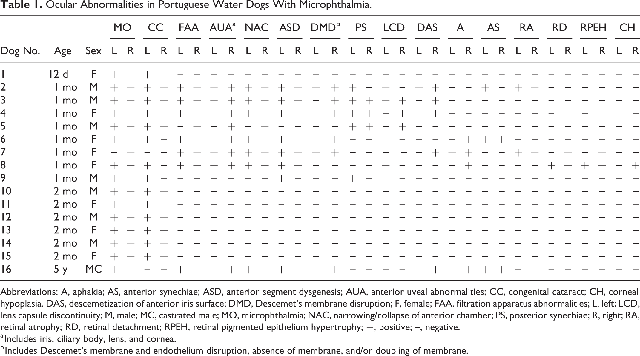
Abbreviations: A, aphakia; AS, anterior synechiae; ASD, anterior segment dysgenesis; AUA, anterior uveal abnormalities; CC, congenital cataract; CH, corneal hypoplasia. DAS, descemetization of anterior iris surface; DMD, Descemet’s membrane disruption; F, female; FAA, filtration apparatus abnormalities; L, left; LCD, lens capsule discontinuity; M, male; MC, castrated male; MO, microphthalmia; NAC, narrowing/collapse of anterior chamber; PS, posterior synechiae; R, right; RA, retinal atrophy; RD, retinal detachment; RPEH, retinal pigmented epithelium hypertrophy; +, positive; –, negative.
a Includes iris, ciliary body, lens, and cornea.
b Includes Descemet’s membrane and endothelium disruption, absence of membrane, and/or doubling of membrane.
Besides ocular lesions, 1 puppy was diagnosed with omphalitis and lissencephaly, and the single adult dog was diagnosed with chondrodysplasia (2/16; 13%). Gross evaluation revealed that all globes, except 1, were small based on the assessment of the prosector, measuring from 0.5 cm to 1.55 cm in diameter. In 3 cases, the left and right globes were equally small. In 8 cases, the left globe was smaller compared to the right globe. In the remaining 5 cases, the right globe was smaller compared to the left globe. Based on measurements taken during gross examination or measurements of paraffin-embedded tissue, the difference in diameter between the globes that differed in size ranged from 0.1 to 1 cm. The anterior and posterior segment lengths were not measured at any point during this study.
Histologic features are presented in Table 1. The 2 most common abnormalities were microphthalmia (31/32 eyes; 97%) and congenital cataract (26/32 eyes; 81%, Figs. 1, 2, 5). Other common histologic lesions included the lack of both ciliary cleft and trabecular meshwork with or without an identifiable scleral venous plexus (15/32 eyes; 47%) (Figs. 1, 3). An abnormal relationship of the iris and ciliary body to each other and to the cornea and/or lens (which included narrowing of the anterior and/or posterior chambers, axially displaced ciliary body plicae, or anterior displacement of the ora ciliaris retinae and peripheral retina) was identified in 15 of 32 eyes (47%) (Figs. 1, 3, 5). A Descemet’s membrane abnormality (discontinuity, absence, and/or doubling) was identified in 11 of 32 eyes (34%) (Fig. 4). Posterior synechia (adhesions between iris and lens) was noted in 9 of 32 eyes (27%) (Figs. 1, 2, 5). Anterior lens capsule discontinuity and/or lens protein leakage (each in 8/32; 25%) (Figs. 1, 2, 5), descemetization of the iris surface (8/32; 25%, characterized by Descemet’s membrane splitting and lining the surface of the iris) (Fig. 4), aphakia (6/32; 19%), and anterior synechia (adhesions between iris and cornea) varying from focal (either peripheral or axial) to broad (10/32; 31%) (Fig. 4) were also identified. Retinal changes, including retinal layering abnormalities (multifocal lack of all retinal nuclear layers in dog Nos. 2 and 7 or lack of retinal ganglion cells in dog Nos. 7 and 16) (total 4/32; 13%), retinal rosette formation peripherally (3/32; 9%), and retinal detachment with retinal pigmented epithelial cell hypertrophy (2/32; 6%), were relatively uncommon. Last, corneal hypoplasia (defined by a small cornea with corneal diameter composing less than two-thirds of the globe’s diameter) was present in 2 of 32 eyes (6%). The lesions of the single adult dog sampled (individual 16) were similar to those of the younger animals that had microphthalmia and cataract as well as other lesions.
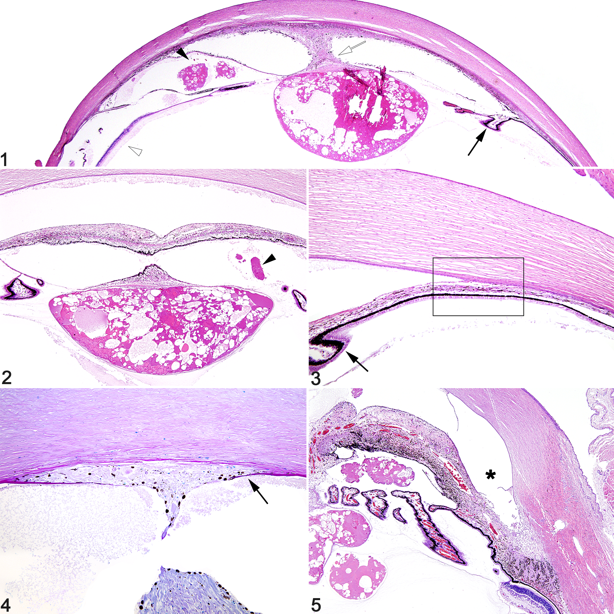
One eye from 1 dog (1/32; 3%; dog No. 7) displayed histologic evidence of secondary glaucoma characterized by loss of retinal ganglion cells and increased globe size compared to the fellow eye from same dog. Rare secondary changes included keratitis with hypopyon (dog No. 6) and intraocular hemorrhage (dog No. 7) (1 eye each; 3%). Sections of 18 globes were stained with PAS; these had absence and/or doubling of Descemet’s membrane (11/32 eyes; 34%, Fig. 4), descemetization of the iris surface (8/32 eyes; 25%, Fig. 4), and lens capsule discontinuity (8/32 eyes; 25%).
Of note, coloboma formation was not identified in any of the globes. In addition, there was no evidence of extensive retinal dysplasia or persistent fetal vasculature such as hyperplastic tunica vasculosa lentis or persistent hyperplastic primary vitreous, suggesting the retina and vitreous had developed relatively normally. Eyelid defects were also absent.
Our gross and histologic findings confirm the clinical findings of microphthalmia with lens abnormality and often multiple additional anterior segment defects in the Portuguese Water Dog. Both sexes were affected with no apparent sex predisposition. Both eyes were affected in all animals. Two dogs had other systemic abnormalities (lissencephaly and chondrodysplasia; 2/16 dogs; 13%), which is less frequent than is reported in humans; 4,5,11 however, due to the young age of many of these animals, it may be that other systemic abnormalities were not apparent at the time of euthanasia.
Secondary ocular changes were rare and included glaucoma, keratitis with hypopyon, and hyphema. Despite the morphologic abnormalities suggesting that aqueous humor outflow would be impeded, we rarely identified lesions of glaucoma in these globes; however, a detailed evaluation of the retina and optic nerve head for evidence of glaucoma was often not possible. We may, therefore, have underdiagnosed glaucoma. In addition, because many cases were under 2 months of age, it is possible glaucoma had not developed yet.
Our finding that cataract is the most frequent histologic abnormality associated with microphthalmia concurs with human studies. 7 All cases in the present study were categorized as complex (all either had cataract or were aphakic with noncolobomatous/nonoptic fissure closure defect microphthalmia) under the human classification system. In our cases, the anterior segment was the most affected part of the globe. Localized anterior segment lesions were cataract and aphakia. Multiple anterior segment lesions involved the lens, ciliary body, iris, and cornea, as well as compression of the anterior segment as evidenced by narrowed anterior and posterior chambers. This is in contrast to human studies, in which posterior segment anomalies, including decreased posterior segment length, were most common in the noncolobomatous/nonoptic fissure closure defect microphthalmia group. 11 In our study, 8 dogs (15/32 eyes; 47%) had instead lesions compatible with features described in anterior segment dysgenesis (ASD) in humans, including lack of ciliary cleft and trabecular meshwork, lens capsule discontinuity, anterior or posterior synechia, and Descemet’s membrane abnormalities. 4
The anterior segment of the eye comprises all of the structures lying between the front surface of the cornea and the front surface of the vitreous. Human anterior segment dysgeneses are a heterogeneous group of syndromes that are attributed to developmental abnormalities of the structures of the anterior segment and are often associated with other ocular and systemic abnormalities. Human anterior segment dysgeneses have been divided into those conditions related only to neural crest cell abnormalities (ASDnc), which affect the anterior uvea and cornea but not the lens, and those that affect the lens and consequently also lead to many lesions that are also seen in the anterior segment dysgeneses due to neural crest abnormalities (called “non–neural crest anterior segment dysgenesis,” ASDnon-nc). 4 Because of the interplay between the developing lens and the rest of the anterior segment structures, any abnormality in lens development frequently leads to abnormality in other structures. 4 As in the various types of human anterior segment dysgenesis, there was a variation in the ocular lesions seen in this group of examined Portuguese Water Dogs. This makes it difficult to directly compare this constellation of lesions in the Portuguese Water Dog to a specific human entity; however, those dogs with lens and other anterior segment abnormalities suggest a non–neural crest cell abnormality-associated ASD (ASDnon-nc).
One of the forms of ASD has been known as anterior chamber cleavage syndrome, Peters anomaly, or keratolenticular dysgenesis, and it occurs when the lens and/or iris fail to separate from the cornea during development, leading to focal anterior synechia and corneal opacity. 4 Several of the individuals from this series had lesions that were suggestive of this specific anomaly in 1 or both eyes (dog Nos. 2, 4, 6, 7, and 16). Only a few ASD cases have been reported in domestic animals, including but not limited to a Springer Spaniel with Peters anomaly and Basenji dogs with persistent pupillary membranes and associated defects. 2,9
Because dog breeds are by definition inbred, the occurrence of similar lesions within a number of individuals in a given breed is likely due to a common genetic mutation or mutations as opposed to exposure to teratogenic compounds, infectious disease, or random developmental mistakes. A number of genetic defects have been implicated in the various human anterior segment dysgeneses, including but not limited to mutations in PITX2, FOXC1, PAX6, and CYP1B1; however, specific genetic mutations may not be known for every individual human patient who is diagnosed with ASD. 4 The fact that the severity of lesions ranged from only microphthalmia and cataract/aphakia to cases with abnormalities of globe size, lens, and anterior segment structures suggests that either multiple genes are involved or that the mutation in a single gene has variable penetrance.
The canine eye develops in a manner similar to the human eye; the principal difference is in the time of occurrence of certain developmental events. At term, the eye of the normal canine fetus, although small in size, is almost fully developed, including the anterior chamber angle structures. Postnatally, beyond day 35, the peripheral retina and tertiary vitreous mature to their adult structure. 1 Because of the range of ocular lesions in these dogs and because we only analyzed postnatal globes, it is difficult to pinpoint the day during development when lesions were initiated; however, the lack of optic fissure defects, which would cause posterior segment coloboma, indicates lesions developed sometime after gestational day 29. 3
This study defines the histologic features in affected Portuguese Water Dogs and implicates a genetic mutation or multiple genetic mutations that cause extensive and variable ocular dysgenesis. The developmental anomaly likely occurs before the end of gestation after the optic fissure closes. It likely affects lens development and the subsequent formation of the anterior uvea and anterior and posterior chambers, similar to cases of non–neural crest–related anterior segment dysgenesis in humans. This study provides a basis for further investigation into ocular abnormalities in Portuguese Water Dogs.
Footnotes
Acknowledgements
We thank the histopathology technicians at the New York State Animal Health Diagnostic Center for performing the histology and histochemistry. We also thank the anatomic pathology residents and faculty at the Cornell University College of Veterinary Medicine for prosection of select necropsy cases.
Declaration of Conflicting Interests
The author(s) declared no potential conflicts of interest with respect to the research, authorship, and/or publication of this article.
Funding
The author(s) received no financial support for the research, authorship, and/or publication of this article.