
Abstract
Congenital ocular disease occurs uncommonly in cattle, with multiple abnormalities reported only sporadically in the literature. This report describes a case of anterior segment dysgenesis resulting in glaucoma in a 4-month-old Texas Longhorn steer. On clinical exam, bilateral buphthalmia was present and intraocular pressures exceeded 47 mm Hg in both eyes. On histopathologic examination, the iridocorneal angle and filtration apparatus were distorted due to collapse of the ciliary cleft and anterior displacement of the anterior portion of the ciliary body. No evidence of inflammation or other causes of glaucoma were recognized.
A 4-month-old Texas Longhorn steer was evaluated for bilateral buphthalmos and corneal edema (Fig. 1). The owner reported that bilateral corneal edema (bluish discoloration) had been noted since the calf was 2 months of age. Physical exam revealed a good body condition, normal mentation, and vital signs within normal limits. Ophthalmic examination confirmed bilateral buphthalmos with no lagophthalmos. Bilaterally, the pupils were fully dilated and nonresponsive to light, and posterior cataracts were present. In addition, Haab’s striae and mild optic nerve cupping were noted. Tonometry was performed and revealed intraocular pressures (IOP) of 49 mm Hg in the left eye and 47 mm Hg in the right eye (reference range, 16–36 mm Hg). 5 Three weeks later, tonometry was repeated, and IOPs of 59 mm Hg in the left eye and 52 mm Hg in the right eye were obtained. Due to the apparent congenital nature of this glaucoma, various management options were considered of low potential for therapeutic response and not logistically feasible. Because the welfare of the animal was believed to be impaired by the elevated IOP, it was euthanized and the eyes were removed immediately upon death.
Differential Diagnoses
Differential diagnoses for buphthalmia include glaucoma and retrobulbar mass, including abscess or neoplasia. Elevated intraocular pressures indicated glaucoma antemortem, and no masses were observed at postmortem examination. Therefore, differential diagnoses for glaucoma were considered, which include congenital abnormalities of the iridocorneal drainage angle, primary open-angle glaucoma with defects of the trabecular meshwork and/or scleral tissues, primary closed-angle glaucoma with an abnormally narrowed or closed iridocorneal angle, goniodysgenesis or pectinate ligment dysplasia or glaucoma secondary to anterior uveal inflammation with posterior or anterior synechiae or occlusion of the trabecular network by inflammatory or neoplastic cells, inflammatory debris, or preiridial fibrovascular membranes.
Microscopic Findings
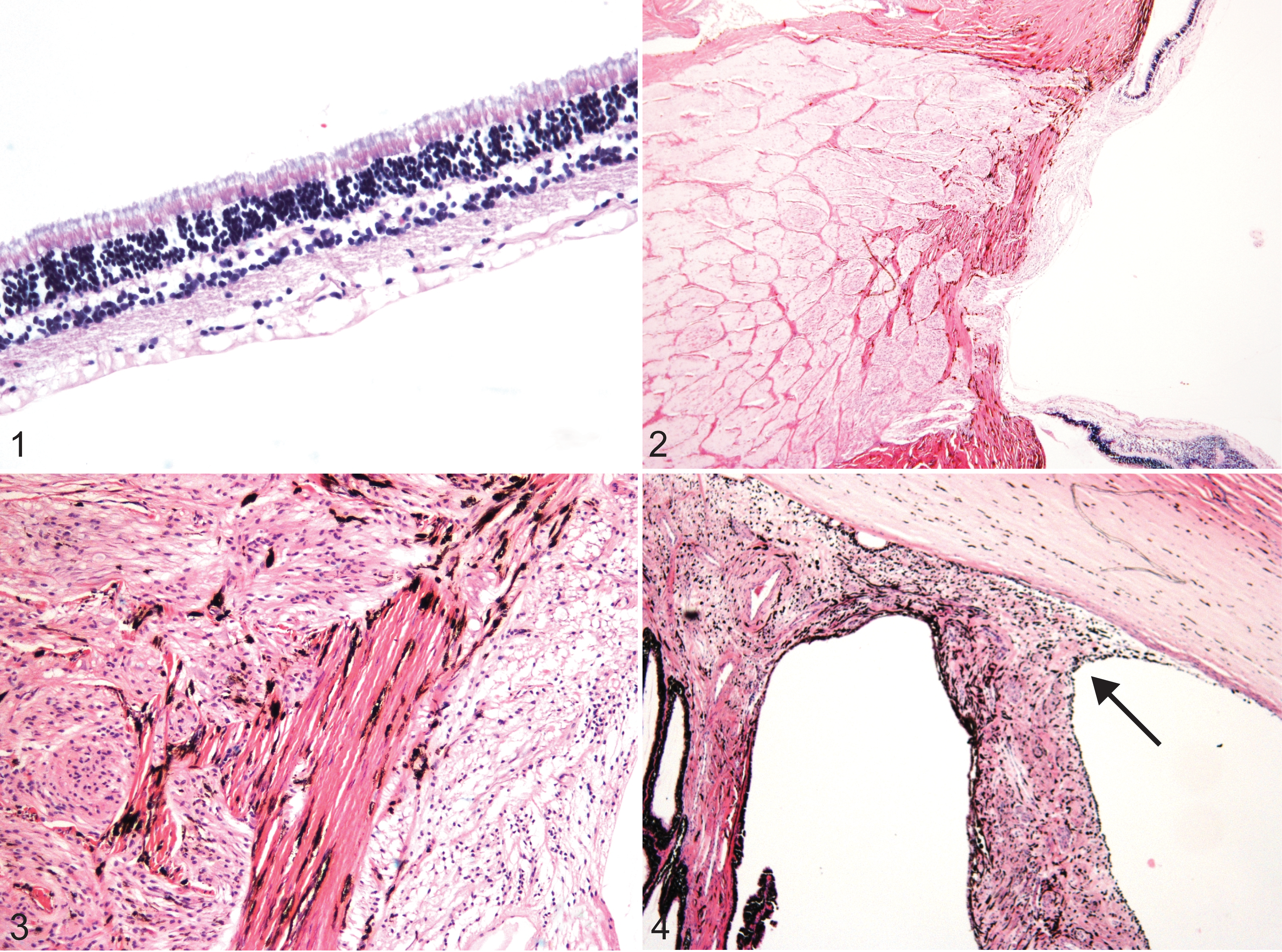
Upon sectioning, the lenses were grossly luxated and loose. The left eye was fixed in 10% buffered formalin, processed routinely for sectioning, and then stained with hematoxylin and eosin (H&E). On histopathologic examination, the retina was largely devoid of ganglion cells (Fig. 2), and there was atrophy (Fig. 3a) and gliosis (Fig. 3b) of the optic nerve head. The iridocorneal angle and filtration apparatus were distorted due to collapse of the ciliary cleft and anterior displacement of the anterior portion of the ciliary body (Fig. 4). There were multiple breaks in Descemet’s membrane, characterized by the formation of a new thinner Descemet’s membrane across the breaks (Haab’s striae). No evidence of inflammation or other causes of glaucoma were recognized.
Diagnosis
Diagnoses of anterior segment dysgenesis, abnormal development of the iridocorneal angle, lens luxation, congenital glaucoma, and Haab’s striae were made.
Discussion
The incidence of spontaneous glaucoma in cattle is rare, although congenital, primary, and secondary glaucomas have been reported in dairy cattle.5,8 Primary glaucoma is an autosomal dominant trait in young Holstein Friesians and has also been seen in the Jersey breed, in both young and mature animals. 8 In addition to natural cases, cattle have been used successfully as a model of experimental corticosteroid-induced glaucoma in research. 11
In a report of bovine ocular disease in India, 3 of 1302 surveyed animals (0.23%) were diagnosed with glaucoma. 10 In another survey of spontaneous ophthalmic disease in 500 Friesian cows, one 9-year-old adult cow was identified with secondary chronic glaucoma. 8 This cow presented with a blind, buphthalmic globe, with IOP ranging from 39 to 45 mm Hg. 8 Histologically, chronic glaucomatous changes, including corneal edema, striae, a subluxated cataractous lens, and optic disk atrophy, were noted. 8
Anterior segment dysgenesis (ASD) is well known in humans and has also been reported in a variety of animal species, including dog, cat, cattle, horse, llama, rabbit, and mouse.2,4,7,12 Lesions associated with ASD in humans and animals include a wide range of anomalies, such as corneal opacities, persistent pupillary membranes, malformations of the iridocorneal angle, microphthalmia or buphthalmia, microphakia, and congenital cataracts, which can cause varying degrees of visual impairment.4,7 Dysgenesis of components of the anterior segment have been rarely reported in cattle and include absent anterior chamber and closure of anterior chamber angles occurring in several calves over multiple years in a single herd of Brahman × Santa Gertrudis × Herefords, iris hypoplasia in albino Hereford cattle, and anterior staphyloma and rudimentary lens in a Holstein calf.1,6,9 Because ASD often describes malformations of the tissues responsible for drainage of aqueous humor from the eye (and thus regulation of IOP), it is often associated with an increased risk of developing glaucoma.3,4,7
This report describes a case of congenital primary glaucoma in a steer, as evidenced by a lack of inflammation and the presence of multiple developmental ocular abnormalities. The high incidence of inflammatory ocular disease in cattle makes the differentiation of primary and secondary causes of glaucoma essential for accurate assessment.
Footnotes
Acknowledgement
The authors thank Dr Jonathan Pucket for his assistance with the ophthalmic examination.
Declaration of Conflicting Interests
The author(s) declared no potential conflicts of interest with respect to the research, authorship, and/or publication of this article.
Funding
The author(s) received no financial support for the research, authorship, and/or publication of this article.