
Abstract
To clarify the morphologic features of the ocular disease recently occurring among Japanese Black cattle in southern Kyushu, 6 globes from 3 Japanese Black cattle, between 11 and 20 months old (cow Nos. 1 to 3), were pathologically examined. cow Nos. 1 and 2 were sired by the same Japanese Black bull, and cow No. 3 was sired by the ancestor (sire) of the former bull. The ocular lesions were pathologically similar to each other, except for the left eye of cow No. 1. The ocular lesions of 5 globes were characterized by microphthalmia, hypoplasia, and/or dysplasia of the lenses; persistence of the primary vitreous; and retinal dysplasia with total nonattachment. The left globe from cow No. 1 had no lens and severe hypoplasia and nonattachment of the retina. Because dysplastic retinal lesions that formed crescentic folds and a central column were the most characteristic features of the eyes, the falciform retinal fold with congenital nonattachment was the most likely disease entity. Although the cause of the ocular disease could not be clarified with the present study, an inherited ocular defect of the bull and its ancestor was suspected.
Congenital ocular disorders are not unusual in cattle, and several types of ocular defects, including microphthalmia, coloboma, and retinal dysplasia, have been reported previously. 8, 23 Various causes have been suspected, including hypovitaminosis A, 17 fetal viral infections, such as bovine viral diarrhea-mucosal disease (BVD-MD) or Akabane viruses, 17, 18, 21 and unidentified genetic factors. 4, 7, 16, 22 The identification of the causative factors might be very important in preventing recurrence of the diseases and economic loss.
Recently, an outbreak of ocular disorders with congenital blindness was recognized among pure bred Japanese Black cattle in southern Kyushu, Japan. Several clinical analyses of 6 affected cattle by using ultrasonographic examinations revealed multiple ocular lesions that consisted of microphthalmia, microcornea, corneal opacity and pigmentation, unattached retina, and persistent hyaloid vessels. However, the histopathologic features of the ocular diseases have not been elucidated. The purpose of this study was to clarify the pathologic findings and to evaluate the similarity of the ocular changes among Japanese Black cattle in southern Kyushu, Japan.
Ocular lesions were examined in 3 Japanese Black heifers, between 11 and 20 months old (cow Nos. 1–3). All 3 cattle were kept in different herds of purebred Japanese Black cattle in southern Kyushu and had congenital blindness, with similar gross ocular lesions. Two affected heifers, cow Nos. 1 and 2, were sired by the same Japanese Black bull (HH). The remaining calf was sired by the other Japanese Black bull (HZ), which was the sire of HH. The growth and the general condition of these cattle were excellent. The cattle were presented to the Animal Hospital of Miyazaki University for clinical examination, including ultrasonography of the eyes and brain scanning by computer tomography (CT). General clinical examinations revealed that all 3 calves had complete visual deficit. By ultrasonography and CT examinations, microphthalmia, absence of the lens, retinal detachment, and persistent hyaloid vessels were suspected in all 3 calves. In cow Nos. 1 and 2, both eyes showed severe medial deviation. The corneas were not visible, because of the medial deviation, and the conjunctival epithelium had undergone a metaplastic change that probably involved squamous metaplasia with pigmentation (Fig. 1). In cow No. 3, the position of the left eyeball was intact, whereas the right eye showed medial deviation. All cattle were then euthanatized and immediately necropsied.
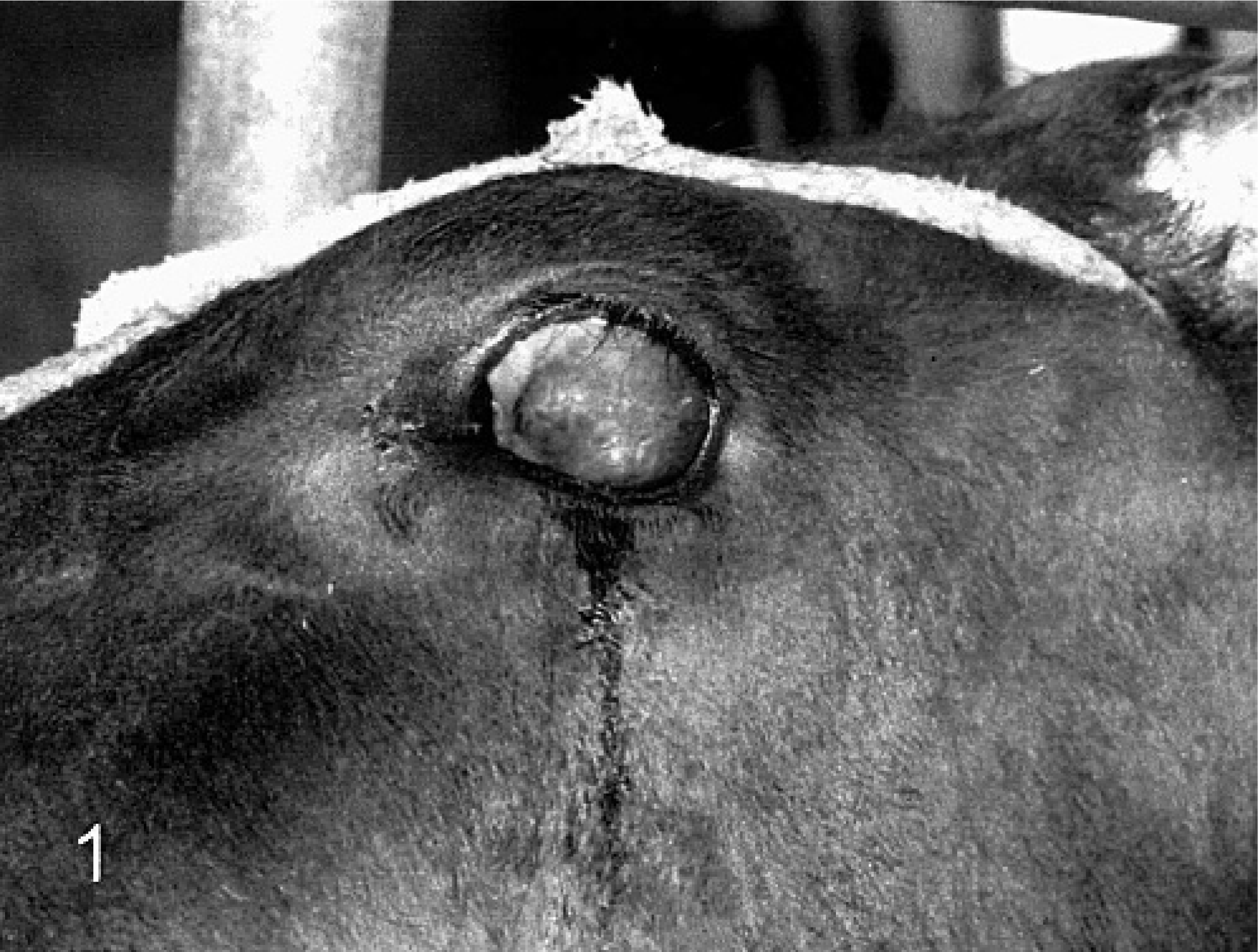
Eye and periocular tissue; Japanese Black cow No. 1. The corneas are not visible because of the medial deviation, and the conjunctival epithelium has undergone a metaplastic change that probably involved squamous metaplasia with pigmentation.
All 6 globes from the 3 calves were fixed in methanol-Carnoy solution. One hour after prefixation, all eyes were sliced and then fixed again in methanol-Carnoy solution for 24 hours. The fixed samples were immersed in pure methanol, pure chloroform, and toluene, and then were embedded in paraffin. Other organs, such as the brains and optic nerves, were fixed in 10% formalin and processed by using general procedures. Paraffin sections 2-μm thick were made from each eye and were stained with hematoxylin and eosin (HE). Immunohistochemistry was done by using an Envision polymer reagent (Dako-Japan, Kyoto, Japan). For primary antibody, mouse monoclonal antibody against bovine rhodopsin (1°1000; LSL Co., Ltd, Japan) was used. After reaction with the primary antibody, the sections were incubated with Envision polymer reagent (Dako-Japan) at 37°C for 40 minutes. The reaction products were visualized by using 3,3′-diaminobenzidine (Sigma, St Louis, MO, USA), and the sections were counterstained with hematoxylin.
All 6 globes from the 3 cattle were small, measuring 1.5–2.5 cm in diameter. In 5 of the globes, the gross lesions were almost identical, whereas the sixth eye (cow No. 1, left eye) had some different features but was essentially a variation of the other 5. The right eye of cow No. 1 and both eyes from cow Nos. 2 and 3 were characterized by a thin layer of fibrous connective tissue that covered the anterior chamber and extended in a stalk to the head of the optic nerve (Fig. 2R). The iris was diffusely thickened, and the lens were not grossly recognizable. The sclera was covered by pigmented epithelium, without the optic retina, and there was no vitreous body. The left eye of cow No. 1 was characterized by a thin central fold of retina that extended to the anterior pole of the globe. In most areas, the retina was detached from the pigmented epithelium. The inner space of the irregularly shaped retina was filled with a small amount of vitreous. The anterior chamber was hard to recognize and, as with all the other eyes, the lens was not grossly detectable (Fig. 2L). There were no significant gross lesions in other organs or tissues.
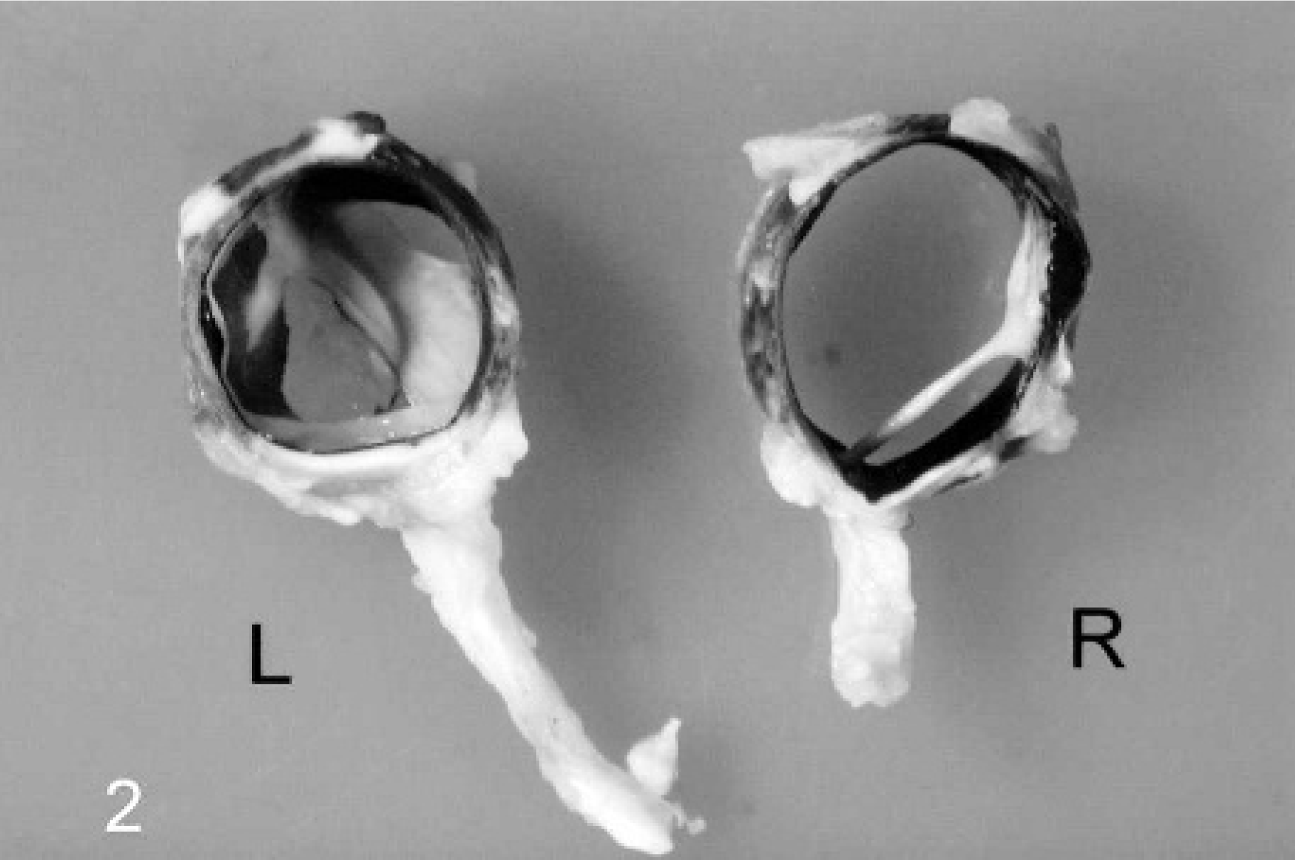
Eye; Japanese Black cow No. 1. Gross lesions of the cut surface of the eyes after fixation. The left eye (L) has a thin central fold of the retina that extended to the anterior pole of the globe. The anterior chamber is absent. In the right eye (R), the anterior chamber was covered by a thin fibrous tissue with a central cord extending to the head of the optic nerve.
The histopathologic features of 5 globes of the 3 heifers (both eyes from cow Nos. 2 and 3, and the right eye of cow No. 1) were similar to each other and those of the left eye of cow No. 1 were different, which corresponded to the gross findings. The 5 similar eyes contained irregular cysts formed by pigmented anterior uveal epithelium, small and thin lenses, and dysplastic retinas. The dyplastic retina extended to the head of the optic nerve, forming a column (Fig. 3) that corresponded to the grossly visible fibrous stalk. The column was composed of a central artery (presumably a persistent hyaloid artery), around which was arranged neuroectodermal tissues, including irregularly arranged granular layers of the retina, a few ganglion cells, nerve fibers, and glial cells and their processes. The fibrous layer that covered the anterior chamber grossly corresponded, histologically, to a thin layer of dysplastic retina attached to cystic ciliary body (covered by a single layer of highly pigmented epithelium) and hypoplastic lens tissues. The irregular, small lenses consisted of a lens capsule that contained a small number of swollen fibers (Fig. 4). In the right eye of cow No. 2, there were 2 lenses, and one was embedded within the adjacent dysplastic retina. In the left eye from cow No. 1, well-developed ciliary processes covered the iris, and no lens could be identified histologically. The sclera was lined with a single layer of pigmented epithelium, but there was severe segmental hypoplasia of the optic retina, characterized by a marked decrease of retinal cells and irregular lamina formation, with limited or no rhodopsin immunoreactivity (Fig. 5). The posterior chamber of the other 5 eyes contained a large number of retinal rosettes surrounding cystic spaces filled with mucinous material resembling vitreous (Fig. 6). The central area of the retinal rosettes showed intense immunoreactivity for bovine rhodopsin. There were no significant histologic lesions in other organs or tissues.
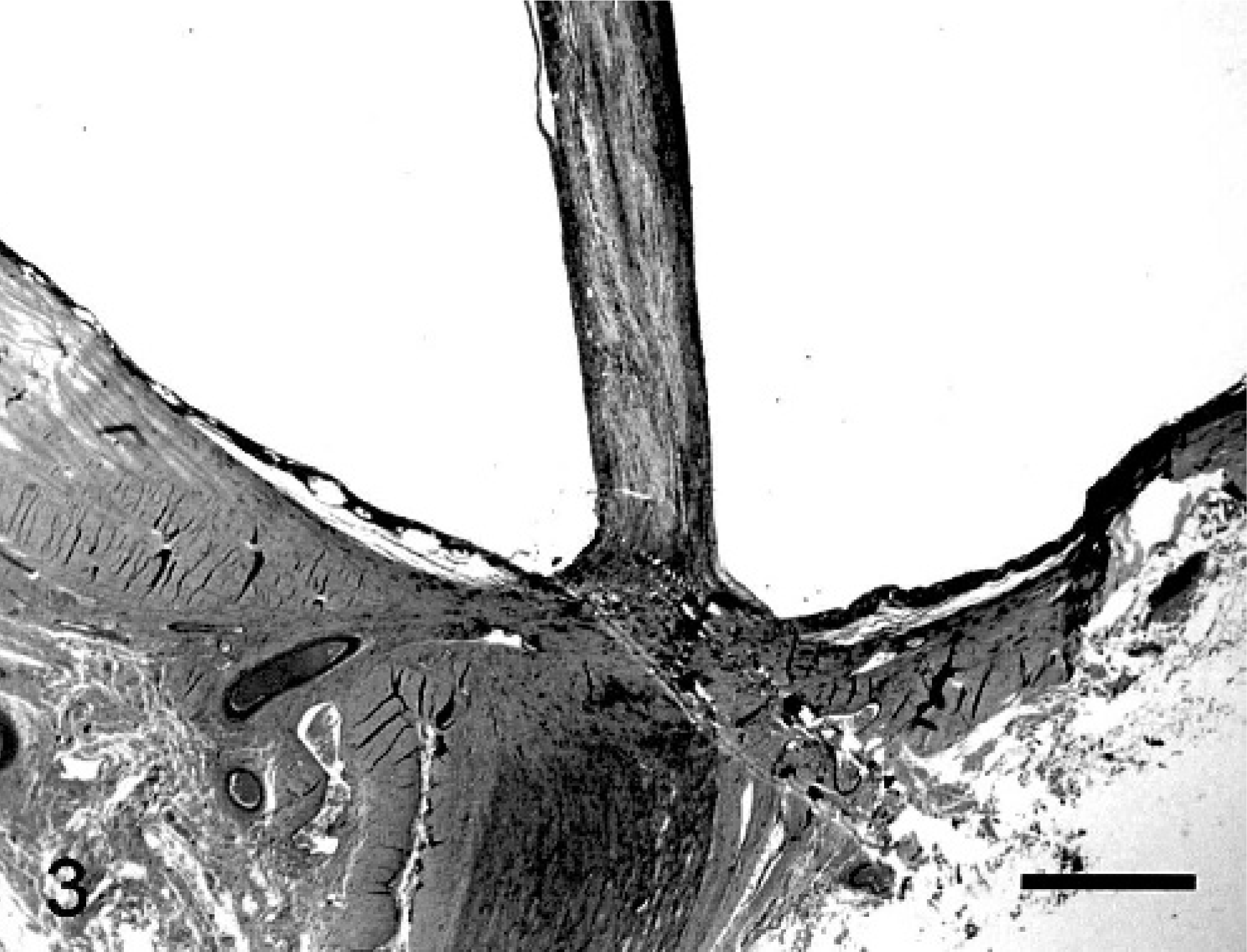
Eye; Japanese Black cow No. 2. The posterior part and optic nerve of the left eye of cow No. 2. The central cord attached to the head of optic nerve consists of central artery and neuroectodermal tissues. The sclera was covered by pigmented epithelium without the optic retina. HE. Bar = 300 μm.
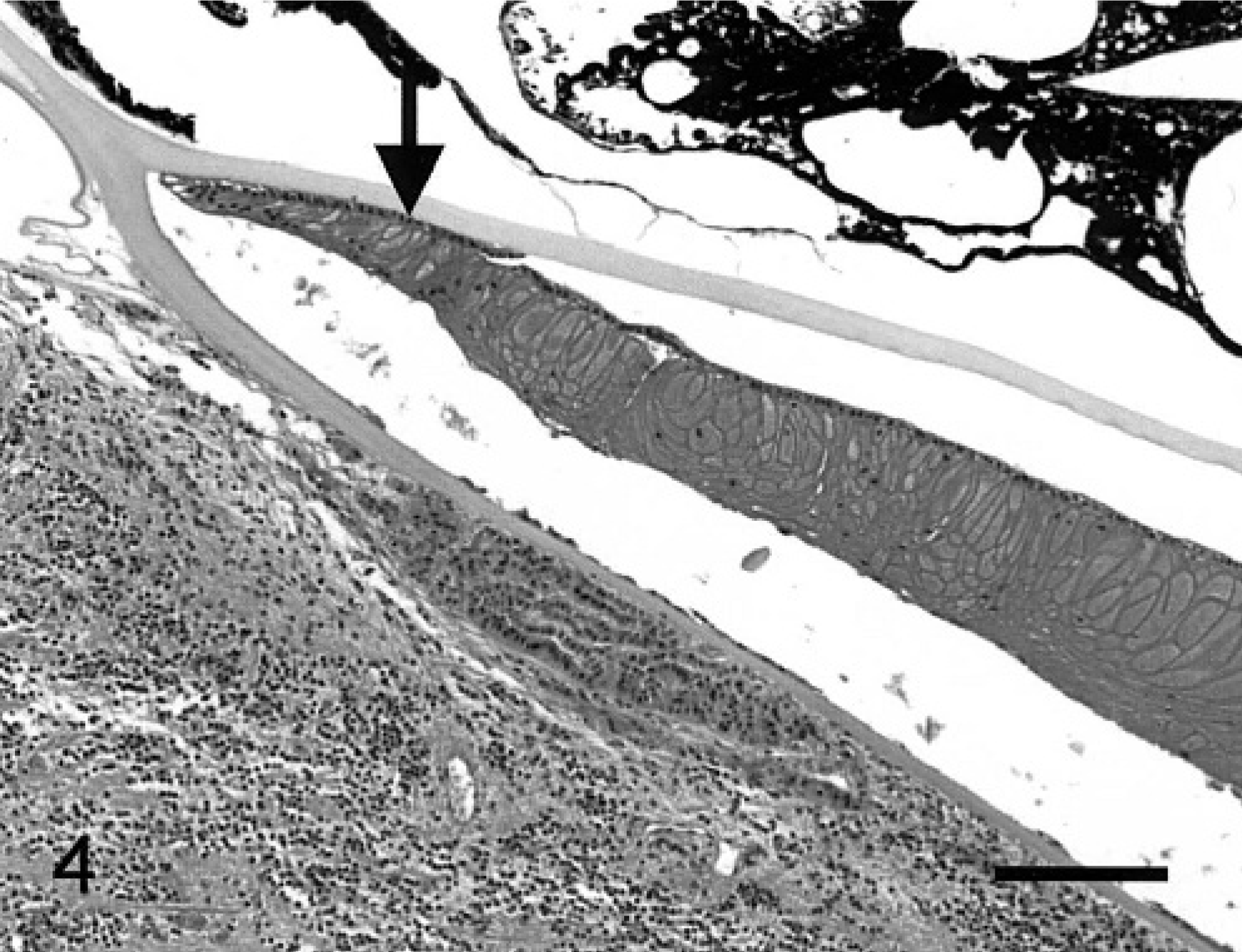
Eye; Japanese Black cow No. 1. There were cysts formed by pigmented anterior uveal epithelium, the lens consisted of a capsule and irregularly arranged swollen fibers (arrow), and a severely dysplastic retina. HE. Bar = 200 μm.
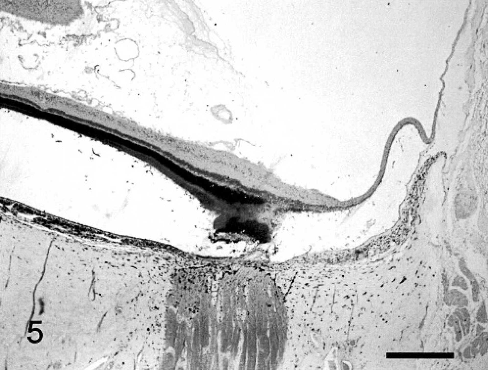
Eye; Japanese Black cow No. 1. The posterior part and optic nerve of the left eye of cow No. 1. The optic retina partly had well-developed laminar structures, showing intense rhodopsin immunoreactivity. Severe hypoplasia characterized by a marked decrease of retinal cells and irregular laminar formation, with limited or no rhodopsin immunoreactivity was recognized. Immunohistochemistry for bovine-rhodopsin. Bar = 300 μm.
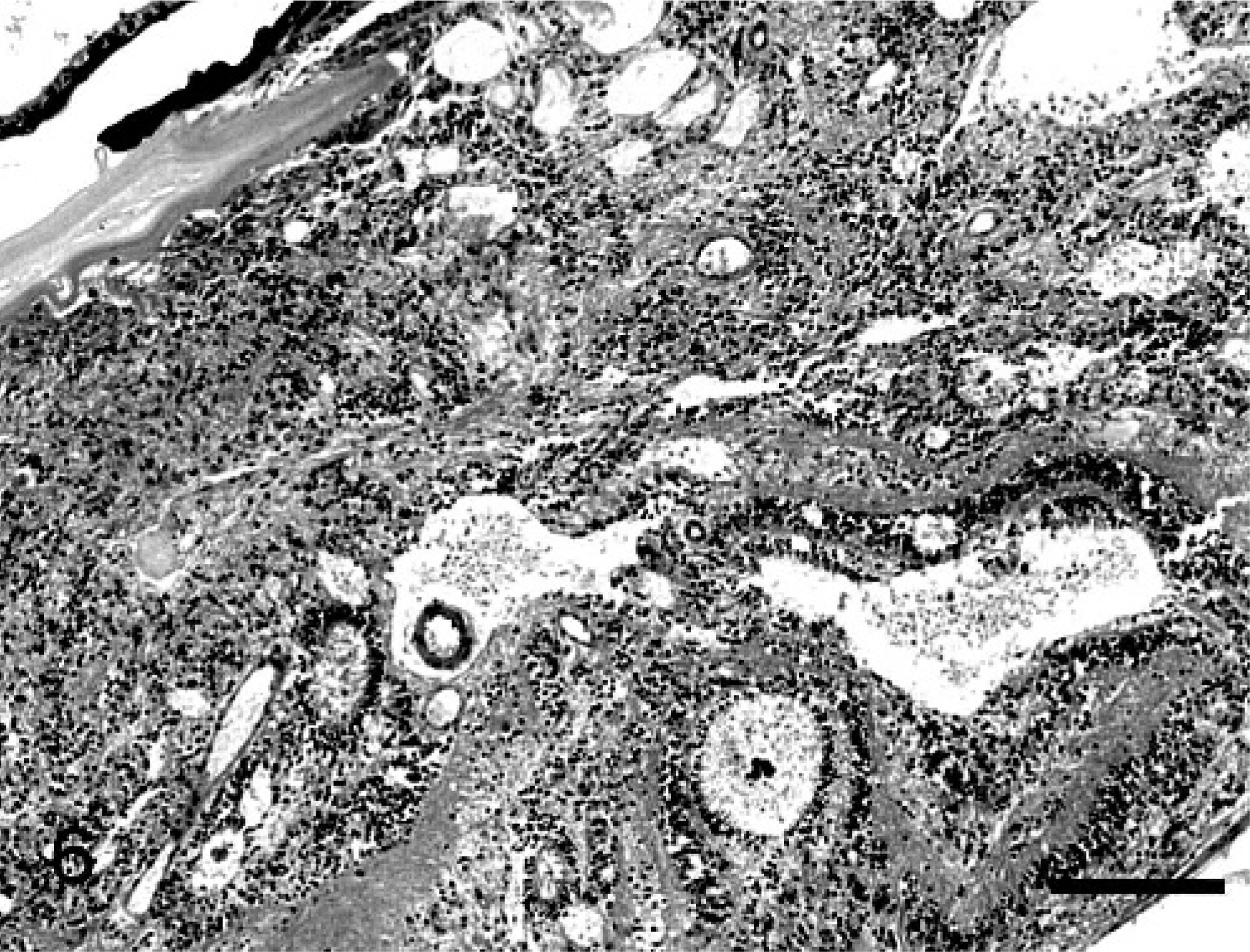
Eye; Japanese Black cow No. 3. Dysplastic retinal tissue adjacent to the lens capsule of the right eye. There were many retinal rosettes and cystic spaces. HE. Bar = 200 μm.
The ocular lesions of the 3 cattle examined were pathologically similar to each other, except for a moderate variation in the left eye from cow No. 1. Microphthalmia, hypoplasia, and/or dysplasia of the lenses; severe hypoplasia of vitreous body; persistence of the hyaloid artery; and retinal dysplasia, with nonattachment commonly characterized the ocular lesions of 5 of the globes. The remaining globe, left eye from cow No. 1, had no lens and a hypoplastic and unattached retina. The homogeneity of the ocular changes of 5 globes from the 3 cattle examined might indicate a consistent disease entity. The ocular lesions of the present cases were not consistent with those of typical congenital ocular diseases previously recognized in cases of hypovitaminosis A 19 or fetal BVD 17, 21 or Akabane 18 virus infections. In addition, the absence of concurrent lesions, such as hydrocephalus, cerebellar atrophy, and optic nerve degeneration, may indicate that these nutritional factor or viral infections are less likely the causative factors. Inherited genetic defect of the bull purchased for breeding seems the possible cause of the present ocular disease, because cow Nos. 1 and 2 were sired by the same Japanese Black bull, and this bull was a half sibling of cow No. 3.
Although the histopathologic features of bovine ocular anomalies are not always reported in detail, partial and multiple ocular defects have been found, together with anomalies in other viscera, such as cerebellar hypoplasia or hydrocephalus. 5, 9, 10 Ocular coloboma has been reported previously in Charolais cattle, and autosomal dominant inheritance was suggested as the possible cause. 1, 4, 22 On the other hand, Kaswan et al. 7 reported multiple hereditary ocular anomalies in calves characterized by microphthalmos, microcornea, microcoria, heterochromia iridis, microlentia, cataracts, retinal dysplasia, and retinal detachment. The authors suspected a genetic defect as the probable cause, because the affected calves were from a herd of Brahman × Santa Gertrudis cows bred with a Hereford bull. 7
Because the most distinct characteristics were the dysplastic retinal lesions, forming crescentic folds and a central column, a falciform retinal fold with congenital nonattachment was the most likely disease entity for the present ocular changes. The falciform fold and the detachment of the retina are found with several inherited ocular human disorders, including Norrie's disease, primary retinal dysplasia, congenital retinal nonattachment, and familial exudative vitreoretinopathy. 2, 3, 6, 11, 20 In some of these inherited human disorders, the causative genetic changes and the manner of inheritance have been well elucidated. 2, 3 Further, there are several reports concerning congenital ocular disorders in cattle, 8, 23 but few concerning bovine retinal dysplasia with falciform folds and nonattachment. The bovine multiple hereditary ocular anomalies reported by Kaswan et al. 7 featured similar posterior changes, including persistence of the hyaloid artery and retinal folds in the lateral globe, while the retinal lesions were not completely identical to those of the present calves. The dysplastic changes characterized by irregular arrangement and retinal rosettes were more prominent in the present calves.
For the cases reported here, the pathogenesis of the ocular changes in the lenses, secondary vitreous body, and retinal dysplasia remains unclear. Organization of the basic structures of the eyes, including the lens and the retina, are completed at the early embryonic stage. Thus, the present ocular lesions might occur at the very early embryonic stage. The incomplete induction of the lens placode might result in the abnormal lens development, and irregular apposition of the 2 layers of the optic cup might cause retinal nonattachment and retinal dysplasia found in the present calves. Furthermore, because the hyaloid artery plays a major role in supplying blood for the development and the growth of the secondary vitreous body and lens, some functional defects of the artery, such as incomplete blanching or regression might occur in the affected calves, resulting in the defect in the vitreous body and lenses with dysplastic retinal formation. Such vascular defects have been recognized in the cochlea of the mouse model for Norrie's disease in humans. 15 The mice with knock-out of the Norrie gene product have been found to have persistent hyaloid vessels, indicating that the ND gene product might be important for the process of regression of these vessels. 14 Furthermore, in human familial exudative vitreoretinopathy characterized by a falciform retinal fold, 13 the vitreoretinal vascular abnormalities are thought to be significant in its pathogenesis. 12 Similar pathogenesis might be also possible in the present bovine ocular disease. Although immunohistochemistry for alpha-smooth-muscle actin and factor VIII-related antigen to visualize vessels (data not shown) was attempted, no significant vascular changes to explain the ocular lesions were recognized. Molecular biologic studies for the molecules related to induction of the lens placode and Norrie gene are now in progress, and those will provide useful information to elucidate the pathogenesis of the present congenital ocular lesions.
In conclusion, the present paper described the pathologic features of some cases of congenital ocular disease occurring in Japanese Black cattle. Because this bovine disorder still occurs sporadically in this area, further epidemiologic and genetic investigations on its progress will reveal the manner of inheritance, causative defects of the gene, and the pathogenesis of the unique ocular disorders.
Footnotes
Acknowledgement
This work was supported by a grant from the Ministry of Agriculture, Forestry, and Fisheries of Japan.