
Abstract
Tg.rasH2 mice are predisposed to hemangiosarcomas. Following the spleen, the uterus is the second most commonly affected organ in the female mice. Female mice are also predisposed to spontaneous vascular proliferative lesions on the serosal surface of the uterus, in which there is proliferation of normal vessels that are lined by well-differentiated endothelial cells. The hemangiosarcomas and vascular proliferative lesions can occur independently. In our facility, we have recorded a total of 47 uterine hemangiosarcomas in 3,985 female Tg.rasH2 mice assigned to various groups in 38 studies. Of these 47 cases, we have seen 22 (46.8%) cases where there was a clear progression of the serosal uterine vascular proliferative lesion into a hemangiosarcoma. In the remaining 25 (53.2%) cases, the uterine hemangiosarcomas involved myometrium and endometrium, but there was no serosal vascular proliferation. Based on the retrospective analysis of our data, we demonstrate that the vascular proliferative lesions noted on the serosal surfaces can progress to hemangiosarcomas and therefore these vascular proliferative lesions should be considered as preneoplastic lesions.
Keywords
Introduction
The 2-year rodent carcinogenicity assays involving conventional rats and mice have been conducted for over 3 decades. As an alternative to the 2-year rodent carcinogenicity bioassays, 26-week short-term carcinogenicity bioassays were approved using transgenic mouse strains, including Tg.rasH2. 1 The Tg.rasH2 model, which can be used for both genotoxic and nongenotoxic compounds, has gained popularity and its use has increased over the years. Currently, more than 75% of all mouse studies are being conducted in Tg.rasH2 mice. 2 The Tg.rasH2 model predicts neoplastic findings relevant to human cancer risk assessment, produces fewer nonbiologically significant neoplastic outcomes, and is thus preferable to a 2-year rodent study. 3
Uterine vascular proliferative lesions in Tg.rasH2 mice, which have been previously diagnosed as vascular anomalies, have been reported by several authors along with uterine hemangiosarcomas. 4 -9 The term anomaly literally means “abnormality” or “deviation from standard, normal, or expected.” There is nothing abnormal about the morphological appearance of the vessels. The proliferations of vessels on the serosal surface of the uterus are normal and are lined by normal vascular endothelium. Grossly, these proliferative lesions can be seen on the uterine serosal surface as reddish foci, reddish discoloration, or raised nodules, whereas hemangiosarcomas noted in the uterus are usually seen as masses that occupy most of the circumference of the uterus. These so-called vascular proliferative lesions have been recorded by us and other authors in various tissues of Tg.rasH2 mice such as brain, kidneys, nasal cavity, spinal cords, urinary bladder, cervix, ovary, oviduct, and uterus. 4 -9 However, per our records, the uterus was the most commonly affected organ with serosal vascular proliferative lesions. 8 In addition, the uterus was the second most commonly affected organ with hemangiosarcoma, following splenic hemangiosarcomas in Tg.rasH2 mice. 7 However, the relationship between vascular proliferative lesions on the serosal surface of the uterus and the hemangiosarcomas in the uterus has not yet been established.
In this article, we performed retrospective analysis of 38 studies conducted at our facility, which demonstrated that these vascular proliferative lesions can progress to hemangiosarcomas. Therefore, these vascular proliferative lesions should be considered as preneoplastic findings.
Methods and Materials
Animals
CByB6F1-Tg(HRAS)2Jic (+/− hemizygous c-Ha-ras) mice, obtained from Taconic Biosciences (Germantown, New York), were used in all studies. The knockin Tg element (human prototype c Ha-ras gene with its own promoter/enhancer) is injected into C57BL/6 x BALB/c F2 zygotes, which were crossed back to C57BL/6J forming C57BL/6JJic-Tg(HRAS)2Jic. The CByB6F1-Tg(HRAS)2Jic (+/−hemizygous c-Ha-ras) is the offspring from a cross of the C57BL/6JJic-Tg(HRAS)2Jic hemizygous male mice with the BALB/cByJJic female mice. Each mouse was genotyped by Taconic to verify the presence of the transgene before being placed on study. Animals were randomized by body weight into groups using a computer program. On the first day of treatment, animals were 7 to 9 weeks of age and weighed at least 17 or 15 g (males and females, respectively). Individual body weights for each dose group of each sex were within ±20% of the mean at the start of the study.
Housing and Environmental Conditions
Housing and environmental conditions were similar in all studies. Animals were multiple housed during acclimation and single housed in polycarbonate cages following randomization, with hardwood bedding chips in environmentally controlled rooms. Animals were verified to be free of illness prior to being placed on a study. All animals had ad libitum access to water and powdered feed (Harlan TEKLAD Global Diet, Madison, Wisconsin). The environmental conditions at the animal facility, including the type of feed, bedding, cleaning detergents, the lighting cycle, humidity, or temperature ranges, were the same for all studies.
Regulatory Requirements
The numbers of animals, procedures, and experimental design for each study were reviewed and approved by the BioReliance Institutional Animal Care and Use Committee. All procedures followed the specifications recommended in The Guide for the Care and Use of Laboratory Animals and were conducted in an Association for Assessment and Accreditation of Laboratory Animal Care-accredited facility. All procedures involving, but not limited to, quarantine and acclimation, randomization, application of unique identification system, housing, provision of food and water, administration of test article, recording of clinical signs, necropsy, and tissue processing were followed in strict accordance with the good laboratory practice regulations, standard operating procedures, and protocol for each study.
Retrospective Analysis
The data were obtained from Tg.rasH2 mice assigned to 38 studies conducted at our facility between 2004 and 2015, following similar study designs. Animals in all of these studies were dosed by gavage, dosed feed, intravenous route, or subcutaneously, depending on the study design. The test article in each study was different. Each study included 25 mice/sex/dose group. Generally, there were 4 dose groups in each study, designated as vehicle, low dose, mid dose, and high dose. However, in some studies, per protocol, there were either 2 vehicle dose groups or the low- and/or the mid-dose groups were eliminated. The differences in the number of animals belonging to each of the dose groups are due to the variations in the number of animals assigned to each study as per the protocol. Each study was peer reviewed. In addition, there was a positive control group in each study that was administered urethane intraperitoneally. All animals that died, were moribund sacrificed, or were sacrificed at the termination of the study were subjected to complete necropsy. All protocol-required tissues were processed, and the slides were stained with hematoxylin and eosin. Tumor data statistics were performed on each study based on a modified version of the Food and Drug Administration Draft Guidance for Industry. 10,11
Statistical Methods
The Cochran-Armitage trend test, 12,13 a 2-sided test, was used to detect any dose-related increasing or decreasing trends among the proportions of combined incidence of vascular proliferation, uterine hemangiosarcomas, and uterine hemangiosarcomas with or without evidence serosal vascular proliferation as presented in Tables 1 and 2.
Incidence and Percentages of Uterine Vascular Proliferation in 38 Studies.
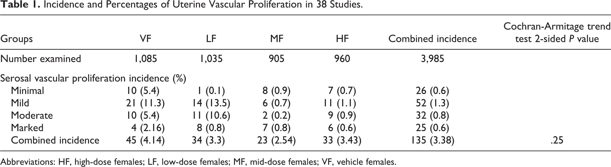
Abbreviations: HF, high-dose females; LF, low-dose females; MF, mid-dose females; VF, vehicle females.
Incidence and Percentages of Uterine Hemangiosarcomas in 38 Studies.
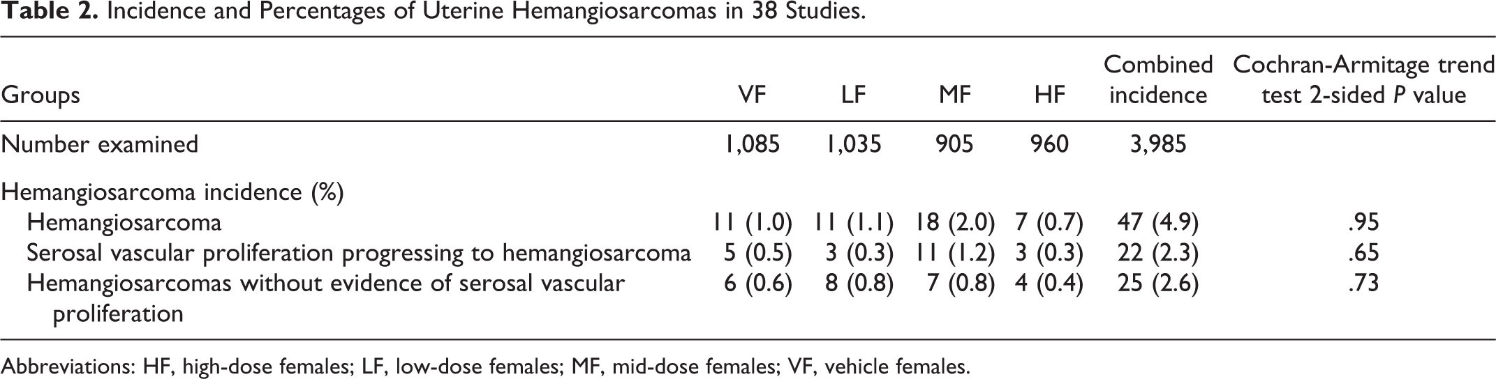
Abbreviations: HF, high-dose females; LF, low-dose females; MF, mid-dose females; VF, vehicle females.
Results
Table 1 shows the number of animals examined in each dose group and the incidence and percentages of vascular proliferative lesions noted on the serosal surface of the uterus in vehicle females (VF), low-dose females (LF), mid-dose females (MF), and high-dose females (HF) assigned to 38 studies. These vascular proliferative lesions were separated by the degree of severity, from minimal to marked, which was determined by the increasing number of proliferating vessels and the surface area covered by these proliferating vessels over the serosa. The proliferating vessels were normal and were lined by normal endothelial cells. The lesions were considered to be vascular proliferative lesions as long as they were restricted to the serosal surfaces and that there was no evidence of invasion of the myometrium and/or endometrium.
Table 2 shows the number of animals examined in each dose group and the incidence and percentages of uterine hemangiosarcoma diagnosed in VF, LF, MF, and HF assigned to 38 studies. This table also shows the number of animals belonging to each dose group that simultaneously showed vascular proliferative lesions on the serosal surfaces that gradually invaded the myometrium and subsequently the endometrium, thus progressing to a hemangiosarcoma. As the vessels started invading the myometrium and the endometrium, the endothelial cells lining the vessels became anaplastic and demonstrated pleomorphism, anisokaryosis, and often a moderate degree of mitosis. Further, the table also shows the number of animals in which a hemangiosarcoma was diagnosed, which primarily involved the endometrium and myometrium that had no simultaneous evidence of vascular proliferation on the serosal surface of the uterus. When the combined incidence of serosal vascular proliferative lesions (Table 1), uterine hemangiosarcomas, and uterine hemangiosarcomas with or without the evidence of serosal vascular proliferative lesions (Table 2) was compared between the groups, there were no statistically significant differences. In general, the vascular proliferation of minimal to mild intensity tended to remain as vascular proliferation and did not progress to hemangiosarcoma. On the other hand, the vascular proliferation of moderate to marked degree tended to progress to hemangiosarcoma, but not always.
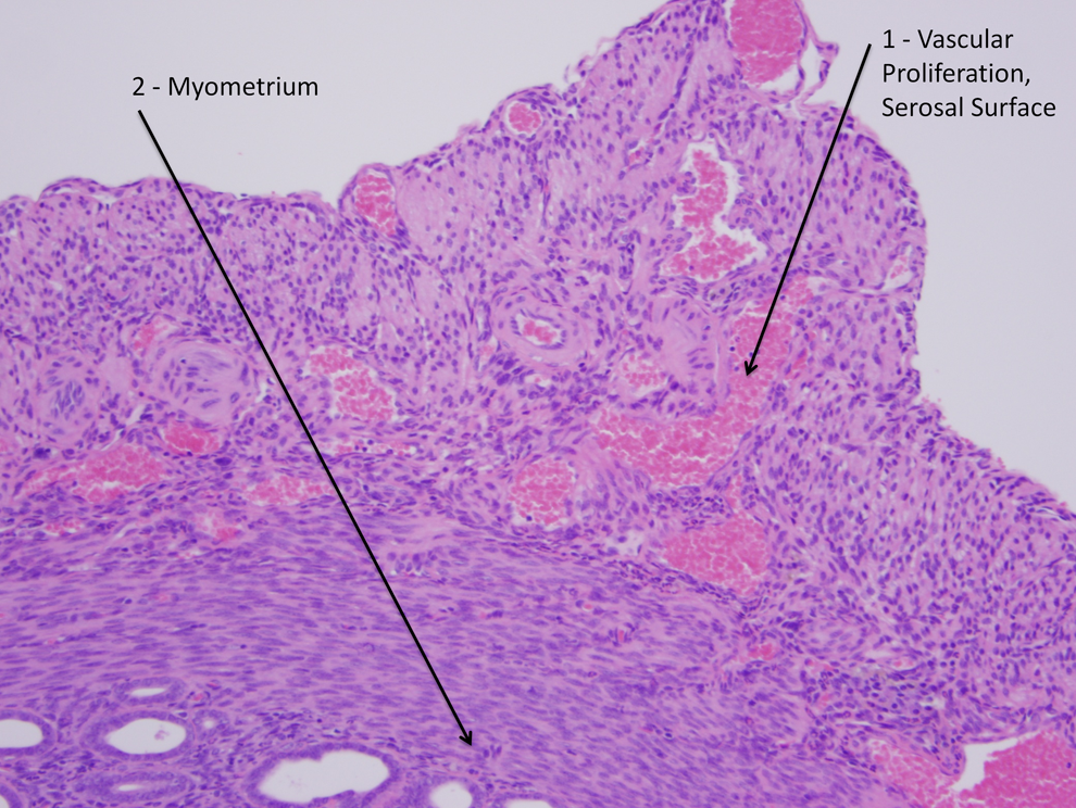
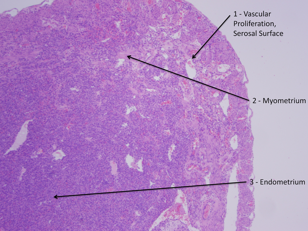
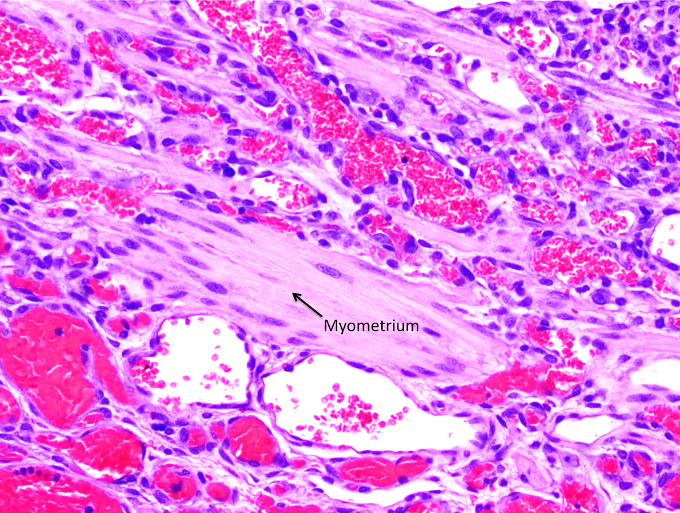
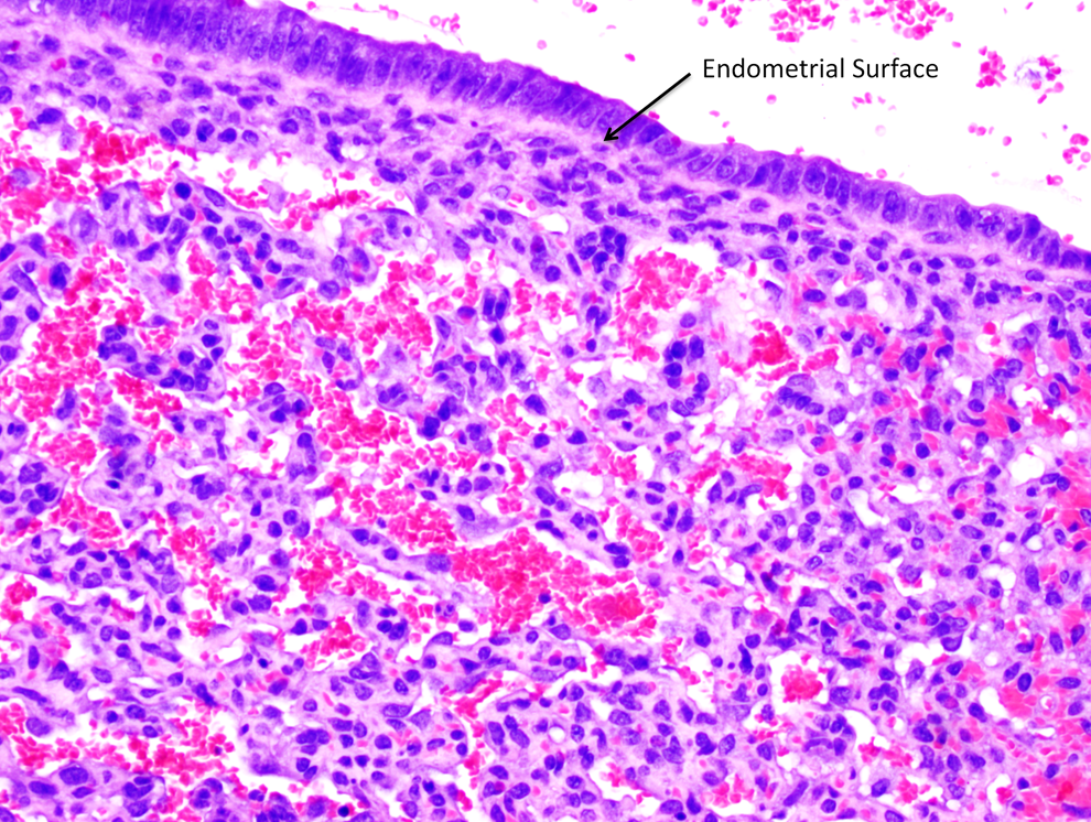
Figure 1 shows a normal uterus, arrows 1, 2, and 3 pointing to the serosal surface, myometrium, and endometrium, respectively. Figure 2 shows vascular proliferation of mild degree restricted to the uterine serosal surface (arrow 1) and that there is no invasion of the myometrium (arrow 2). The proliferating vessels were often thick and the vessel walls contained amorphous to occasionally hyalinized material and were lined by well-differentiated normal endothelial cells. Figure 3 shows the progression of vascular proliferation from the serosal surface (arrow 1) to the myometrium (arrow 2) and eventually to the endometrium (arrow 3). Figure 4 shows a hemangiosarcoma invading the myometrium (arrow). This particular tumor did not show vascular proliferation on the serosal surface. Figure 5 shows a hemangiosarcoma that had no serosal vascular proliferation but directly invaded the endometrium (arrow). The neoplastic endothelial cells showed anisokaryosis, pleomorphism, and a moderate degree of mitosis in the vessels invading the myometrium and endometrium.
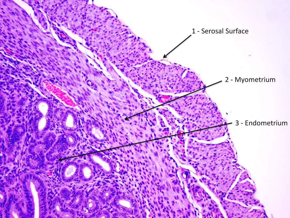
Normal uterus serosal surface without vascular proliferation, ×10.
Vascular proliferation, mild, restricted to the serosal surface, ×10.
Vascular proliferation, marked, progressing to hemangiosarcoma by invading the myometrium and endometrium, ×4.
Hemangiosarcoma invading the myometrium without evidence of serosal vascular proliferation, ×10.
Hemangiosarcoma invading the endometrium without evidence of serosal vascular proliferation, ×20.
The results of this analysis showed that from all dose groups, a total of 22 of the 47 mice (with a hemangiosarcoma) showed vascular proliferative lesions on the serosal surface that gradually progressed to hemangiosarcoma as they invaded the myometrium and endometrium, which accounts for 46.8% of all uterine hemangiosarcomas. While this gradual progression occurred, the vessels on the serosal surfaces were still normal and were lined by normal endothelium, but the vessels invading the myometrium and endometrium showed endothelial cells with neoplastic characteristics. The remaining 25 animals lacked such progression, and the hemangiosarcomas that were present in the myometrium and endometrium had no accompanying serosal vascular proliferative lesions. The tumor data statistics performed on each individual study did not show statistically significant increase in the incidence of hemangiosarcomas in any of the test article–treated dose groups.
Discussion and Conclusions
In the 38 studies conducted at our facility, the uterus was not considered to be a target organ either because of hemangiosarcomas or vascular proliferative lesions on the serosal surfaces. The tumor data statistics performed on each study did not show a statistically significant increase in any of the studies for uterine hemangiosarcomas. Our retrospective analysis also showed that in 46.8% of the cases, the vascular proliferative lesions did progress to hemangiosarcomas, whereas in the remaining 53.2% of the cases, no such correlationship was noted. This also indicates that while in certain number of cases the vascular proliferative lesions can progress to hemangiosarcomas, in the remaining cases the hemangiosarcomas can independently develop in the uterine myometrium and endometrium without any evidence of vascular proliferation on the serosal surfaces. When the combined incidence of serosal vascular proliferative lesions, uterine hemangiosarcomas, and uterine hemangiosarcomas with or without the evidence of serosal vascular proliferative lesions was compared between the groups, there were no statistically significant differences.
Based on our retrospective analysis, we propose that the term vascular anomaly should not be used as it implies abnormality, even though these vessels that proliferate along the uterine serosal surface are normal and are lined by normal vascular endothelium; based on this, the lesion should be called “vascular proliferation.” Our retrospective analysis also shows that these vascular proliferative lesions should also be considered as preneoplastic findings and should be taken into account along with uterine hemangiosarcomas in order to determine whether the uterus could be a potential target organ either for preneoplastic or neoplastic lesions or for the combined incidence of nonneoplastic and neoplastic lesions.
Vascular proliferative lesions have been noted in numerous tissues such as brain, spinal cords, kidneys, liver, nasal cavity, cervix, ovary, oviduct, urinary bladder, and uterus; however, of all these organs, the uterus showed the highest incidence. 8 Similarly, the uterus also happens to be the second organ, after the spleen, to develop hemangiosarcomas in female Tg.rasH2 mice. 7 The progression of these vascular proliferative lesions to hemangiosarcomas has not been noted by us in organs other than the uterus. Thus, the uterus appears to be a unique organ that shows such a transformation.
It is not a common practice in regulatory toxicology and carcinogenicity studies to perform the special immunohistochemical stains, as it requires client permission and substantial reasoning to perform special stains with protocol amendments. Nevertheless, prior immunohistochemical staining performed by others 14 has shown that the hemangiosarcomas and hemangiomas in mice are composed of endothelial progenitor cells expressing Cluster of Differentiation 34 (CD34), Vascular Endothelial Growth Factor Receptor 2 (VEGFR 2), and Cluster of Differentiation 31 (CD31) but not factor VIII-related antigen, which is the most common antigen shown to be positive for hemangiosarcomas in other species.
Thus, in summary, the vascular proliferative lesions should be considered as preneoplastic lesions in Tg.rasH2 mice and should be taken into account while evaluating the carcinogenic potential of a test article in any given study. For nonproliferative and proliferative lesions in the rat and mouse female reproductive system, neither the terms vascular anomaly nor the term vascular proliferation are defined. 15 In conclusion, this lesion appears to be unique to the Tg.rasH2 female mice and should be simply called vascular proliferation. In addition, vascular proliferation should be considered as a preneoplastic lesion and added to the International Harmonization of Nomenclature and Diagnostic Criteria (INHAND) document as a new lesion and diagnosis.
Footnotes
Author Contributions
Paranjpe, M. contributed to conception and design, contributed to acquisition, analysis, and interpretation, and drafted manuscript; Belich, J. contributed to design, contributed to analysis, drafted manuscript, and critically revised manuscript; Richardson, D. contributed to design, and contributed to analysis; Vidmar, T. contributed to design, contributed to analysis, and critically revised manuscript; Mann, P. contributed to conception, contributed to interpretation, drafted manuscript, and critically revised manuscript; McKeon, M. contributed to conception, contributed to interpretation, drafted manuscript, and critically revised manuscript; Elbekai, R. contributed to conception and design, contributed to acquisition and interpretation, drafted manuscript, and critically revised manuscript. All authors gave final approval and agree to be accountable for all aspects of work ensuring integrity and accuracy.
Declaration of Conflicting Interests
The author(s) declared no potential conflicts of interest with respect to the research, authorship, and/or publication of this article.
Funding
The author(s) received no financial support for the research, authorship, and/or publication of this article.