
Abstract
Since 2003, the Tg.rasH2 model has been accepted by regulatory agencies worldwide for 26-week short-term carcinogenicity assays as an alternative to the standard 2-year assays in conventional mice. However, over the decade, the number of actual studies conducted with alternative mouse models has remained low. The primary cause for low acceptance of this model has been lack of a historical database for the incidence of spontaneous lesions. Recently, we published the historical control database on spontaneous tumors in the Tg.rasH2 mice. The purpose of this publication is to present a large database pertaining to the non-neoplastic spontaneous lesions noted in Tg.rasH2 mice from studies conducted at our facility. Lesions that are considered unique in Tg.rasH2 mice are skeletal muscle myopathy, vascular anomalies involving various organs, and mesenteric arterial thrombosis. Other notable lesions are extramedullary hematopoiesis of spleen, subacute inflammatory foci in the liver, and infiltration of histiocytes in the lungs.
Keywords
Introduction
The Tg.rasH2 mouse is a hemizygous transgenic mouse carrying multiple copies of human c-Ha-ras gene with its own promoter and enhancer. This mouse model was endorsed and validated by the International Life Science Institute Health and Environmental Science Institute (ILSI HESI) through an international collaborative project (Robinson and MacDonald 2001) as an alternative to the conventional 2-year carcinogenicity bioassays in mice. Under the ILSI HESI project, 26-week carcinogenicity studies in Tg.rasH2 mice were conducted for 21 compounds at different facilities in the United States, Japan, and Europe (Takaoka et al. 2003). These compounds included genotoxic and nongenotoxic human carcinogens, rodent carcinogens/putative human noncarcinogens, and noncarcinogens. Data from these studies gave considerable confidence that this model produces low false positive and false negative rates (Pritchard et al. 2003; Usui et al. 2001; Morton et al. 2002; Saitoh et al. 1990; Yamamoto et al. 1998).
The Tg.rasH2 model has since been accepted as an alternative to the 2-year traditional model for carcinogenicity assessment of genotoxic and nongenotoxic agents by the regulatory agencies as described in the ICH S1B document (International Committee on Harmonization 1998). Despite its acceptance by the regulatory agencies in the United States, Europe, and Japan, the use of this model remains relatively limited compared to the conventional 2-year mouse models. The low acceptance of the 26-week short-term carcinogenicity assays has been primarily due to the lack of a historical control database (Long et al. 2010; Storer et al. 2010). There have been few published reports on the incidence of spontaneous tumors in Tg.rasH2 mice (Nambiar et al. 2012; Paranjpe et al. 2013; Usui et al. 2001), and few reports pertaining to non-neoplastic lesions such as skeletal muscle myopathy and vascular anomalies (De Jonghe et al. 2000; Kanno et al. 2003; Morton et al. 2002; Tsuchiya et al. 2001). There also have been published reports on the non-neoplastic lesions in the parent strains of Tg.rasH2, C57BL/6, and BALB/c mice (Frith et al. 1983; Frith and Ward 1988; Pettan-Brewer and Treuting 2011). However, to the best of our knowledge, there has been no comprehensive database published on the incidence of non-neoplastic lesions in Tg.rasH2 mice. As with any toxicology study, it is important for the pathologist to have complete knowledge of the morphology and incidence of spontaneous neoplastic and non-neoplastic lesions in any animal species or strain that is being used for carcinogenicity studies. We now present a large database summarizing the historical incidence of spontaneous non-neoplastic lesions in 1,420 mice (710 male and 710 female) assigned to 26 studies conducted at our facility.
Materials and Methods
Studies and Experimental Design
The database was constructed based on data from Tg.rasH2 mice assigned to 26 studies conducted at our facility, following the same study design. The incidence of non-neoplastic lesions was collected from 710 male and 710 female mice receiving negative (vehicle) control by oral gavage (21 studies), dosed feed (3 studies), or intravenous injection (2 studies) for 26 weeks. Animals were assigned to groups using a computer-generated randomization program. On the first day of treatment, animals were 6 to 10 weeks of age and weighed at least 20 or 15 g (males and females, respectively).
The numbers of animals, procedures, and experimental design for each study were reviewed and approved by the BioReliance Institutional Animal Care and Use Committee (IACUC). All procedures followed the specifications recommended in The Guide for the Care and Use of Laboratory Animals and were conducted in an Association for Assessment and Accreditation of Laboratory Animal Care (AAALAC)-accredited facility. All procedures involving but not limited to quarantine and acclamation, randomization, application of unique identification system, housing, provision of food and water, administration of test article, recording of clinical signs, necropsy, and tissue processing were followed in strict accordance with the good laboratory practice regulations, standard operating procedures, and protocol for each study.
Animals
CByB6F1-Tg(HRAS)2Jic (+/− hemizygous c-Ha-ras) mice, obtained from Taconic Farms (Germantown, NY), were used in all studies. The knock-in Tg element (human prototype c-Ha-ras gene with its own promoter/enhancer) is injected into C57BL/6 × BALB/c F2 zygotes, which are crossed back to C57BL/6J forming C57BL/6JJic-Tg(HRAS)2Jic. The CByB6F1-Tg(HRAS)2Jic (+/− hemizygous c-Ha-ras) is the offspring from a cross of the C57BL/6JJic-Tg(HRAS)2Jic hemizygous male mice with the BALB/cByJJic female mice. Each mouse was genotyped by Taconic to verify the presence of the transgene before being placed on study.
Housing and Environmental Conditions
Housing and environmental conditions were similar in all studies. Animals were single housed in polycarbonate cages with hardwood bedding chips in environmentally controlled rooms. Animals were verified to be free of illness prior to being placed on a study. All animals had ad libitum access to feed (Harlan TEKLAD Global Diet, Madison, WI) and water.
Study Termination and Pathological Examination
Animals dosed with the negative control were euthanized by CO2 overdose 26 weeks after the first day of dosing. An extensive necropsy was performed on all animals at study termination or on the day of death for animals found dead or euthanized in moribund condition prior to study termination. All tissues listed in Table 1 were collected, fixed in 10% neutral buffered formalin, embedded in paraffin, sectioned, stained with H&E, and evaluated microscopically. All non-neoplastic lesions were generally graded for severity from 1 to 4 (minimal to marked). Histopathological evaluation of 23 studies was performed by a single pathologist and the remaining 3 studies were evaluated by another pathologist. All studies were peer reviewed.
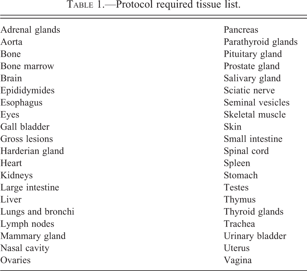
Protocol required tissue list.
Results
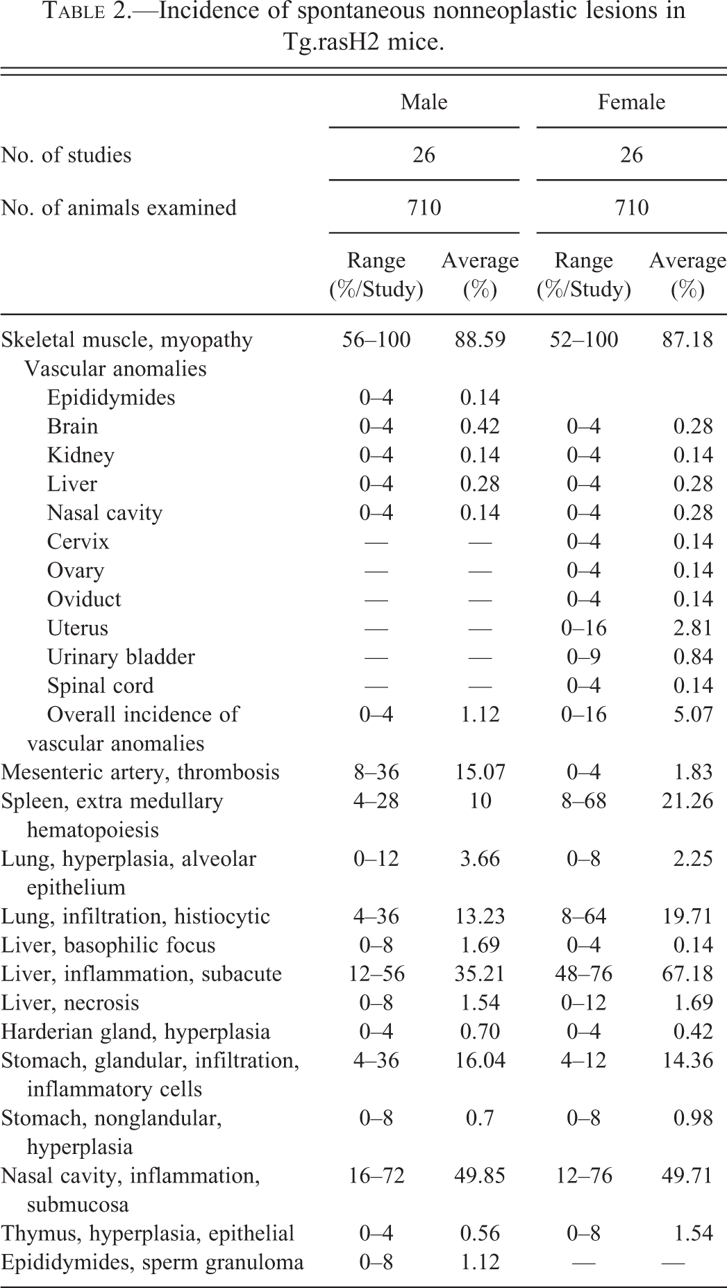
Incidence of spontaneous non-neoplastic lesions in 710 male and 710 female Tg.rasH2 mice expressed as range (%/study) and average (%) is presented in Table 2. Even though lesions were graded microscopically as minimal to marked with increasing severity, they are not presented separately by severity in Table 2 for simplicity.
Incidence of spontaneous nonneoplastic lesions in Tg.rasH2 mice.
Skeletal Muscle Myopathy
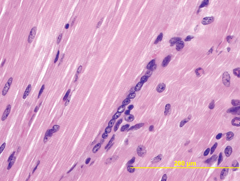
Skeletal muscle from the left thigh is examined in all mice in 26-week carcinogenicity studies using the Tg.rasH2 mice. Microscopically, the degree of severity can be extremely variable; however, there is usually no difference in the incidence and severity between the male and female mice. The incidence of skeletal muscle myopathy is similar in male and female mice. The primary lesions in these skeletal muscles are degenerative and necrotic changes. There is often rarification and/or vacuolation of individual myofibers as the affected muscles stain poorly compared to the normal myofibers (Figure 1). Occasionally, myofibers are necrotic as there are loss of striations, coagulation, hyalinization, and increase in eosinophilia. Such necrotic fibers are often surrounded by mild population of mixed inflammatory cells that include neutrophils, lymphocytes, and rarely macrophages (Figure 2). The striking alteration is the regenerative changes often noted in the adjacent myofibers. These fibers demonstrate increased basophilia and there is typical nuclear rowing indicative of regeneration (Figure 3).
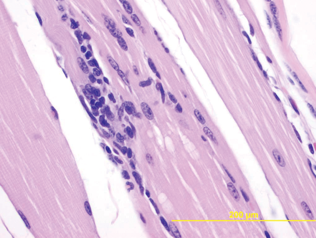
Degeneration of skeletal muscle. 400×.
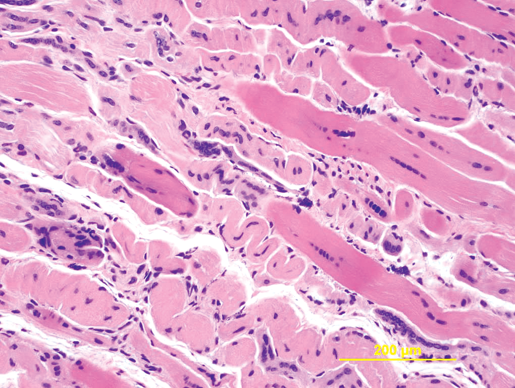
Necrosis of skeletal muscle. 200×.
Nuclear rowing in the skeletal muscle. 400×.
Vascular Anomalies
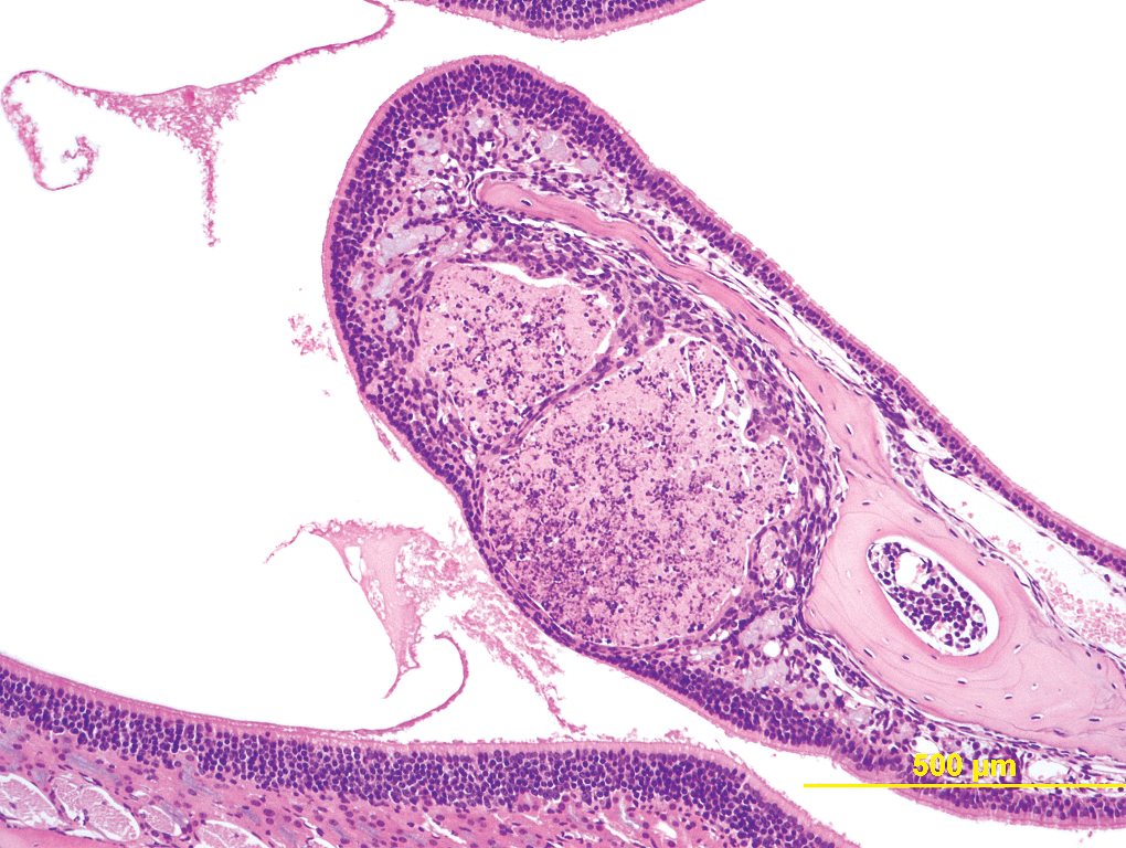
Vascular anomalies are usually noted grossly as red foci or reddish discoloration. They are evident grossly on the surface of the uterus and urinary bladder, but are difficult to detect grossly in other tissues. Microscopically, these lesions are characterized by thick walled, blood containing vessels lined by well-differentiated endothelial cells. The highest incidence of these lesions is in the uterus (2.81%) followed by urinary bladder (0.84%). The urinary bladder lesions are also present only in the female and not the male mice. The overall incidence of vascular anomalies is much less in males (1.12%) compared to females (5.07%). However, the incidence of vascular anomalies, excluding the uterus and urinary bladder, is otherwise very low in both female and male mice. Figure 4 shows a micrograph of a normal uterus. Figure 5 shows uterus with vascular anomaly, and Figure 6 shows vascular anomaly and a uterine hemangiosarcoma in the same animal.
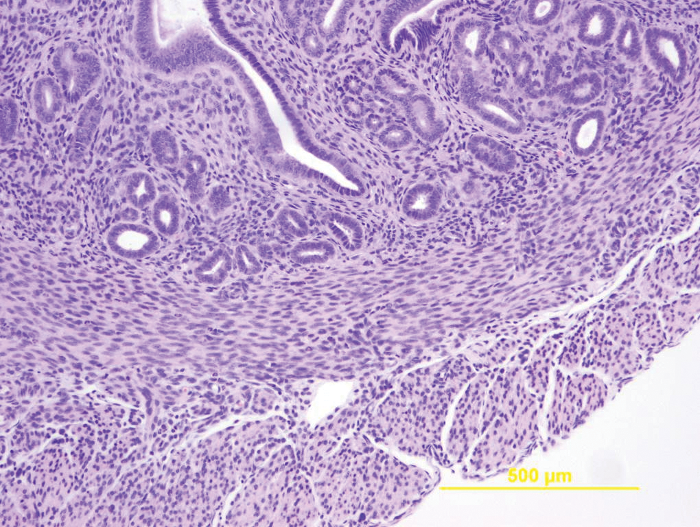
Normal uterus. 100×.
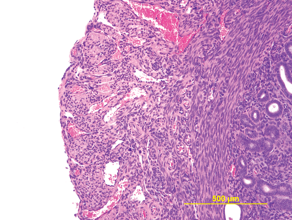
Uterus with vascular anomaly. 100×.
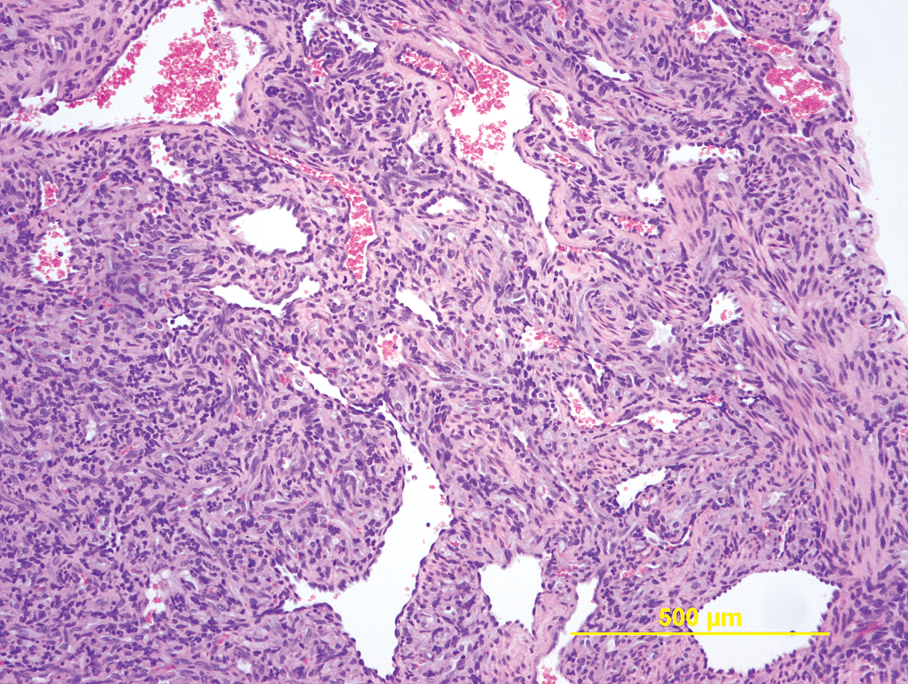
Uterus, vascular anomaly progressing to a hemangiosarcoma. 100×.
Thrombosis of Mesenteric Arteries
The incidence and severity of thrombosis of the mesenteric arteries are significantly higher in male mice compared to female. Mesenteric artery is not a protocol required tissue but these arteries are almost always present on the same slide of the mesenteric lymph nodes, which is a protocol required tissue. If the mesenteric arteries were specifically examined, the overall incidence of mesenteric arterial thrombosis may be actually higher than presented in Table 2. The lesions are not usually noted grossly, but they are usually noticed during trimming because of their brittle nature. Microscopically, the thrombi are well organized and often contain mineral that causes the brittle nature at the time of trimming. Occasionally, the thrombi may show evidence of early ossification. Around these thrombi there is usually proliferation of connective tissue and infiltration of mixed inflammatory cells. A typical mesenteric arterial thrombosis is shown in Figure 7.
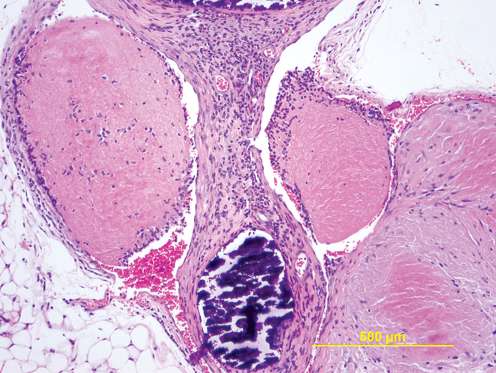
Mesenteric artery, thrombosis. 100×.
Extramedullary Hematopoiesis (EMH) in Spleen
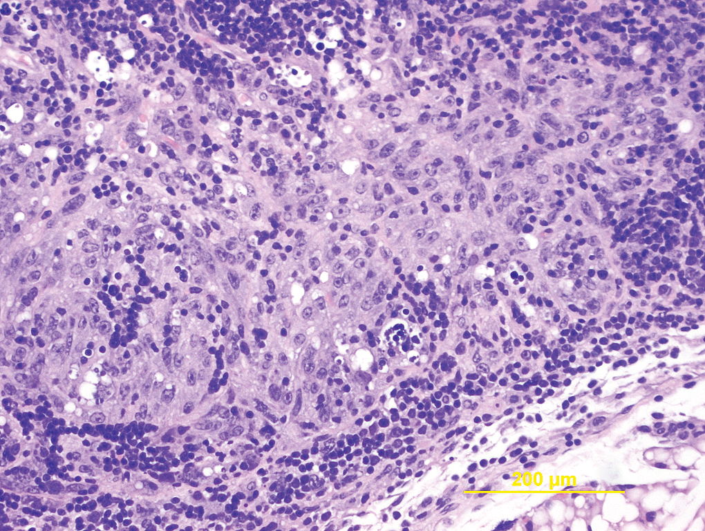
The incidence and severity of EMH are higher in females compared to male mice. Enlarged spleens may be seen grossly when EMH is of moderate to marked severity. Figure 8 shows EMH of mild severity with hyperchromatic nucleated red blood cells (RBCs). Figure 9 shows marked EMH with loss of follicular structures and replacement of splenic parenchyma by nucleated RBCs; this particular mouse with marked splenic EMH also had a hemangiosarcoma in the skin/subcutis.
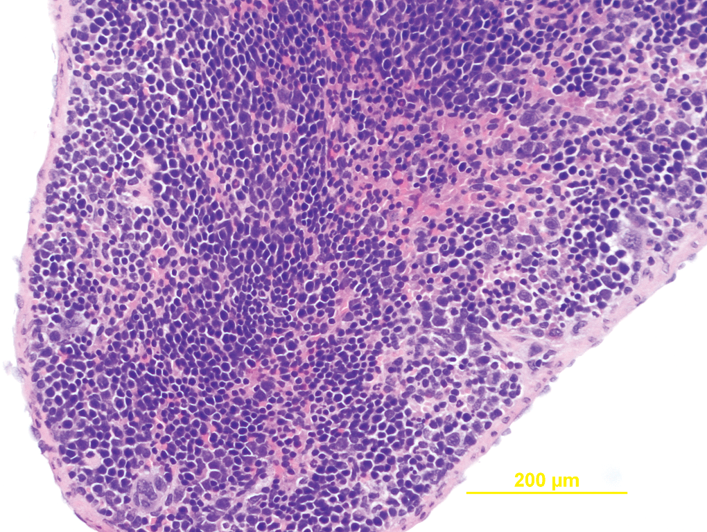
Spleen extramedullary hematopoiesis, minimal to mild. 200×.
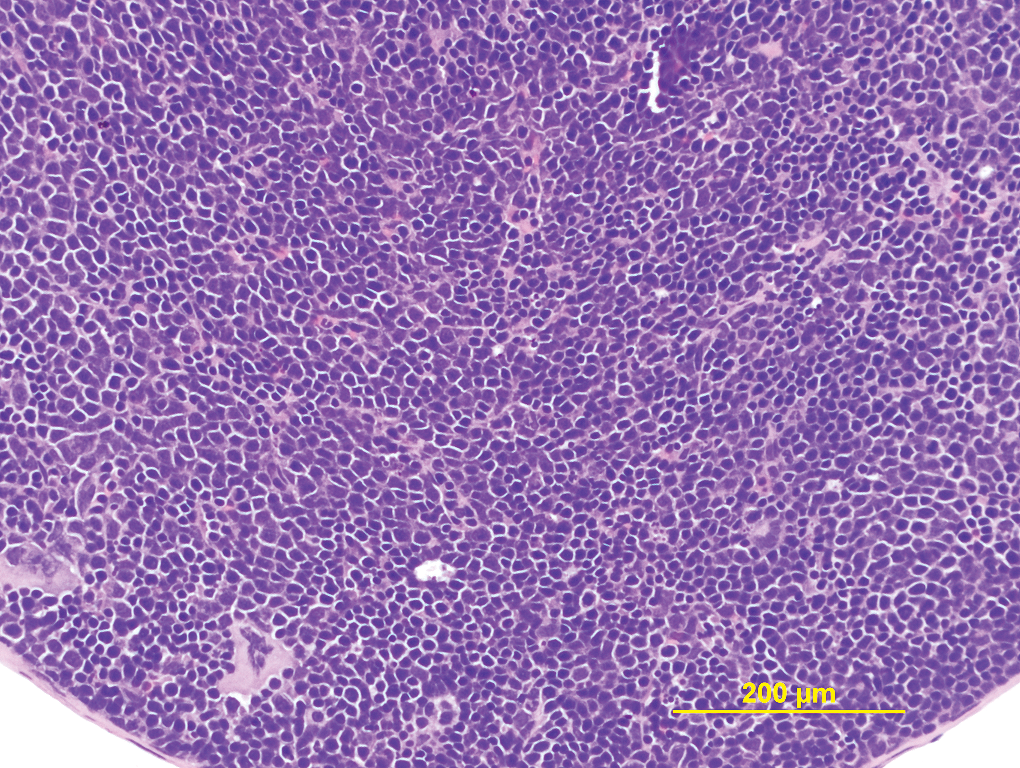
Spleen extramedullary hematopoiesis, marked. 200×.
Infiltration of Histiocytes in the Lungs
This is a common spontaneous non-neoplastic lesion in the lungs, the incidence and severity of which is lower in male compared to the female mice. Grossly, these lesions are seen as foci, as against the nodules seen grossly with pulmonary adenomas. The infiltrating histiocytes are round to oval and usually have an abundant intensely acidophilic cytoplasm. The infiltrating histiocytes may contain eosinophilic crystals and free crystals can also be seen in the alveoli. Often, there is an accompanying infiltration of neutrophils with few giant cells. The lesions are usually focal and only rarely focally extensive or diffuse. Pulmonary adenomas and carcinomas are common spontaneous tumors of Tg.rasH2 mice, and there is often extensive infiltration of histiocytes that surround these tumors. The infiltration of acidophilic histiocytes is shown in a microscopic photo (Figure 10).
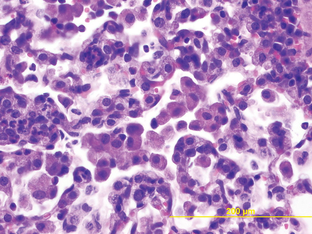
Lung, infiltration of acidophilic histiocytes. 400×.
Alveolar Epithelial Hyperplasia in the Lungs
Incidence of alveolar epithelial hyperplasia in males is slightly higher than in female mice. These lesions are usually not seen grossly. Microscopically, alveolar epithelial hyperplasia is characterized by proliferation of well-differentiated epithelial cells that can form small papillary projections, but the alveolar structure is usually very well maintained. The lesions are not well circumscribed and there is no compression of the surrounding parenchyma. These features are helpful in differentiating them from alveolar adenomas, which are common spontaneous tumors of Tg.rasH2 mice. The adenomas are usually well circumscribed, have a solid pattern that obliterates the underlying alveolar structures, and compress the surrounding alveolar parenchyma.
Subacute Inflammatory Foci in the Liver
The incidence of subacute inflammatory foci male mice is almost half of the incidence noted in the female mice. The severity of foci in both sexes is usually minimal and only rarely mild or greater. Usually when <5 foci are observed microscopically in the entire section, they are graded as minimal. Foci >5 but <10 are graded as mild. The increasing grade of the foci is determined by the increase in the number of foci rather than the size of the foci. These foci are randomly distributed throughout the parenchyma and usually contain few neutrophils and lymphocytes, and only occasionally macrophages. About 50% of these foci in both sexes contain isolated single necrotic cells. Figure 11 shows a typical focus of subacute inflammation without a necrotic cell whereas Figure 12 shows a similar focus with a necrotic cell.
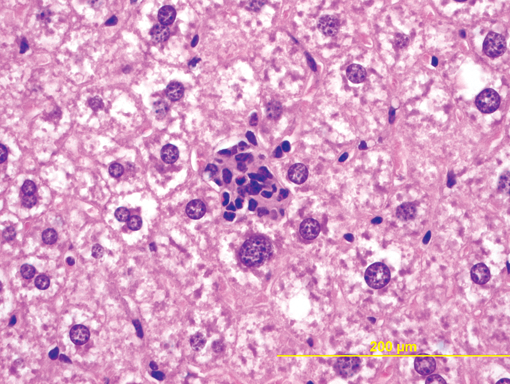
Liver, subacute inflammation without necrosis. 400×.
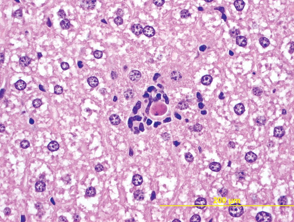
Liver, subacute inflammation with necrosis. 400×.
Focal Necrosis of Liver
Small foci of necrosis are occasionally seen in the livers of mice of both sexes with incidence more or less similar in males and females. At times, this lesion may be seen grossly as a pale focus. Most of the foci diagnosed microscopically in the vehicle-treated Tg.rasH2 mice were graded minimal. These foci are randomly distributed and are characterized by clusters of few hepatocytes that have undergone a coagulative type of necrosis with swelling of the cytoplasm and loss of nuclei or presence of pyknotic nuclei. Few scattered neutrophils may be present around the focus.
Basophilic Foci in the Liver
The incidence of basophilic foci in male Tg.rasH2 mice is much higher than in female mice. These basophilic foci are randomly distributed, have relatively indistinct margins, and usually do not compress the surrounding parenchyma. The hepatocytes within these foci are arranged in normal cords and the cells are more basophilic than normal. A typical basophilic focus is shown in Figure 13.
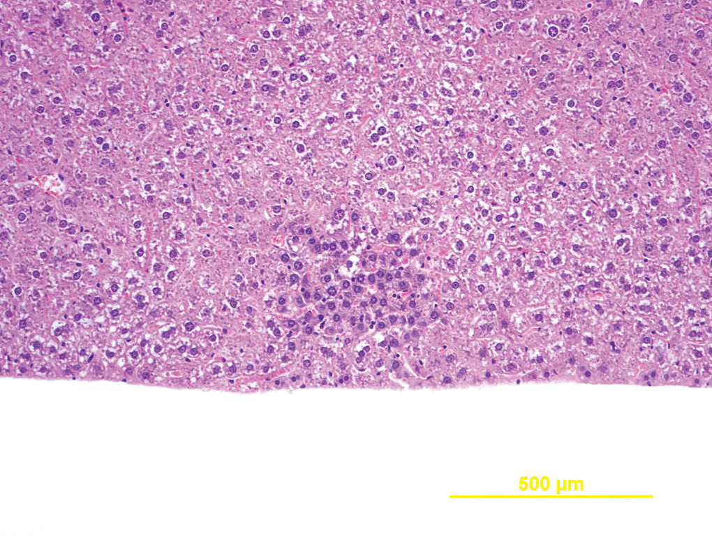
Liver, basophilic focus. 100×.
Harderian Gland Hyperplasia
There is a low incidence of Harderian gland hyperplasia in both sexes. Focal hyperplastic lesions in the Harderian glands are not well circumscribed and do not compress the surrounding tissue, as is seen with Harderian gland adenomas. The cells are usually well differentiated but may be slightly more basophilic and may form few papillary projections.
Glandular Stomach, Infiltration of Inflammatory Cells
Infiltration of mixed inflammatory cells in the submucosa of the glandular stomach is commonly noted in male and female mice with similar incidence. The lesions are usually of minimal severity and only rarely of mild or moderate severity. Only lesions that are at least of moderate severity are seen grossly as a thick wall, usually during trimming and not at necropsy. Microscopically, there is focal infiltration of mainly neutrophils and few lymphocytes in the submucosa and the overlying glandular epithelial cells may contain eosinophilic hyaline inclusions. Increases in the number of goblet cells are seen in lesions that are at least of moderate severity. A typical glandular stomach with infiltration of mixed inflammatory cells is shown in Figure 14.
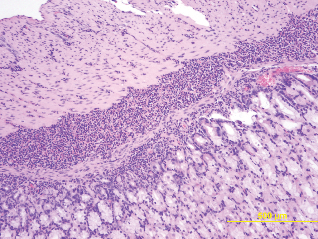
Stomach, glandular, infiltration of inflammatory cells. 100×.
Nonglandular Stomach, Hyperplasia
Hyperplasia of the nonglandular stomach occurs at low incidences in both male and female mice. These may be seen grossly as raised foci but usually only at the time of trimming. The lesions are characterized by proliferation of the squamous epithelium, which may form small papillary projections that invade the submucosa. There is usually deposition of increased amount of keratin on the surface.
Nasal Cavity, Submucosal Inflammation
We routinely examine nasal cavity sections at 3 levels. This lesion is not detected grossly and is mostly observed microscopically at minimal severity and only rarely at mild severity. The submucosal glands usually contain inspissated material with necrotic debris and few degenerate neutrophils. The area surrounding these glands may contain mixed inflammatory cells, particularly neutrophils and lymphocytes. Few of the adjacent submucosal glands may contain hypertrophied epithelial cells with eosinophilic inclusions. Occasionally, eosinophilic crystals may be seen in the glands and the mucosal epithelium may be minimally hyperplastic. Not all features, however, are noted simultaneously. The incidence of these lesions is high in both male and female mice and almost close to 50% in each sex. Figure 15 shows submucosal glands with necrotic debris and degenerate neutrophils and Figure 16 shows eosinophilic inclusions in the hypertrophic epithelium and several eosinophilic crystals on the surface.
Nasal cavity, subacute inflammation. 100×.
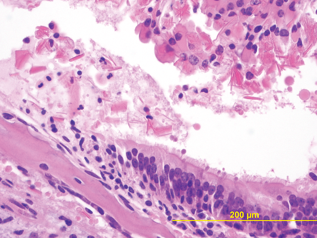
Nasal cavity, eosinophilic inclusions and crystals. 400×.
Thymus, Epithelial Hyperplasia
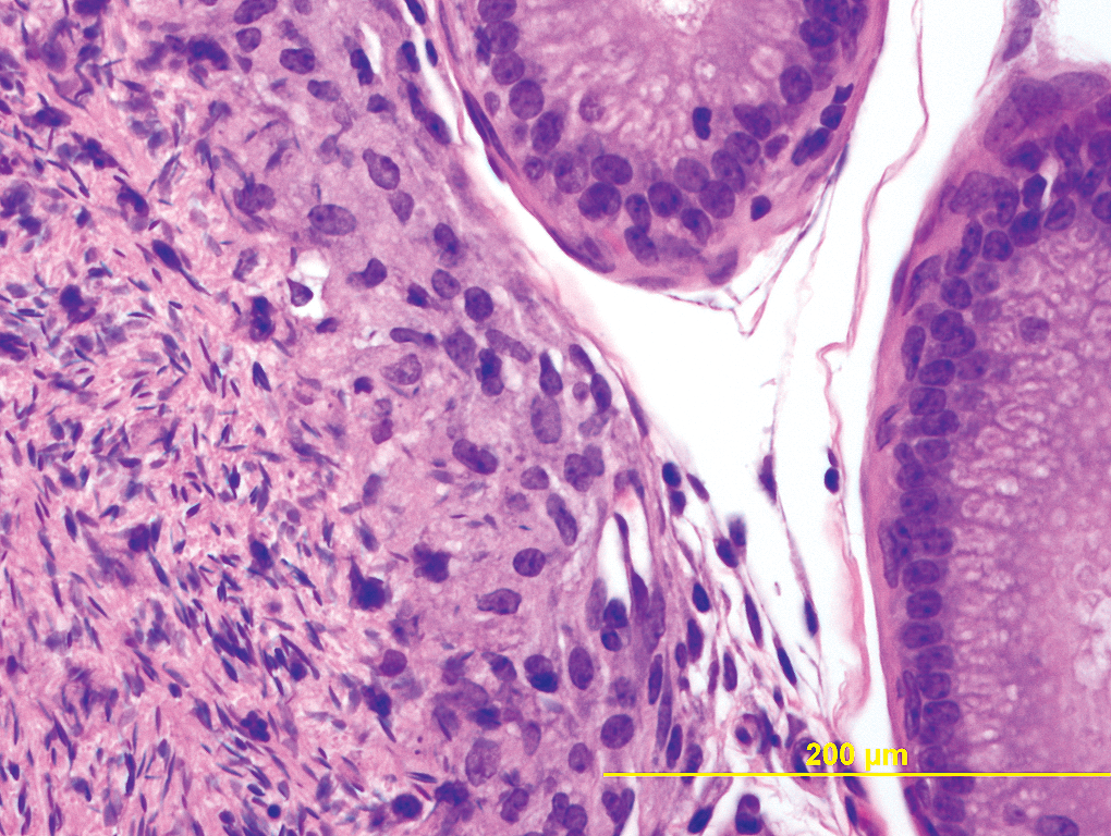
Epithelial hyperplasia of the thymus is a rare lesion, with a higher incidence in females than males. Clusters of proliferating epithelial cells can be readily identified microscopically and the cells may appear squamoid or spindeloid. The lesions are mainly of minimal to moderate severity. A typical hyperplastic lesion is shown in Figure 17.
Thymus, epithelial hyperplasia. 200×.
Sperm Granulomas in the Epididymis
Sperm granulomas are also seen at low incidence rate of 1.12%. The lesions are usually noted grossly as raised foci or small nodules of pale or tan color. Microscopically the granulomas are usually focal, well circumscribed, but nonencapsulated. They also contain abundant macrophages with engulfed spermatozoa. At the periphery, few multinucleated giant cells may be present. The edge of a typical sperm granuloma with macrophages engulfing the spermatids is shown in Figure 18.
Epididymis, sperm granuloma. 400×.
Discussion and Conclusions
It is a good practice to record the incidence and severity of background non-neoplastic lesions consistently in every study. This not only helps in constructing the database for spontaneous non-neoplastic lesions, but it also provides a baseline for comparison should there should be an increase in the incidence of these lesions in the test article–treated groups.
Myopathy of the skeletal muscles similar to what we observed has been recorded by other authors in Tg.rasH2 mice (De Jonghe et al. 2000; Kanno et al. 2003; Morton et al. 2002; Tsuchiya et al. 2001). These authors observed various changes in the skeletal muscles such as myocyte degeneration, fragmentation, atrophy, alignment of nuclei in rows in the central cytoplasm and inflammatory changes, which are similar to what we have noted. The incidence of these spontaneous lesions in those studies ranged from 81 to 100%, which is once again very similar to what we have noted. In all of our studies, the clinical signs and gross lesions were not associated with myopathy. At our facility, we have also performed several studies with other transgenic mouse strains such as Tg.AC and p53, but we have not observed similar myopathy in these strains.
We have observed vascular anomalies with incidence of 1.12% in males, which is much lower than that in the female mice at 5.07%. Apart from the involvement of uterus and urinary bladders in females, the involvement of other tissues is rare in both males and females. The lesions we noted represent thick-walled vessels that contain blood and are lined by well-differentiated endothelial cells. Lesions similar to those descri-bed above have been diagnosed as a vascular anomaly by other authors in Tg.rasH2 mice (Morton et al. 2002). These authors recorded vascular anomalies in various organs such as subcutis, salivary glands, skeletal muscles, lungs, and uterus, which is similar to what we observed. The incidence involving specifically the uterus only was not stated (Morton et al. 2002). We have recorded hemangiosarcomas and hemangiomas as common spontaneous tumors of the Tg.rasH2 mouse with incidence being 5.91% in males and 7.18% in females and incidence of splenic hemangiosarcoma in males and females being identical at 3.66% (Paranjpe et al. 2013). Uterus is the most commonly affected organ other than spleen in the female mice (Paranjpe et al. 2013). Others have also shown that this strain of mouse is in general predisposed to splenic hemangiosarcomas or hemangiosarcomas in different organs (Morton et al. 2002; Nambiar et al. 2012; Usui et al. 2001). From our studies, the incidence of uterine hemangiosarcoma and vascular anomalies of uterus has been 1.40% and 2.81%, respectively. It appears that there may be a relationship between the vascular anomalies and hemangiosarcomas in Tg.rasH2 mice. In at least a few of our cases, we have noted the vascular anomaly and a hemangiosarcoma in the same animal, which is similar to what has been observed by others (Morton et al. 2002). This suggests that these vascular anomalies may lead to the hemangiosarcomas; however, evaluation of more such cases may be needed to make this positive connection.
The mesenteric arterial thrombosis appears to be unique to the Tg.rasH2 mouse. It is also common to see thrombosis in both benign and malignant vascular tumors, hemangiomas and hemangiosarcomas. Given the fact that the Tg.rasH2 mouse strain is predisposed to these vascular tumors, it is very important that any lesion with a thrombus in the section should be carefully evaluated and possible existence of a vascular tumor should be ruled out.
EMH of spleen is commonly seen in most rats and mice including the parent strains of the Tg.rasH2 mice, the C57BL/6, and BALB/c mice (Frith et al. 1983; Frith and Ward 1988; Pettan-Brewer and Treuting 2011). In our experience, EMH of marked severity in either sex is usually associated with the presence of a hemangiosarcoma, or some other malignant tumor in the same mouse in organs other than spleen. The exact cause and effect relationship between EMH of marked severity and a malignant tumor is not known, but it is most likely secondary to loss of blood from the tumor in the same animal.
Infiltration of intensely acidophilic histiocytes in the lungs can pose a diagnostic challenge when the infiltration of histiocytes is noted in and around lung adenomas, complicating microscopic evaluation. A recut of the block does not help as the lesion is usually not present in the recut. Similar lesions in Tg.rasH2 mice (Takaoka et al. 2003) and in other strains of mice have been diagnosed as acidophilic macrophage pneumonia (Pettan-Brewer and Treuting 2011; Murray and Luz 1990). The incidence of alveolar epithelial hyperplasia parallels the incidence of pulmonary adenomas noted in male and female mice, with hyperplasia generally of lower incidence than adenomas (Paranjpe et al. 2013).
The incidence of subacute inflammatory foci in the livers of rodents have been variously named as microgranulomas or simply as infiltration of inflammatory cells and they are thought to be secondary to the enterohepatic showering of bacteria (Eustis et al. 1990). It is important also to differentiate these foci from small foci of EMH that are occasionally seen in the liver, particularly when there is EMH of moderate or greater severity in the spleen of the same mouse. We have also noted small random necrotic foci in the livers of Tg.rasH2 mice at a low incidence. Also it is important to judge the random distribution of these foci in the control mice as against mostly the zonal nature of necrotic foci in the test article–treated mice. The basophilic foci in the livers noted by us have also been recorded by others (Kanno et al. 2003; Takaoka et al. 2003). Different types of foci have been described in rodents such as basophilic, eosinophilic, mixed, vacuolated, and clear cell and the experimental models suggest that these foci are preneoplastic (Eustis et al. 1990). In our studies, we have not seen any progression of these basophilic foci toward the tumors. We have seen only 2 hepatocellular adenomas in the 710 male mice that we examined and none in the 710 female mice (Paranjpe et al. 2013). Low incidence of hepatocellular adenoma has been recorded by others also in male mice but not in the female mice (Nambiar et al. 2012).
Infiltration of inflammatory cells in the submucosa of glandular stomach noted by us is similar to what has been noted by others in Tg.rasH2 mice (Kanno et al. 2003). We have also recorded hyperplasia of the nonglandular stomach with very low incidence in males and females., similar to the low incidence of papilloma and squamous cell carcinoma noted by us and others. (Nambiar et al. 2012; Paranjpe et al. 2013).
Incidence of subacute inflammatory lesions in the nasal cavities we have recorded is very similar in both males and females at about 50% in both. However, in the test article–treated mice, the lesions we have seen are more of an exudative nature. As against the submucosal inflammation that is restricted mainly to the submucosa in the control mice, the exudative lesions in the test article–treated mice are characterized by accumulation of inflammatory exudate in the turbinate cavities. We have seen with increasing severity of the submucosal or exudative inflammatory lesions, increasing mucosal hyperplasia. The latter is thought to be because of irritation caused by the inflammation rather than the primary proliferative response. The incidence of nasal cavity benign and malignant tumors in our database is low at 0.42% and 0.70% in males and females, respectively (Paranjpe et al. 2013). While interpreting the lesions in the nasal cavities, the submucosal inflammatory lesions, exudative inflammatory lesions, any hyperplastic lesions, and then tumors should all be taken into consideration to possibly provide an explanation for the pathogenesis of tumors.
The incidence of epithelial hyperplasia in the thymus we have noted parallels the incidence of thymomas (Paranjpe et al. 2013). The hyperplastic and neoplastic lesions appear at slightly higher frequencies in females than in males.
In conclusion, we have thus far presented the largest database on the spontaneous non-neoplastic lesions in the Tg.rasH2 mice. The non-neoplastic lesions that appear to be unique to the Tg.rasH2 mouse are the skeletal muscle myopathy, the vascular anomalies, and the mesenteric arterial thrombosis. The vascular anomalies are most commonly noted in the uterus. There may be some association between the vascular anomalies and the progression to hemangiosarcomas, the most common spontaneous tumor of the Tg.rasH2 mouse. However, more studies may be needed to establish this relationship.
Footnotes
The author(s) declared no potential conflicts of interest with respect to the research, authorship, and/or publication of this article.
The author(s) received no financial support for the research, authorship, and/or publication of this article.