
Abstract
The Tg.rasH2 mouse is a hemizygous transgenic mouse, approved by regulatory agencies for carcinogenicity assessment. However, the absence of a historical database for the incidence of spontaneous neoplasms has subsequently led to reluctance by some pharmaceutical companies to adopt the use of this short-term carcinogenicity assay. Our laboratory has generated a database summarizing the mortality, body weights, and the incidence of spontaneous tumors in 1420 male and female mice assigned to 26 studies conducted at our facility. In addition, we present the incidence of tumors in positive control mice treated with urethane from these studies. Mortality in the vehicle-treated Tg.rasH2 mouse was low (average of 1% in each study). The most common spontaneous tumors in the Tg.rasH2 mice were alveolar bronchiolar adenoma of the lungs (10.14% in males and 5.77% in females) and hemangiosarcoma of the spleen (3.66% in both males and females). The incidence of all other tumors was generally very low. In the positive control, urethane-treated animals, the incidence of alveolar bronchiolar adenomas and alveolar bronchiolar carcinomas in the lungs was 93.69% and 42.88% in males and 92.43% and 72.79% in females, respectively. In addition, the incidence of splenic hemangiosarcomas in urethane-treated males was 89.18% and 92.25% in females. The 6-month Tg.rasH2 assay is more precise, faster, and more economical than the conventional 2-year mouse assays because of the low incidence of background tumors, very high survival, shorter duration, and the lower number of animals used.
Introduction
Cancer risk assessment of pharmaceuticals and environmental carcinogens remains heavily dependent on rodent carcinogenicity studies. Historically, 2-year bioassays in 2 rodent species, rats and mice, served as the standard for carcinogenicity assessment. These conventional rodent assays are costly, not always relevant for human risk assessment, and require large amount of test article and a large number of animals. General dissatisfaction with the conventional carcinogenicity assays, and the need to reduce the duration of the assays and the number of animals used, fueled the endorsement of short-term carcinogenicity assays in genetically engineered mice by the International Conference on Harmonization (ICH) in 1998. Validation of the use of short-term carcinogenicity studies using transgenic mice, including the Tg.rasH2 (CByB6F1-Tg(HRAS)2Jic [+/− hemizygous c-Ha-ras]) mouse, was carried out by the International Life Science Institute Health and Environmental Science Institute (ILSI HESI) through an international collaborative project. 1
Developed at the Central Institute for Experimental Animals, the Tg.rasH2 mouse is a hemizygous transgenic mouse carrying multiple copies of human c-Ha-ras gene with its own promoter and enhancer. Under the ILSI HESI project, 26-week carcinogenicity studies in Tg.rasH2 mice were conducted for 21 compounds at different facilities in the United States, Japan, and Europe. 2 These compounds included genotoxic and nongenotoxic human carcinogens, rodent carcinogens/putative human noncarcinogens, and noncarcinogens. Data from these studies gave considerable confidence that this model produces low false positive and false negative rates.3,4 In addition, data from these and other studies also showed that the Tg.rasH2 mouse model can be used for both genotoxic and nongenotoxic carcinogen identification.3,5–7
Despite extensive initial research, validation studies and the acceptance of this 6-month carcinogenicity model as an alternative to the 2-year traditional model by the regulatory agencies as described in the ICH S1B document 8 and acceptance by the regulatory agencies in the United States, 9 Europe, 10 and Japan, 11 the use of this model is still very limited compared to the conventional 2-year mouse models. The causes of low acceptance of the 6-month short-term carcinogenicity assays have been discussed in recent publications and they include lack of knowledge of unexpected tumor findings, lack of knowledge of tumor identification and characterization, unfounded perception that the transgenic animals are oversensitive, and in general, a lack of historical control database.12,13
A historical control database, used in conjunction with statistical analysis, is a cornerstone in interpreting the biological significance of a tumor response and helps reduce false positive and false negative results. 14 In treatment groups, tumors may show statistically significant, albeit only small, increases in incidence relative to controls. In these instances, defining the biological significance of these tumors may only be achieved by comparing the occurrence of these tumors to the incidence seen in the historical database. Irrespective of the results of statistical analysis, a drug- or chemical-induced effect may be verified or nullified, based on where the incidence falls within the historical range. Alternatively, the historical database may be used to verify the validity of the study, by determining whether the incidence of tumors in both the negative and the positive controls used are consistent with what has been reported previously. In addition, the historical control database is useful in assessing possible differences in tumor response between two negative control groups in a single carcinogenicity study.
Previously published data on spontaneous tumors in Tg.rasH2 mice is limited. In 2001, as part of the ILSI HESI project, a summary of spontaneous tumors in 180 male and 178 female control Tg.rasH2 mice derived from 12 studies conducted at multiple sites was reported. 3 However, since the adoption of the ICH guidelines by regulatory agencies in the United States, Europe, and Japan, limited data have been published on the spontaneous incidence of tumors in Tg.rasH2 mice. 15 We now present a database summarizing the mortality, body weights, and the historical incidence of spontaneous tumors in 1420 mice (710 male and 710 female) assigned to 26 studies conducted at our facility, which, to the best of our knowledge, is the largest to be published. In addition, we also present the incidence of the urethane (ethyl carbamate)-induced tumors in positive control mice generated from these studies.
Methods and Materials
Studies and Experimental Design
The database was constructed based on data from Tg.rasH2 mice assigned to 26 studies conducted at our facility, following the same study design. The incidence of spontaneous tumors was collected from 710 male and 710 female mice receiving negative (vehicle) control by oral gavage (21 studies), dosed feed (3 studies), or intravenous injection (2 studies) for 26 weeks. In addition, data were collected from 555 males and 555 females assigned to the positive control groups in these studies. Animals were assigned to groups using a computer-generated randomization program. Animals in the positive control groups received a total of 3 intraperitoneal injections of 1000 mg/kg of urethane (Sigma-Aldrich, St Louis, Missouri) during the first week of study. On the first day of treatment, animals were 6 to 10 weeks old and weighed at least 20 or 15 g (males and females, respectively). All procedures were performed in strict accordance with the Good Laboratory Practice regulations.
Animals
CByB6F1-Tg(HRAS)2Jic (+/− hemizygous c-Ha-ras) mice, obtained from Taconic Farms (Germantown, New York), were used in all the studies. The knock-in Tg element (human prototype c Ha-ras gene with its own promoter/enhancer) is injected into C57BL/6 x BALB/c F2 zygotes, which are crossed back to C57BL/6J forming C57BL/6JJic-Tg(HRAS)2Jic. The CByB6F1-Tg(HRAS)2Jic (+/− hemizygous c-Ha-ras) is the offspring from a cross of the C57BL/6JJic-Tg(HRAS)2Jic hemizygous male mice with the BALB/cByJJic female mice. Each mouse was genotyped by Taconic to verify the presence of the transgene before being placed on study.
Housing and Environmental Conditions
Housing and environmental conditions were similar in all the studies. Animals were single housed in polycarbonate cages with hardwood bedding chips in environmentally controlled rooms. Animals were verified to be free of illness prior to being placed on a study. All animals had ad libitum access to feed (Harlan TEKLAD Global Diet, Madison, Wisconsin) and water.
Study Termination and Pathological Examination
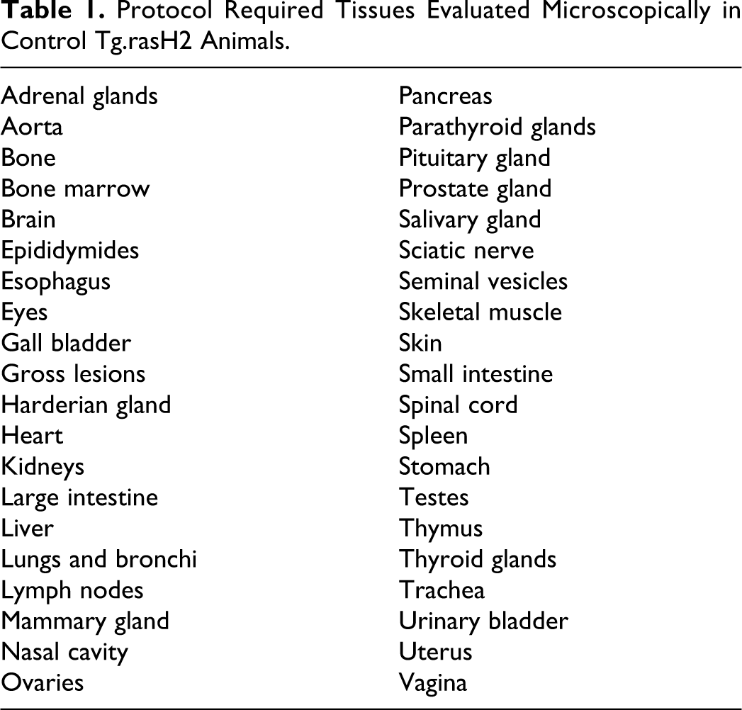
Animals dosed with the negative control were euthanized by carbon dioxide overdose 26 weeks after the first day of dosing. Due to high incidence of mortality, surviving positive control animals were euthanized 14 to 19 weeks after receiving the first dose of urethane. An extensive necropsy was performed in all animals at study termination or on the day of death for animals found dead or euthanized in moribund condition prior to study termination. All tissues listed in Table 1 were collected, fixed in 10% neutral buffered formalin, embedded in paraffin, sectioned, stained with hematoxylin and eosin, and evaluated microscopically. Only the expected target organs (lungs and spleen) in urethane-treated animals were evaluated microscopically. Histopathological evaluation of 23 studies was performed by a single pathologist and the remaining 3 studies were evaluated by a second pathologist. All studies were peer reviewed.
Protocol Required Tissues Evaluated Microscopically in Control Tg.rasH2 Animals.
Results
Mortality
From the 26 studies, 28 male and 28 female control Tg.rasH2 animals, of the 710 animals per sex, were found dead or euthanized due to moribundity prior to termination of the study at 26 weeks (Table 2). Mortality was generally low, with a range of 0 to 3 animals in males and 0 to 4 in females, per study. The average mortality rate from all studies was identical in males and females, at 3.94%. Hemangiosarcomas, either in spleen or in other tissues, were the cause of death in 4 of the 28 males and 10 of the 28 females. The cause of death for 11 of the 28 males and 12 of the 28 females included various tumors such as lymphomas, lung adenomas and carcinomas, stomach carcinomas, and mesotheliomas, among others. The cause of death could not be determined for 7 of the 28 males and 5 of the 28 females. Gavage accident was the cause of death in 2 of the 28 males. For all remaining animals, the cause of death was non-tumor lesions.
Mortality and Survival Data of Tg.rasH2 Vehicle Control Treated Mice.
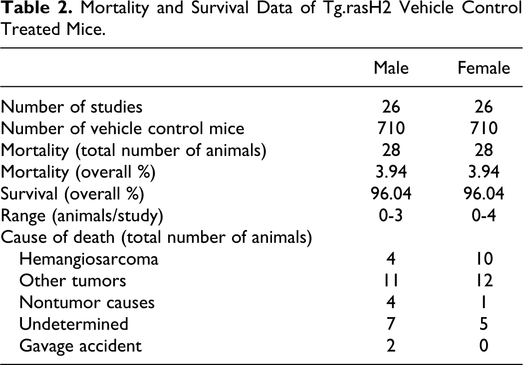
Mortality in the urethane-treated Tg.rasH2 animals was consistently high in all the studies. From 1110 male and female animals examined, 28 animals died within 2 weeks of receiving the first urethane dose. Only 1 of these animals had an identifiable cause of death (lung adenoma). The remaining 27 animal deaths were likely caused by direct urethane toxicity. Mortality in the remaining urethane-treated mice increased sharply after week 10 of the study and all animals treated with urethane in all the studies were either found dead or sacrificed by week 19 due to moribundity. The typical signs in moribund animals included dyspnea, rapid and shallow breathing, or respiratory difficulty due to development of pulmonary tumors. In animals with large splenic tumors or other abdominal tumors there was often distension of the abdomen due to peritoneal effusion. The cause of death in over 90% of these animals was the combination of lung tumors and splenic hemangiosarcomas.
Body Weights
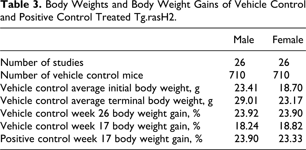
The average body weights of Tg.rasH2 male and female animals were similar in the vehicle control and positive control groups on the first day of dosing. The body weights of the control animals increased consistently from the first week of dosing until study termination (Figure 1). With the exception of the second week of each study, the average body weights of male and female positive control animals increased to a greater degree when compared to the age-matched control animals. The body weight gains of the positive control males and females were also higher than control animals at termination of the positive control animals during week 17 (Table 3, week 19 data not shown). The average body weights of Tg.rasH2 control male and female animals at the termination of the 6-month studies were 29.01 and 23.17 g, respectively. The body weight gains noted in these animals were similar in males and females (Table 3).
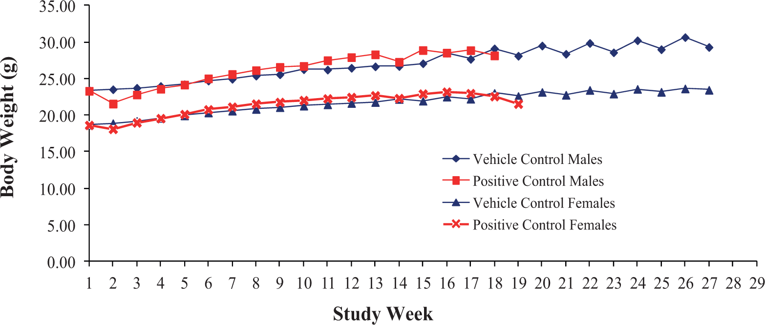
Average body weights of Tg.rasH2 animals treated with control or urethane (positive control) for up to 26 weeks of study.
Body Weights and Body Weight Gains of Vehicle Control and Positive Control Treated Tg.rasH2.
Spontaneous Lung Tumors in Tg.rasH2 Mice
The historical incidence of tumors from 710 male and 710 female Tg.rasH2 mice treated with vehicle (negative control) for 26 weeks is presented in Table 4. The incidence of tumors varied slightly between males and females, with a higher prevalence in males than females. Single alveolar bronchial adenoma of the lungs is the most common spontaneous tumor in both sexes of control mice (10.14% incidence in males and 5.77% incidence in females). The incidence of multiple adenomas in control male mice is 1.55% and 0.70% in control female mice while the incidence of alveolar bronchiolar carcinomas is 0.56% and 1.27%, respectively. When all primary pulmonary tumors (single adenomas, multiple adenomas, and carcinomas) are taken into account in the control male and female mice, their combined incidence is 12.25% in males and 7.74% in females (Table 4).
Incidence of Spontaneous Tumors in Tg.rasH2 Mice.
Abbreviation: NA, not applicable.
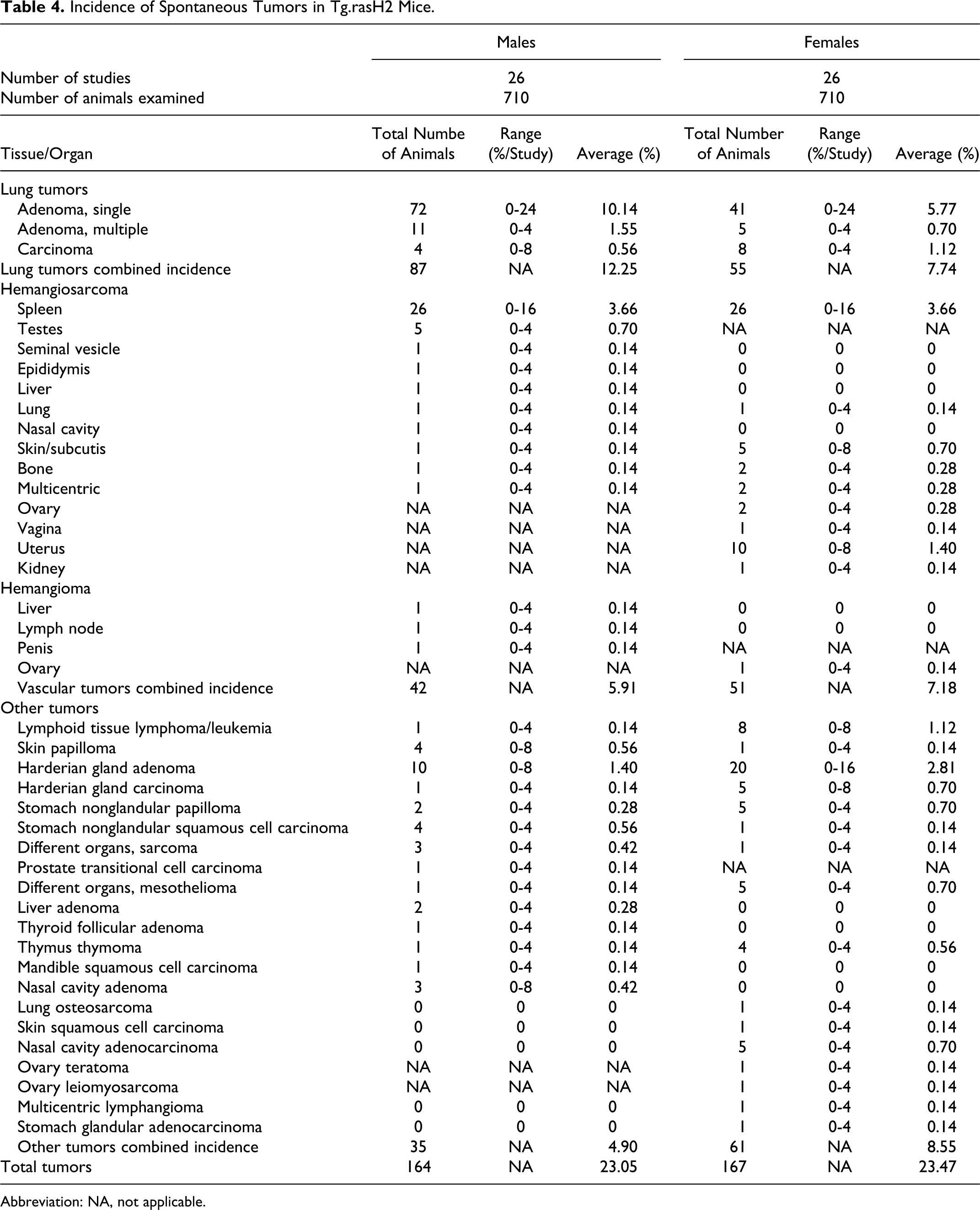
Grossly, the pulmonary adenomas are 3 to 8 mm in diameter and are noted as raised nodules on the lung surface. Tumors <1 mm in diameter are difficult to detect grossly, particularly, if they are embedded deeper in the parenchyma. Microscopically, typical alveolar bronchiolar adenomas are discrete lesions that compress the surrounding parenchyma (Figure 2). These tumors are solid in pattern and obliterate underlying alveolar structure. The tumor cells are cuboidal to columnar and form papillary projections or compact acinar structures. The cells are well differentiated, have uniform round to oval nuclei and abundant cytoplasm. The cellular outlines are usually distinct. Mitosis is usually not a feature of these tumors.
Representative micrograph of a spontaneous lung alveolar bronchiolar single adenoma at ×100.
Grossly, alveolar bronchiolar carcinomas are larger masses occupying part of the lobule, the entire lobule, and occasionally even multiple lobules, often with necrotic areas. Microscopically, alveolar bronchiolar carcinomas occupy part of the lobule, the entire lung lobule, or even multiple lobules and obliterate underlying alveolar architecture. Proliferating epithelial cells form multiple layers and often grow in solid clusters or sheets of cells. The cells have indistinct cellular outlines and there is marked cellular atypia characterized by pleomorphism. The nuclei may be round to oval and may acquire polygonal to spindle shape when the tumors are highly undifferentiated. Mitosis is usually moderate. Invasion of surrounding parenchyma and small islands of neoplastic cells detached from the primary tumor are commonly seen in the surrounding lung tissue. Occasionally, there is marked scirrhous reaction with foci of necrosis; secondary inflammatory changes are commonly noted. Metastasis of the carcinomas, even to the regional lymph nodes, is rare.
Spontaneous Splenic Hemangiosarcoma in Tg.rasH2 Mice
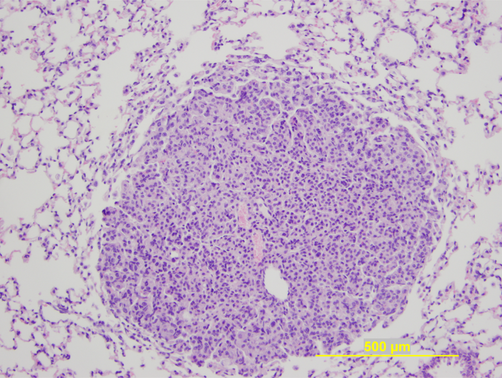
Spleenic hemangiosarcomas are the second most common spontaneous neoplastic lesion in the control Tg.rasH2 mice with identical incidence of 3.66% in both males and females (Table 4). In both females and males, only 1 study had an incidence of spleenic hemangiosarcomas greater than 8%. Grossly, the splenic hemangiosarcomas present as nodules, 2 to 7 mm in diameter, either red or tan in color, or depressed foci of similar color and dimensions. Microscopically, malignant endothelial cells in hemangiosarcomas often line vascular spaces that contain blood. The cells have polygonal to spindeloid hyperchromatic nuclei and sparse cytoplasm. The cellular outlines are usually distinct. However, in poorly differentiated tumors, the cellular outlines are indistinct as the tumors acquire solid patterns. The mitotic rate is usually mild to moderate. Metastasis or even implantation of these tumors in the peritoneal cavity is rare. Figure 3A and B provides representative microscopic images of splenic hemangiosarcomas.
A, Representative micrograph of a spontaneous splenic hemangiosarcoma at ×100. B, Representative micrograph of a spontaneous splenic hemangiosarcoma at ×400.
Spontaneous Hemangiomas and Hemangiosarcomas in Organs Other Than the Spleen in Tg.rasH2 Mice
Tg.rasH2 mice also occasionally develop hemangiomas and hemangiosarcomas in organs other than the spleen. These tumors present grossly as blood-filled nodules or masses. The microscopic features of these hemangiosarcomas are very similar to the ones described for the spleen above. Hemangiomas on the other hand are usually well circumscribed and are composed of blood-filled vessels that are lined by well-differentiated endothelial cells. The organs commonly affected include testes and uterus.
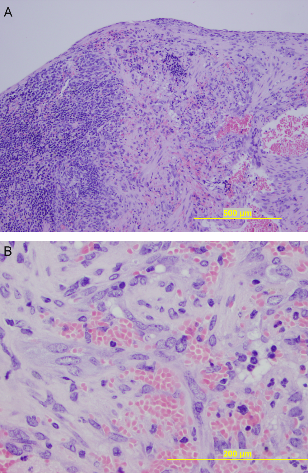
With the exception of the uterus and skin, the incidence of hemangiosarcomas in tissues other than the spleen ranged from 0% to 4% in each study. Although the average incidence is low, hemangiosarcomas of the reproductive tissues (testes, ovaries, and uterus) and skin are the most common in tissues other than spleen (Figure 4A and B). Hemangiomas have been observed in male animals and in the ovaries of female animals, with a range of 0% to 4% per study.
A, Representative photograph of an ovary with a grossly identifiable spontaneous hemangiosarcoma (left) and a normal ovary (right). B, Representative photograph of an epididymis with a grossly identifiable hemangiosarcoma (bottom) and a normal epididymis (top).
The combined incidence of all hemangiomas and hemangiosarcomas (splenic and nonsplenic) in males is 5.91%; whereas that in the female mice is 7.18% (Table 4).
Other Spontaneous Tumors in Tg.rasH2 Mice
There are only a few nonpulmonary and nonvascular spontaneous neoplastic lesions with a combined incidence of 4.90% and 8.55% in males and females, respectively (Table 4). Of these, only harderian gland adenoma has an incidence greater than 1% (1.4% in males and 2.81% in females). The occurrence of all other tumors is generally sporadic and their incidence is low.
Incidence of Tumors in Urethane-Treated Mice
To confirm the responsiveness of the Tg.rasH2 animal model to carcinogens, each study was conducted with a positive control group that was administered 3 doses of urethane at 1000 mg/kg/day, intraperitoneally. The lungs and spleens are the only organs examined based on historical data of target tissues in urethane-treated mice. The incidence of tumors in urethane-treated positive control mice is presented in Table 5.
Incidence of Lung and Splenic Tumors in Positive Control (Urethane) Treated Tg.rasH2 Mice.
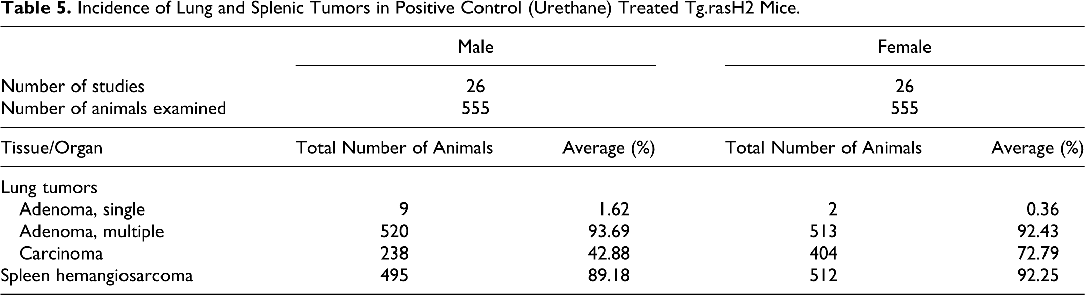
As shown in Figure 5, pulmonary tumors in the lungs grossly present as multiple nodules or masses in most urethane-treated animals. The incidence of multiple alveolar bronchiolar adenomas in urethane-treated animals is high, with a similar incidence in males and females (93.69% and 92.43%, respectively). On the other hand, the incidence of alveolar bronchiolar carcinomas in urethane-treated male mice is 42.88%, which is much lower than the incidence in the female mice (72.79%). Splenic hemangiosarcomas grossly present as nodules, often filled with blood (Figure 6). The incidence of hemangiosarcomas was similar in males and females, ranging from 80% to 100% in each study, with an average incidence of 89.18% in males and 92.25% in females.
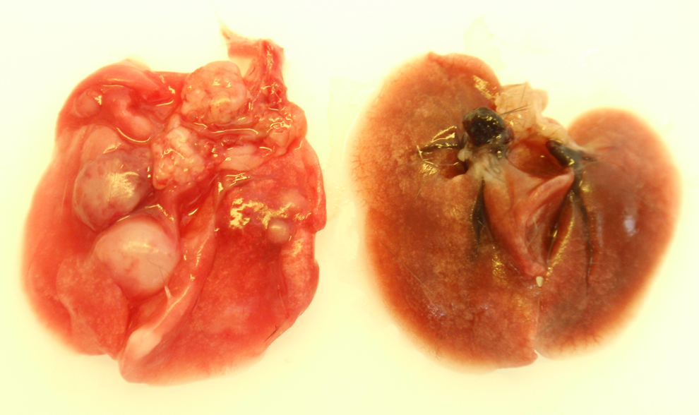
Representative gross photograph comparing the lungs from Tg.rasH2 mouse treated with urethane (left) or vehicle control (right). The lung on the left shows several nodules.
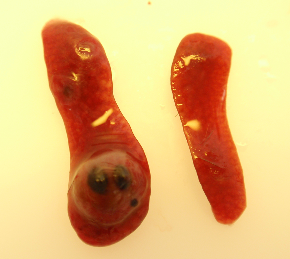
Representative gross photograph comparing the spleen from Tg.rasH2 mouse treated with urethane (left) or control (right). The spleen on the left is enlarged and shows one large nodule.
Discussion and Conclusions
As a humanized model for cancer risk assessment, the 6-month bioassay using Tg.rasH2 mice provides a carcinogenicity assessment system with many advantages over the conventional 2-year rodent assay. The 6-month bioassay provides significant cost savings and flexibility during drug development by shortening study duration (6 months in Tg.rasH2 vs 2 years in conventional mice), using fewer animals (210 in 6 months Tg.rasH2 vs up to 600 in 2-year conventional mice) and reducing overall necessary resources. Data from earlier Tg.rasH2 studies gave considerable confidence that this model produces low false positive and false negative rates.3,4 In addition, the combination of a 2-year rat study with a transgenic 6-month study has produced zero false negatives, while a combination of two 2-year chronic rodent bioassays in rats and mice produced a high number of false positives. 16
Despite the obvious advantages of this short assay, there is still some reluctance to its use, largely due to the absence of a historical database, lack of knowledge of unexpected tumor findings, lack of knowledge of tumor identification, and characterization and fear of false positives due to an inaccurate perception that transgenic animals are oversensitive.12,13 Herein, we have presented a large database summarizing the mortality, body weights, and the historical incidence of spontaneous tumors in 1420 male and female mice assigned to 26 studies conducted at our facility under identical conditions, representing the largest historical control data from a single laboratory.
The overall incidence of spontaneous tumors in our 6-month Tg.rasH2 studies is considerably lower compared to the conventional 2-year mouse models. The percentage of total spontaneous tumors in our studies is 23.05% in males and 23.47% in females. In comparison to the Tg.rasH2 mice, the percentage of total spontaneous tumors in 2-year B6C3F1 mouse studies is approximately 70% in both males and females17,18; whereas, the percentage of total spontaneous tumors in CD-1 male and female mice is around 78%. 19 Since spontaneous tumors interfere with the evaluation of the carcinogenic response, there is discussion of possibly completely eliminating 2-year bioassays by replacing the traditional chronic rodent carcinogenicity assays with a carcinogenicity battery that would include a transgenic study in mice along with a 12-month rat bioassay. 16
The starting age of the mice on the study is critical. In our studies, the starting age is between 6 and 10 weeks, although we prefer the starting age not to be more than 9 weeks. At termination of the 26-week study, the age of the mice is usually 34 to 36 weeks. This age at sacrifice is optimal since at the age of 40 weeks, mortality in Tg.rasH2 mice significantly increases, more so in females than in males, due to rapid development of spontaneous tumors. 6 The average mortality rate from all our 26 studies was very low and identical in males and females (3.94%); thus, over 95% of the animals survived to terminal sacrifice at 6 months. Our survival rates are similar to recently published data for Tg.rasH2 mice, wherein over 95% of the mice reached terminal sacrifice. 15 Survival of an adequate number of animals in carcinogenicity studies is very important in the statistical evaluation of the true carcinogenic potential. In comparison to the Tg.rasH2 model, the survival rate of control B6C3F1 mice in 2-year carcinogenicity studies conducted by the National Toxicology Program (NTP) is low, ranging between 60% and 77% in males and females17,18; while in CD-1 mice in the 2-year carcinogenicity studies, it is around 40% in both males and females. 19
The average body weights of Tg.rasH2 male and female mice at 26 weeks were 29.01 and 23.17 g, respectively. The average body weight gains in males and females were 23.92% and 23.90%, respectively. Our results are similar to those published before for Tg.rasH2 mice. 15 With the exception of the second week of each study, the average body weights of male and female positive control animals increased to a greater degree when compared to the age-matched control animals. The increase in the body weights of positive control mice compared to the age-matched vehicle control mice is due to formation of tumors in the lungs and spleen and effusions in the pleural and peritoneal cavities secondary to tumor formation.
In all our studies, we diagnosed lung alveolar bronchiolar adenomas as single or multiple (more than 1 tumor) in any given animal. We considered the multiplicity of the pulmonary tumors as an important criterion in distinguishing the aggressiveness of the tumors as well as an important feature in evaluating the effect of the test articles. This was particularly evident in urethane-treated animals in which most of the lung adenomas were multiple whereas most of the lung adenomas in vehicle control mice were single. Single adenomas were seen in only a few urethane-treated animals, mainly in animals that were removed early from the experiment either because of natural death or moribundity. Although the basic gross and microscopic features of alveolar bronchiolar carcinomas are in general similar in all Tg.rasH2 mice, the alveolar bronchiolar carcinomas in the urethane-treated female mice are more aggressive grossly and microscopically as compared to the urethane-treated male mice. Similarly, the incidence of alveolar bronchiolar carcinomas in urethane-treated female mice is much higher compared to the urethane-treated male mice. In the vehicle control mice, the incidence of single adenomas, multiple adenomas, and the combined incidence of all primary lung tumors (adenomas and carcinomas) in male mice is much higher than that in the female mice. On the other hand, the incidence of pulmonary carcinomas is almost double in female control mice compared to the male control mice. Our results are generally comparable to the ones noted by others, although the percentage of incidence varied slightly in each study.3,2,15
Grossly, spleen hemangiosarcomas in the control animals present as a single nodule or a single depressed focus of red or tan color, up to 5 mm in diameter. However, it is common to see either multiple nodules or multiple depressed foci in the spleens of urethane-treated animals. Often, there is concurrent enlargement of the spleen, usually in urethane-treated mice. The splenic enlargement is generally due to extramedullary hematopoiesis noted in nontumor portions of spleen. We also note that there is often gross enlargement of spleen due to microscopic evidence of extramedullary hematopoiesis in animals with a hemangiosarcoma in nonsplenic tissues. Interestingly, the incidence of splenic hemangiosarcomas in both males and females in our studies is identical at 3.66%. Once again our results are in general agreement with others.3,2,15 Tg.rasH2 mice also develop rare spontaneous hemangiomas and hemangiosarcomas in isolated organs other than spleen as shown in our studies. It is noteworthy that these tumors are more common in the testes and uterus.
In addition to the vascular tumors discussed, Tg.rasH2 mice develop other rare tumors, notably harderian gland adenomas. There has been very limited published data available pertaining to the presence of these tumors.3,2,15 Our database has identified a much larger spectrum of rare tumors, including nonsplenic hemangiosarcomas, hemangiomas, sarcomas, and mesotheliomas, although the incidence of each of these tumors is very low. The larger spectrum of rare tumors we identified is most likely because of the larger number of animals included in our database. Identifying rare tumors in a database is pivotal since rare tumors are usually assessed using less stringent statistical tools. A nonsignificant increase in the incidence of rare tumors may be of concern. Only when a large database of spontaneous tumors is available from the same laboratory that performs the study in question can the true significance of these rare tumors be determined. It can be concluded that such an apparent increase is truly biologically insignificant only if the incidence falls within the historical control database. Thus, in determining whether or not an apparent increase is biologically meaningful, any small change in the incidence noted in the historical control database may become important. Borderline increases in the incidence of tumors can be interpreted more appropriately when the historical database is taken into account. Caution should be exercised in using historical control data published from other laboratories, as stated by the US Food And Drug Administration. 20 Since interlaboratory differences in tumor incidence have been previously reported,2,21 it would not be appropriate to use a database generated at a different facility. Interlaboratory differences in the database often result from differences in study design (including the age of the animals, duration of dosing, and husbandry practices) and pathologists’ variability in diagnostic criteria and nomenclature used.
The purpose of the positive control in the Tg.rasH2 studies is to prove the validity of the assay. All previously published data on Tg.rasH2 mice used N-methyl-N-nitrosourea (MNU) as the positive control material.2,3,15 We have identified several advantages in the use of urethane over MNU. Urethane is a well-classified genotoxic–mutagenic rodent carcinogen and a possible human carcinogen. It is also a long-acting anesthetic and the signs and symptoms produced post dose verify the appropriate administration of the positive control. In addition, the dose formulation of urethane can be easily prepared with a 21-day stability when stored at 2 °C to 8 °C. 22 In retrospect, MNU is highly volatile, has a short stability period and should be used immediately after preparation. 23 Thus, depending on the method of preparation and handling of MNU, tumor response may be vary between studies. It is expected that the positive control material should produce the same types of tumors as those noted spontaneously. Thus, another major disadvantage of using MNU is that the most common tumors induced by MNU treatment in the Tg.rasH2 mice are stomach squamous cell papilloma/carcinoma, lymphoma, and skin papilloma, which are not the most common spontaneous tumors of the Tg.rasH2 mice. Lung adenomas and splenic hemangiosarcomas, the most common spontaneous tumors in the Tg.rasH2 mice, are induced at a relatively low rate by MNU.2,15 From our database, it is clear that over the past decade of using urethane, we have produced pulmonary and splenic tumors consistently in every study. Using urethane as the positive control material, the incidence of alveolar bronchiolar adenomas and alveolar bronchiolar carcinomas in lungs is 93.69% and 42.88% in males and 92.43% and 72.79% in females, respectively. In addition, the incidence of splenic hemangiosarcomas in urethane-treated males is 89.18% and 92.25% in females. When the combined incidence of all pulmonary and splenic tumors is taken into account the tumorigenic response in the positive control mice is almost 100%. This shows that the Tg.rasH2-urethane system is robust and proves the validity of assay beyond any doubts. The consistent high incidence of tumors in the urethane-treated groups has allowed us to reduce the number of animals in the positive control group from 25 to 10 animals/sex. The early onset of these tumors has also allowed us to reduce the length of study for these urethane-treated animals, from 17 weeks to 10 weeks. 24 Production of a high number of lung and spleen tumors with urethane allows us to examine only these 2 tissues microscopically; when MNU is used, a number of different tissues usually need to be examined microscopically.3,2,15
In conclusion, although statistical analysis remains a pivotal mechanism in analyzing results from short-term and long-term carcinogenicity studies, evaluating the incidence of tumor occurrence against an established historical control is useful in a variety of situations when statistical analysis alone could not establish the biological significance. The usefulness of a database is dependent on the sample size. Herein, we have presented the findings from our historical control database for the Tg.rasH2 mouse, which represents the largest set of data published for this strain. The low incidence of background tumors in the 6-month Tg.rasH2 model compared to the high incidence in a 2-year conventional mouse model allows us to more effectively draw conclusions about any treatment-related effects or lack thereof. The use of urethane as a positive control agent consistently produces pulmonary tumors (adenomas and carcinomas) and splenic hemangiosarcomas proving the validity of the assay. Since the establishment of the database in 2004, we have noted a very low variability in the incidence of spontaneous tumors between each of the studies and there have been no changes or differences in tumor incidence or trend, demonstrating the robustness of the animal model and the genetic stability of the strain, and confirms the reproducibility of the results.
Footnotes
Author’s Note
The authors Paranjpe and Elbekaei contributed equally for this article.
Declaration of Conflicting Interests
The author(s) declared no potential conflicts of interest with respect to the research, authorship, and/or publication of this article.
Funding
The author(s) received no financial support for the research, authorship, and/or publication of this article.