
Abstract
We aimed to evaluate the outcome of different treatment modalities for extremity venous thrombosis (VT) in neonates and infants, highlighting the current debate on their best tool of management. This retrospective study took place over a 9-year period from January 2009 to December 2017. All treated patients were referred to the vascular and pediatric surgery departments from the neonatal intensive care unit. All patients underwent a thorough history-taking as well as general clinical and local examination of the affected limb. Patients were divided into 2 groups: group I included those who underwent a conservative treated with the sole administration of unfractionated heparin (UFH), whereas group II included those who were treated with UFH plus warfarin. Sixty-three patients were included in this study. They were 36 males and 27 females. Their age ranged from 3 to 302 days. Forty-one (65%) patients had VT in the upper limb, whereas the remaining 22 (35%) had lower extremity VT. The success rate of the nonsurgical treatment was accomplished in 81% of patients. The remaining 19% underwent limb severing, due to established gangrene. The Kaplan-Meier survival method revealed a highly significant increase in both mean and median survival times in those groups treated with heparin and warfarin compared to heparin-only group (P < .001). Nonoperative treatment with anticoagulation or observation (ie, wait-and-see policy) alone may be an easily applicable, effective, and a safe modality for management of VT in neonates and infants, especially in developing countries with poor or highly challenged resource settings.
Introduction
Neonatal and infantile venous thrombosis (VT) although being rare has dramatically increased during the last decade by over 70%. 1 This pathology poses a major challenge to neonatologists, pediatricians, and surgeons with an increased morbidity, mortality and health-care costs. 2 The increased incidence may be attributed to the more aggressive care of neonates and infants with serious and life-threatening disorders. 1 –3 Prolonged use of intravenous (IV) catheters may be indicated by the occurrence of superficial thrombophlebitis and, less often, deep VT (DVT). Various indirect factors such as prolonged sepsis, trauma, and congenital heart diseases may trigger spontaneous thrombosis in the pediatric population. 4,5 In critically ill neonates and infants, central venous catheters (CVCs) are commonly used for parenteral nutrition/giving medication. These catheters are usually inserted in central or peripheral veins. However, the most frequently encountered complications associated with CVC insertion is the development of VT. The incidence of CVC-related thrombosis in neonates and infants depends on the type of the catheter used, the used diagnostic tests, and the index of suspicion for development of thrombosis. 6,7 Development of spontaneous/catheter-related VT may be associated with prothrombotic genetic factors compared to the general population. 8,9 Consequently, venous thromboembolism (VTE), despite being rare, may lead to gangrene of the extremities in those neonates and infants. Conservative management of this pathology is the best option for treating most of the cases with an ongoing debate regarding which treatment modality is the best. 10,11 The aim of this work was to evaluate the outcome of different treatment modalities for treatment of post-injection/infusion peripheral venous gangrene in neonates and infants, highlighting the current debate.
Methods
This 9-year retrospective study was performed from January 2009 until December 2017, after being approved by our institutes’ researcher board ethical committees. Infants and neonates were considered for enrollment if they were referred from the neonatal intensive care unit for having manifestations of upper or lower extremity threatening gangrene post-IV injection/infusion. Those patients’ files were thoroughly reviewed. Patients were classified into 2 groups; the first group (GI) included those who underwent the administration of unfractionated heparin (UFH) alone, whereas the second group (GII) included those who had the same regimen of UFH, plus the addition of warfarin (vitamin K antagonists). Clinically, neonates and infants were considered having extremity VT and consequently gangrene if they were presented with an extremity swelling, limb pain, cyanotic/hyperemic/tenderness of the affected arm or leg, or with a fixed color change. Also considered as VT, were the presence of subcutaneous collateral veins, (provided that there is a history of either IV injection/infusion of the affected limb), CVC dysfunction, as well as unexplained thrombocytopenia, hemodynamic disturbance, and arrhythmias. 12 –14 Excluded from this study were those patients with arterial thrombosis, cerebral sinus VT, renal vein thrombosis, hemolytic disorders, and those with preexisting/known prothrombotic risk factors (diagnosed by laboratory tests). Diagnostic imaging modalities included color Doppler ultrasonography, contrast venography, and computerized tomography scanning/magnetic resonance venography as described in the literature. 15 Furthermore, local treatment to the affected limb was adopted by removal of the IV line, cold fomentations, local antiseptic, and limb elevation. Systemic antibiotics after gaining blood culture were carried out. Following exclusion of intraventricular bleeding, patients are allocated to systemic anticoagulation therapy. Additionally, -pretreatment laboratory testing was conducted, including complete blood counting in addition to platelet count, activated partial thromboplastin time (aPTT), prothrombin time as well as the international normalized ratio (INR), alanine aminotransferase, aspartate aminotransferase, bilirubin total and direct as well as creatinine levels. As previously described, 16,17 all patients were conservatively treated with anticoagulants, namely UFH with a dosage of 75 to 100 units/kg over 10 minutes duration. Moreover and according to the recommendations of the American College of Clinical Pharmacy guidelines for heparin administration with prior warfarin intake, the required time to reach a therapeutic level ranged between 3 to 5 days due to the procoagulant transient effect. 18 –20 It was avoided in those children who had a significant risk of bleeding. According to the CHEST guidelines, the initial infusion stated with a dosage of 28 units/kg per hour, with infants (aged 2 months up to < 1 year corrected for gestational age); yet, we considered the individual risk factors when choosing initial dose. 21 –23 The adjustment of heparin maintenance dose was determined to maintain aPTT between 55 and 85 seconds. It was usually assessed within 4 to 6 hours of the loading heparin dosage and 4 hours post any change in the infusion rate. It was measured daily once the needed therapeutic levels were achieved. 22 Furthermore, we adopted the use of an initial warfarin dosage of 0.33 mg/kg aiming to reach a therapeutic INR level of 2.5 (ranging 2.0-3.0), according to the infantile INR nomogram 24,25 (Table 1). Heparin-induced major bleeding was treated by the transient termination of IV heparin infusion, in addition to the immediate administration of protamine sulfate and blood transfusion. The dose of protamine sulfate was determined according to the previously 2 hours of heparin administration. 21
Dose of Anticoagulants.

Statistical Analysis
Data were statistically analyzed using the Statistical Package for the Social Science (SPSS) version 23, IBM Corporation, Armonk, New York. Continuously distributed variables were summarized by the mean and standard deviation (SD) or median and range. In addition, odds ratios (ORs) and 95% confidence intervals (CIs) were calculated. Finally, the Kaplan-Meier survival analysis was used to graphically describe amputation-free survival and a P-value of < .05 was statistically significant.
Results
Sixty-three patients were included in this study. They were 36 males and 27 females with the male to female ratio of 1.3:1. Their age ranged from 3 up to 302 days with a mean of (72.10 ± 61.60). Most patients were premature 79% (n = 50), with a gestational age range from 32 to 34 weeks (mean = 33 ± 8; Table 2). Forty-one (65%) patients had VT in the upper limb (Figure 1), whereas the remaining 22 (35%) had lower limb VT (Figure 2). Out of those 41 patients with upper limb VT, 4 (6.5%) patients had only 1 finger gangrene. Three (5%) patients had gangrene affecting more than 1 finger, and 1 patient (1.5%) had gangrene of the whole hand. On the other hand, 4 (6%) patients with lower limb VT developed gangrene in 1 toe 3% (n = 2), multiple toes in 1.5% (n = 1), and whole foot gangrene in 1.5% (n = 1; Table 2). Seventeen (77.3%) patients underwent central venous catheterization inserted into the femoral vein whereas the remaining 5 (22.7) patients had a peripheral venous catheter. The overall success rate of the nonsurgical treatment adopted on all patients was 81% (n = 51). The remaining 19% (n = 12) patients underwent amputation due to an established gangrene. However, in GI, 46 patients who underwent treatment with UFH alone, 35 of them showed the success of the treatment, whereas in the remaining 11 patients the treatment was not successful necessitating finger, toe, or even limb amputation in. Alternatively, in GII, 16 patients showed a successful response to the combination of UFH and warfarin therapy with only 1 patient who underwent little toe amputation due to the failure of treatment (Table 3 and Figure 3). The average time of conservative treatment showed a range of 7 to 22 days (mean = 15.1 ± 1.6). The total mortality rate was 3 (5%) out of the total 63 patients. Postoperatively, 2 patients died due to severe sepsis whereas the remaining one expired before surgery due to severe complicated multiple congenital anomalies.
Patient’s Demographics and Lesion Characteristics.

Abbreviation: SD, standard deviation
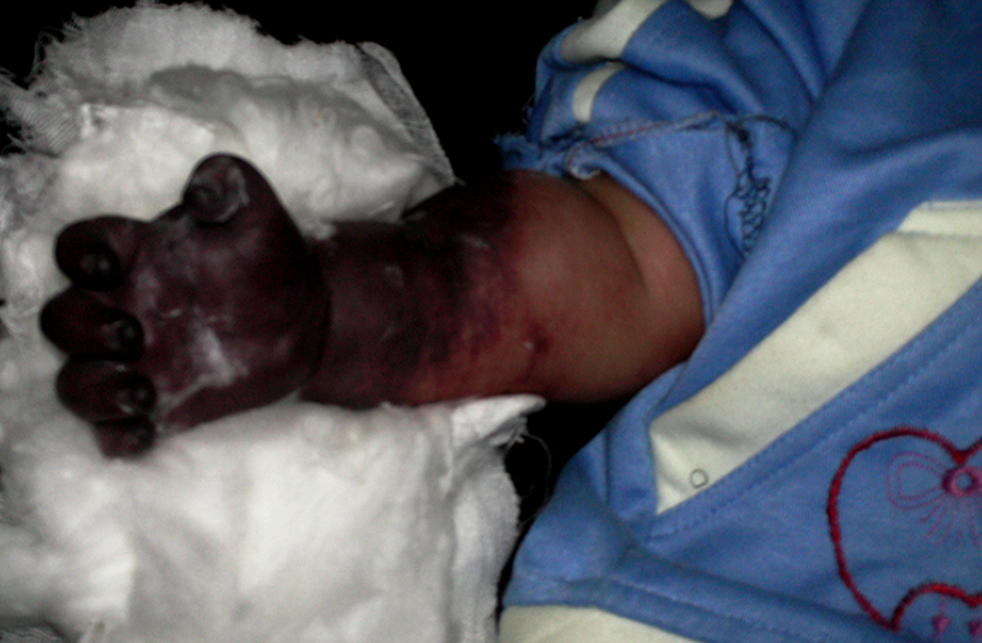
Gangrene of the right hand and right forearm.

Gangrene of the left foot and left leg.
Results of Treatment of Impending Gangrene.


Patients treated conservatively and those undergoing amputation.
Major bleeding events because of UFH administration were encountered among 2 (3%) patients in GI and in 1 (1.5%) patient in GII. Yet, warfarin-induced major bleeding was not encountered in any of our patients. Moreover, the absolute risk for the development of gangrene because of VT combining warfarin with heparin was about 6% compared to using heparin alone at 24%. Moreover, the cross-relation between the methods of treatment and the incidence of amputation revealed that 11 cases out of 46 treated with heparin had an amputation. On the other hand, only 1 patient out of those 17 treated with heparin and warfarin developed gangrene and underwent amputation. The OR was 4.065 and the CI was 0.56 to 29.15 (Table 4). This means that patients treated with UFH group (GI) were more vulnerable to amputation, approximately 5 times compared to those heparin and warfarin-treated group (GII). The mean and median survival time of the 2 groups with different treatment options are shown in (Table 5). Moreover, there was a highly significant increase in both the mean and the median survival times in the heparin and warfarin-treated group compared to the heparin-only group (P < .001), as depicted in (Figure. 4).
Cross Tabulation Between Treatment and Amputation in the Studied Groups.

Abbreviations: AR, absolute risk; CI, confidence interval.
Mean and Median Survival Times in the Studied Groups

Abbreviations: df, degree of freedom; SE, standard error.

Survival curve for the 2 studied groups using the Kaplan-Meier method.
Discussion
The highest risk of developing VT and consequently gangrene does exist among neonates and infants with critical illness. This may lead to changes in hemostatic balance toward thrombosis. 26 –28 Many factors may have contributed to the increased incidence of this condition up to almost 3-folds in the last 2 decades. The accused factors are mainly sepsis, liver dysfunction, peripheral and central venous lines, fluid fluctuations, and systemic inflammation. 1,29 Central venous catheter is one of the most common clinical factors that may be responsible for the development of VT and gangrene. 26 –28 Due to both increased plasma levels of plasminogen activator inhibitor and decreased plasma activity of plasminogen, the activity of the fibrinolytic system in the newborn is reduced compared to adults and older children. This fact may explain the high rate of VT associated with the insertion of intravascular devices in the newborns. 21
Treatment of a newborn with VT is usually difficult with the use of antithrombotic therapy. This may be due to the altered physiology and metabolism of the anticoagulants. 30,31 The current study reported 4 (6.5%) cases who underwent VT and consequently gangrene of the lower limb induced by CVCs. These data are contradicting with a 30% incidence of CVC-associated VT in a previously published report. 27 Moreover, venous thromboses are more likely pronounced in premature infants. 32 We also reported VT in 38.1% premature infants. This is comparable to other literature data, whereas preterm neonates accounted for 34% of the thrombosis. 26 Most of our patients were in the neonatal period (n = 55) 87%, coinciding with previously published reports that showed an incidence of neonatal thrombosis to be 2.4/1000 and 5.1/100.000, respectively. 13,30 It may be due to a higher hematocrit as well as the greater liability of the hemostatic system because of a general decrease in levels of coagulation factors and their inhibitors in neonates. 31,32 Neonatal extremity VT and consequently gangrene is usually treated conservatively aiming to prevent the occurrence of infection at the affected part, allowing the gangrenous area to declare spontaneously in order to optimize any future reconstruction. 33
The anticoagulant most commonly used in the treatment of neonatal VTE is UFH, although LMWH can also be used. The advantages of UFH include the potentially easier reversibility with protamine sulfate. 34 However, bleeding complication is the main risk-associated with heparin administration. The incidence of bleeding complication following anticoagulant administration in our series was reported in 3% and 1.5% in GI and GII, respectively. These results coincide and nearly similar to the previously published reports of 2% incidence of major bleeding using UFH, 28 although it contradicts to other literature data reporting an incidence that reached 24%. 29
The recommendations for treatment of adult VTE cannot apply to the management of neonatal and infantile VTE, as their vascular system, the hemostatic system, and comorbidities create a balance of hemorrhage and thrombosis. The severity of the thrombosis, the possibility of limb impairment, the presence of comorbidities, and the risk of bleeding all influence the decision to treat or to observe. Randomized trials evaluating type and duration of treatment are lacking in the neonatal population, and treatment decisions are largely based on consensus evidence-based guidelines. The anticoagulant therapy reduces the risk of thrombus extension and subsequently pulmonary embolism, while allowing the natural fibrinolytic system to gradually reduce the clot size. 35 Although controversy exists in the literature, as regard the best tool for conservatively treating VT and consequently gangrene, we reported 51 (81%) patients out of 63 patients with a good response to UFH despite not using LMWH because of the financial limitations in our settings. Nevertheless, previously published data from highly developed settings expressing both the Canadian and German experience suggested the use of thrombolysis and anticoagulation in the form of UFH and LMWH as the primary tool for treating this condition. Yet, it is difficult to verify which treatment modality is superior to the other. 10,13,30
In those 2 registries, the most frequently used drug was heparin as the anticoagulant of choice. Other Canadian research did use the LMWH and found out that it might have the advantages over the standard UFH. This may explain that neonates and infants might require LMWH as well as UFH. 36,37 Furthermore, monitoring of the therapeutic effect of LMWH requires the determination of anti-Xa, which is practically unfeasible in our settings.
Conclusion
Catheter-related VT—whether centrally or peripherally located—was frequently encountered in our series, despite their little number in the literature. This may be due to the presence of infection/inadequate nursing care owing to lack of training in proper insertion and hygienic care of the venous catheters in neonates and infants. Therefore, it is strongly recommended to adopt more aggressive programs on teaching nurses and paramedics indulged in the neonatal care on how to properly insert and hygienically care of venous catheters. Additionally, we highly advocate the initial aggressive therapy using heparin with early bridging with warfarin. This regimen is feasible, reliable, effective, and may be safe modality for management of neonatal and infantile extremity VT having good outcome results with a lower incidence of amputation, especially in developing countries with poor or highly limited-resource settings. The study is limited by the variability of vascular care policies and procedures among the different institutions in the study. Another issue is the lack of information on infants and children hospital course, and on associated diagnosis of familial or genetic thrombotic disease. These limitations may be solved by adopting a future prospective research on the topic.
Footnotes
Authors’ Note
AM, OMZ, IH, MAN, TAS, MA, AME, HA, ASD, MAB, MYZ, DEE, BEB, ASH, MN, MMA, KAR made a substantial contribution to the acquisition of the work. AM, OMZ, IH, MAN, TAS, MA, AME, HA, ASD, MAB, MYZ, DEE, BEB, ASH, MN, MMA, KAR performed the analysis and interpretation of data. AM, OMZ, IH, MAN, TAS, MA, AME, HA, ASD, MAB, MYZ, DEE, BEB, ASH, MN, MMA, KAR performed the acquisition of data. AM, OMZ, IH, MAN, TAS, MA, AME, HA, ASD, MAB, MYZ, DEE, BEB, ASH, MN, MMA, KAR revised it critically for important intellectual content AM, OMZ, IH, MAN, TAS, MA, AME, HA, ASD, MAB, MYZ, DEE, BEB, ASH, MN, MMA, KAR final approval of the version to be published. All authors read and revised the manuscript carefully for final publication in response to the reviewers’ comment. All procedures performed in study involving human participants were in accordance with the ethical standards of the institutional and/or national research committee and with the 1964 Helsinki declaration and its later amendments or comparable ethical standards. As this is a retrospective study, an informed consent in this article was not obtained.
Acknowledgments
The authors would like to thank Prof. Tarek D. Hussein, Professor of Zoology, Faculty of Science, Cairo University; for his effort for performing the statistical analysis for this article.
Declaration of Conflicting Interests
The author(s) declared no potential conflicts of interest with respect to the research, authorship, and/or publication of this article.
Funding
The author(s) received no financial support for the research, authorship, and/or publication of this article.