
Abstract
Currently, although the improvement of surgical techniques and the development of chemotherapy drugs have brought a certain degree of development to the treatment of osteosarcoma, the treatment of osteosarcoma has many shortcomings, and its treatment is limited. MiRNAs and exosomes can be used as diagnostic tools, and they play an important role in the occurrence and chemotherapy resistance of osteosarcoma. Therefore, providing a new method for the treatment of osteosarcoma is the key to solving this problem. To systematically summarize the research status of exoskeleton drug-loaded miRNA in osteosarcoma, we identified and evaluated 208 studies and found that exosome-carrying miRNA can be used as an index for the diagnosis and prognosis of osteosarcoma and share a certain relationship with chemosensitivity. In addition, exosomes can also be used as a carrier of genetic drugs able to regulate the progression of osteosarcoma. Based on the above findings, we propose suggestions for the future development of this field, aiming to bring new ideas for the early diagnosis and treatment of osteosarcoma.
Introduction
Osteosarcoma (OS) is a primary malignant tumor that occurs frequently among teenagers. Its clinical manifestations are mainly local pain and swelling. Currently, the main clinical treatment for OS is surgery combined with chemoradiotherapy, and first-line chemotherapeutic drugs used in OS treatment mainly include platinum, doxorubicin, and methotrexate. 1 In addition, cyclophosphamide, bleomycin, and pingyangmycin have also been applied in the clinic. 2 Although the application of these drugs has improved the prognosis of OS, the clinical outcomes of patients with OS are not ideal due to the widespread emergence of drug resistance. Therefore, solving the problem of OS drug resistance represents a main breakthrough in promoting the development of OS treatment. 1 Moreover, explaining the mechanism of OS resistance is helpful for the implementation of individualized precise treatment strategies in OS, which will bring more hope to OS patients.
Exosomes are vesicles with diameters of 40-100 nm that are secreted to the outside of cells after fusion of the cell membrane and the vesicles. Since its discovery in 1983, mechanisms defining the production, composition, transport, and delivery of exosomes have been widely reported, and increasing attention has been paid to its role as a carrier of endogenous information.
3
The ab initio synthesis of exosomes is related to the endosome maturation pathway. Early endosomes can be formed by plasma membrane invagination, and the fusion of early endosomes or endoplasmic reticulum, Golgi budding vesicles, and early endosomes can provide content for early endosomes. Early endosomes (Rab5 as a marker) form late endosomes (Rab7 as a marker) through acidification and material-exchange maturation. Late endosomes can eventually form multivesicular bodies (MVBs), which contain intralocular vesicles (ILVs), which are formed by inward depression of the MVB membrane. Multivesicular and lysosomal or autophagic lysosomal fusion results in material degradation, and fusion with the plasma membrane leads to the secretion of lumen vesicles to the outside of the cell, which are exosomes.
4
Exosomes have been identified in different types of body fluids (blood, lymph, urine, saliva, semen, and milk). They are considered an intercellular information transmitter because they carry cell-derived DNA, mRNA, miRNA, proteins, and lipids. Numerous studies have confirmed that exosomes from different sources play different roles in the treatment of diseases (Figure 1). In OS, exosomes are used as diagnostic markers, and have also been proposed as potential therapeutic targets due to their effects on the occurrence and progression of tumors. Production and transport of exosomal miRNAs. After the completion of transcription, translation, and other expression activities, miRNAs are enclosed into exosomes in the cytoplasm and excreted by the cells, and are then transported to the target cells through the liquid system in the body where they exert their physiological activity.
Noncoding RNAs are a type of RNA that lack a protein-coding function, and mainly comprises housekeeping and regulating noncoding RNAs. Noncoding RNAs are found in exosomes and play a role in cell proliferation, differentiation, migration, and apoptosis by regulating gene expression through different targets. 5 As a widely used regulator of noncoding RNAs, miRNAs play a regulatory role in many diseases. The expression of miRNA in OS cell lines or tissues is significantly different from that in found in normal organisms, and the change in miRNA content can regulate the progression of OS by regulating the expression of downstream targets and activating signaling pathways.
Based on the above characteristics, whether exosomes can be used as miRNA carriers to regulate the occurrence and development of OS and be further applied to the clinical diagnosis and treatment of OS is the key focus of this review. Herein, the role of miRNA, exosomes, and exosomes loaded with miRNA are reviewed and prospectively analyzed in terms of their potential application for the diagnosis and prognosis, progression, and chemotherapy resistance in OS.
Application of miRNA in OS Therapy
Effects of miRNA on proliferation, migration, invasion, and apoptosis of OS cells
Upregulated miRNA.

Notes: Five upregulated miRNAs in OS lines or tissues with clear research mechanisms and extensive research and their target genes, effects, and references.
Downregulated miRNA.

Notes: Five downregulated miRNAs in OS lines or tissues with clear research mechanisms and extensive research and their target genes, effects, and references.
The PI3K/Akt signaling pathway, activated by its upstream or bypass signals, acts on the downstream signaling, to promote cell growth and inhibition of cell apoptosis, which is closely related to the occurrence and development of tumors. miRNA-21 can inhibit PTEN expression, activate the PI3K/Akt signaling pathway, and promote OS cell proliferation. 11 MiRNA-133b inhibits the activation of the PI3K/Akt signaling pathway by targeting FGFR1, thus inhibiting the development of OS. 38
MiRNA-144 can further inhibit the activation of related signaling pathways and induces apoptosis of OS cells by inhibiting the synthesis of mTOR protein. 44
Effects of miRNA on Chemoresistance of OS
At present, the clinical treatment of OS is still a combination of surgery and adjuvant radiotherapy and chemotherapy, but for patients with OS in the middle and late stages, chemotherapy is the only choice to extend overall survival. 45 Therefore, the drug resistance of commonly used chemotherapy drugs has gradually become one of the biggest obstacles in OS treatment. Meanwhile, cisplatin (CDDP) and doxorubicin (DOX) are commonly used to treat OS, but CDDP resistance, DOX resistance, and multidrug resistance are very common. 1
Different types of miRNAs play different roles in the process of OS resistance.46,47 They can play a positive role in promoting OS resistance; although, some miRNAs can also have a negative role in inhibiting or even reversing OS resistance. The above effects are mainly mediated by regulating the expression of target proteins responsible for regulating the resistance of OS. 47
MiRNA-34a is a marker RNA that promotes multidrug resistance in OS in OS by inhibiting the expression of Dll1, CD117, and AGTR1.48-50 MiRNA-199a-3p targets the downregulation of AK4 expression, reducing the multidrug resistance of OS. 51 MiRNA-143 increases OS sensitivity to DOX by downregulating Atg2b and Bcl-2 expression. 52
Another important mechanism of miRNAs in OS resistance is the regulation of autophagy, which affects the sensitivity of OS to drugs. MiRNA-140-5p can upregulate HMGN5 and mediates autophagy by interacting with nucleosomes, thus promoting chemotherapy resistance. 53 At the same time, it can inhibit autophagy mediated by IP3K2, reverse the induction of chemotherapy drugs, and enhance the resistance of OS. 54
MiRNA-155 targets PTEN expression, enhances PI3K/Akt/mTOR signaling pathway activation, inhibits DOX-induced apoptosis and autophagy, and reduces OS sensitivity to DOX. 55 MiRNA-199a-5p can inhibit CDDP-induced autophagy and it enhances drug resistance by downregulating the expression of Baclin1. 56 In addition, a study by Li et al, 57 the authors knocked out miRNA-214 in order to determine whether miRNA-214 plays a role in the response to radiation therapy, and found that both in vivo and in vitro, miRNA-214 can enhance the radiosensitivity of OS, which is achieved by targeting PHLDA2 to inhibit the PI3K/Akt pathway. These studies indicated that, irrespective of anti-chemotherapy or anti-radiotherapy, the regulation of miRNA content can play a therapeutic role in OS.
Application of Exosomes in OS Therapy
Exosomes Can be Used as a Diagnostic Marker
To determine the characteristics of OS exosomes, Jerez et al. 58 analyzed the secretory proteins in the conditioned medium of three human OS cells and isolated and identified exosomes containing soluble proteins and supernatants without exosomes. In this study, they found that tumor-derived exosomes play a dual role: tumor-derived exosomes are involved in the development of tumors, but antigens expressed by exosomes are related to different invasive phenotypes; thus, exosomes can be regarded as antigen targets of tumor cells. Based on the above evidence, it is possible that exosomes can be used as biological indicators of tumor diagnosis and prognosis and may become potential targets of treatment. Additionally, exosomes are widely present in body fluids and are easily separated from peripheral blood and other body fluids; therefore, they are noninvasive diagnostic tools. Exosomes derived from malignant cells and other affected organs present in the blood are called circulating cancer exosomes, which can be identified in liquid biopsies because of their similarities with tumor cells. Brady et al. 59 isolated exosomes from the serum of eight dogs with OS, five healthy dogs, five dogs with traumatic fractures, and OS patients. The protein components of exosomes were analyzed using unmarked mass spectrometry. The exosomes of OS patients and those of OS dogs were highly similar. This indicates that the exosomes from OS share a close similarity between experimental animals and human beings, suggesting that the results from animal models can be further transferred to clinical research. In addition, during the development of OS, the components of exosomes collected at different time points are significantly different. This indicates that the component analysis of exosomes in different disease courses can be used as a tool for early diagnosis and clinical staging of OS, and this effect can be extended from dogs carrying cancer to human patients with OS.
Exosomes Secreted by Different Cells Regulate the Progress of OS in Different Ways
As an important carrier of information interaction in the tumor microenvironment (TME), exosomes can be produced and secreted by multiple cells.3,60 At present, some studies have clarified the mechanisms involving OS cells-associated with exosome transport to shape their supportive TME and promote growth and vascularization. In specific exosomes, miRNA-148a and miRNA-21 are known to help shape the TME.61,62 The TME is conducive to the proliferation and metastasis of OS cells and provides a foundation for the development of OS. Therefore, by regulating the production, secretion, and delivery of exosomes, tumor progression can be regulated by affecting the TME.63,64 At the same time, exploiting the natural targeting of exosomes in gene therapy
65
and drug delivery
66
seems to be a feasible scheme, which will revolutionize the development of OS treatment. Exosomes secreted from different cells have different functions that are activated through different regulatory pathways. There are three main regulatory approaches (Figure 2). Different functions of exosomes secreted by different cells. Exosomes secreted by different cells can regulate the prognosis of OS in different ways: (i) they directly regulate the proliferation and apoptosis of OS cells by regulating the signaling pathways; (ii) the promote OS by altering the tumor microenvironment, promoting bone resorption and tumor angiogenesis; and (iii) they influence the occurrence and development of OS by regulating the immune response.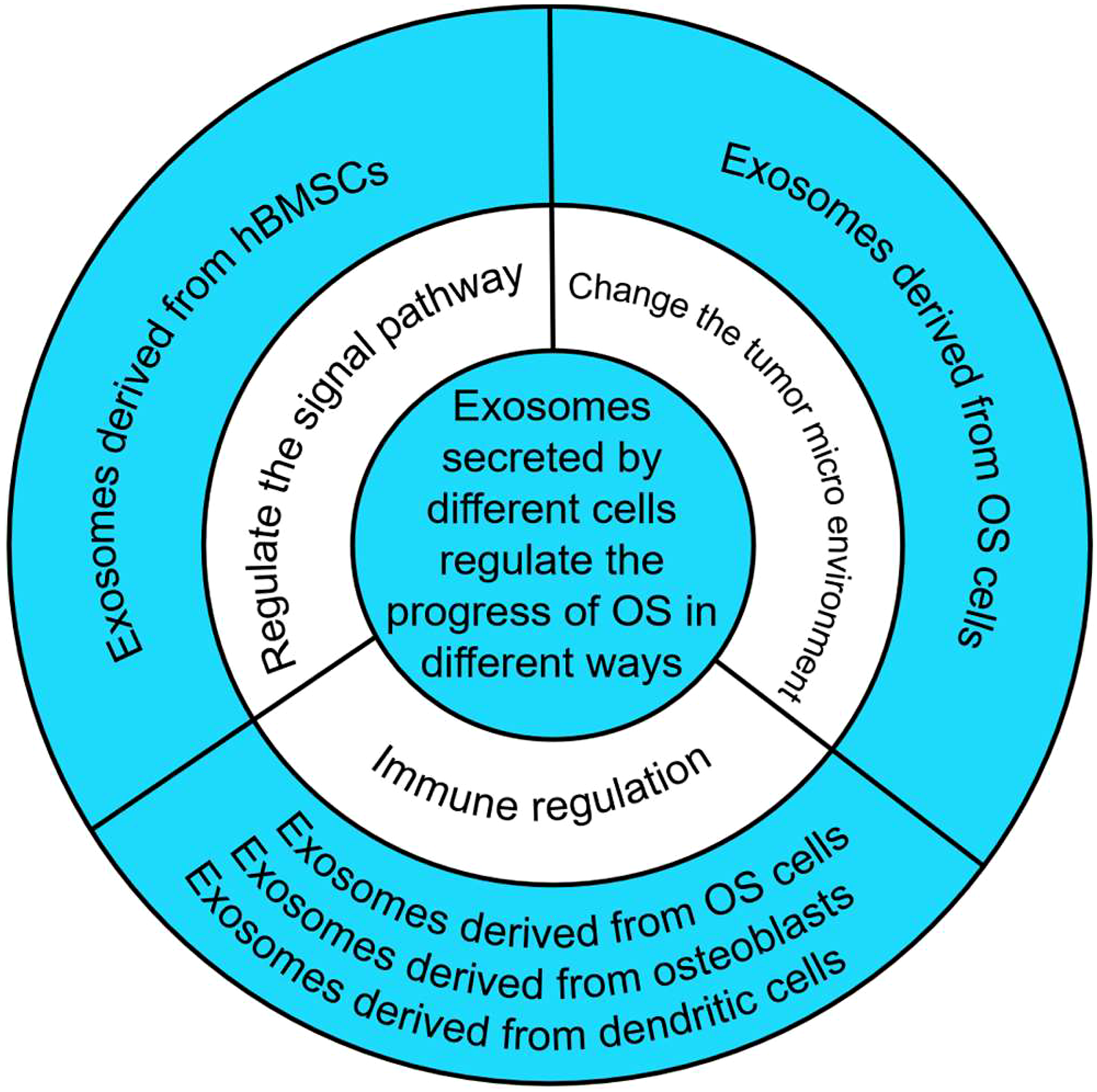
(i) Exosomes can directly regulate the proliferation and apoptosis of OS cells by regulating signaling pathways. HuBMSCs transfer exosomes to cancer cells. Exosomes derived from hBMSCs activate the Hedgehog signaling pathway in OS cell lines, thereby increasing the proliferation of OS cells and promoting OS growth. 67
(ii) By changing the tumor microenvironment, bone resorption and tumor angiogenesis can be promoted, which in turn promotes OS. The tumor microenvironment plays an important role in tumorigenesis and tumor development. The main cell components and activities include fibroblasts and osteoclasts, and immune cell mesenchymal cells and neovascularization. In the development of OS, osteoclast proliferation enhances osteolysis and enhances the destruction of OS to bone. Tumor blood vessels provide the demand for rapid growth of the OS tissue. 68 Raimondi et al. 69 found that OS-derived exosomes promote osteoclast gene expression, induce osteoclast maturation, and enhance absorption activity. It can also stimulate the release of angiogenic factors, induce tumor angiogenesis, and promote the progression of OS.
(iii) OS progression is regulated by immune regulation. Troyer et al. 70 isolated exosomes from dog OS cells and healthy osteoblasts, determined their protein components, and evaluated the direct impact of these two sources of exosomes on the dog immune system. It was found that the exosomes derived from OS contain protein components that inhibit immune function, including TGF-β, α-fetoprotein, and heat shock protein. These exosomes can directly reduce the rate of T cell proliferation, promote the differentiation of CD4+ cells into a T-regulated phenotype (Foxp3+), and reduce the expression of CD25+ on the surface of CD8+ cells, thereby inducing immunosuppression and promoting the immune escape of OS cells. Osteoblast exosomes can also reduce T cell activity, but they are not as marked as OS-derived exosomes and do not promote the T-regulatory phenotype due to the lack of TGF-β. Dong et al. 71 found that osteosarcoma-derived exosomes can induce T cell proliferation and cytotoxic T cell responses by loading tumor-derived exosomes with dendritic cells in vitro, thus inhibiting the growth of OS cells.
Application of Exosomes Loaded miRNA in OS Therapy
miRNAs in Exosomes Can be Used as Biomarkers for OS Diagnosis and Prognosis
There are several free miRNAs in the blood of healthy and tumor patients. Many researchers collected blood samples from OS patients and healthy individuals and detected the content of miRNA in serum by PCR. It was found that 21 miRNAs in the serum of OS patients were altered, including 13 upregulated miRNA (i.e., miRNA-17, 72 and 8 were downregulated (i.e., miRNA-195 73 ). Compared with healthy individuals, both upregulated and downregulated serum miRNAs are associated with tumor volume, distant metastasis, advanced clinical stage, short overall survival period, and poor prognosis. This suggests that serum miRNA can be used as a diagnostic and prognostic indicator of OS. However, the expression of miRNA in the serum is not stable, which raises additional research questions.
Given the lipid or lipoprotein complexes present in the blood circulation, apoptotic bodies, microbubbles, and exosomes can resist RNA degradation enzymes, and circulate stably exist in the blood. 74 Fujiwara et al. 75 purified exosomes from a number of OS cell lines (U2OS, HOS, 143 B, and SAOS2) and MSCs, and analyzed the expression of miRNAs by qRT-PCR. Both miRNA-17-5p and miRNA-25-3p were found to be enriched in exosomes and both were upregulated in OS cells. This indicated that miRNA-17-5p and miRNA-25-3p could not only exist stably in serum but could also be used as diagnostic biomarkers. Li et al. 5 determined that miRNA-744-5p expression in the exosomes of OS patients was higher than that in healthy individuals. In addition, exosome miRNA is clinically important in predicting OS in patients with poor response to chemotherapy. 76 This confirms our conjecture that in the future, the detection of miRNA content in exosomes may be a new biomarker for the diagnosis and prognosis of OS.
Relationship Between Changes of miRNA in Exosomes and the Chemosensitivity of OS
Xu et al. 76 selected 31 healthy individuals as controls, 25 patients with good response to OS, and 28 patients with adverse reactions to OS. The expression of 746 miRNAs was analyzed using TaqMan miRNA array. The authors found that 12 miRNAs were upregulated and 18 miRNAs were downregulated in exosomes of OS patients with poor chemotherapy response compared with those of OS patients with good chemotherapy response. Further analysis showed that the levels of miRNA-124, miRNA-133a, miRNA-199a-3p, and miRNA-385 in serum exosomes of patients with adverse reactions to chemotherapy were significantly decreased, while the contents of miRNA-135b, miRNA-148a, miRNA-27a, and miRNA-9 were significantly increased. This suggested that the differential expression of miRNAs in exosomes was closely related to the adverse effects of chemotherapy. In addition, we also found that the differentially expressed miRNAs in the exosomes were enriched in different biological pathways, among which the Hippo signaling pathway, PI3K-Akt signaling pathway, Ras signaling pathway, ubiquitin mediated proteolysis, choline metabolism, and other biological pathways were highly correlated with differentially expressed miRNA.
Exosomes as Carrier of Gene Therapy in OS Therapy
MiRNA-208a in exosomes derived from BMSCs can enhance the activity of OS cells, promote clone formation and migration ability of OS cells, inhibit the expression of 3'-UTR by targeting PDCD4, and activate the ERK1/2 pathway, thereby enhancing the invasion of OS. 77 Wang et al. 78 identified 13 types of miRNAs using miRNA chip analysis and found that these miRNAs were significantly increased in exosomes derived from cancer-related fibroblasts (CAFs) and corresponding para-cancerous fibroblasts (PAFs). In addition, CAF can transfer miRNA-1228 to human OS cells through exosomes, promoting the proliferation and migration of OS, which is achieved by targeting the inhibition of SCAI expression. Instead, miRNA-675 in exosomes from metastatic OS promotes the proliferation and migration of stromal cells by downregulating CALN1. Moreover, tumor-derived exosomes act as messengers between metastatic cancer cells and stromal cells, and remodel the tumor microenvironment (TME) in OS by transferring miRNAs. 79 In addition, miRNA-135b can be isolated from OS-derived exosomes. Therefore, if OS cells can capture the exosomes secreted by immune cells, we can inhibit the proliferation of OS cells by exploiting natural exosomes. 76
All of these studies suggest that exosome-loaded miRNAs can regulate OS progression by regulating OS proliferation, invasion, or drug resistance. Therefore, we can speculate that artificially overexpressing or inhibiting some miRNAs and using exosomes as carriers to target the proliferation or invasion of OS cells, may represent a potential approach to prevent the progression of OS. Shimbo et al. 80 proved that our conjecture is feasible. They introduced a synthesized double-stranded miRNA-143 into BMSCs and found that the secretion of exosomes containing miRNA-143 increased and miRNA-143 was transferred to the outside of exosomes, which was then captured by OS cells, inhibiting their migration. This suggests that miRNA-143 in exosomes can be effectively transferred to receptor cells and to exert their activity. Therefore, exosomes can be used as carriers for gene therapy to regulate OS progression.
Discussion
Based on the in-depth review of current research, we believe that exosome-loaded miRNA not only can be used as a noninvasive biological marker for the diagnosis, prognosis of OS and the evaluation of resistance to chemotherapy, but these exposomes can also express human miRNAs. Further, these exosomes can be used as an important carrier of targeted therapy to achieve precise regulation of OS progression compared with treatment with miRNA or exosomes alone. Although there are few studies in this area, a novel approach to OS therapy may involve loading exosomes with miRNAs that can inhibit the growth of OS and enhance the sensitivity to chemotherapy drugs in the future.
Nonetheless, the following concerns should be addressed prior to the application of exosomes loaded with miRNAs for the treatment of OS. (1) Removing bioactive substances from exosomes that do not play a therapeutic role, (2) Determining whether ectopic expression and load have side effects and whether there are ethical concerns. (3) Ensuring the integrity of the carrier and miRNAs in the transfer mechanism (4) The results of in vitro and animal experiments can be clinically converted.
If the above four problems can be resolved, then exosomes loaded with miRNA will play a fundamental role in the treatment of OS and will provide additional possibilities for clinical treatment.
Conclusions
Through the evaluation of existing studies, it is easy to demonstrate that exosomes loaded with miRNA have great potential for the diagnosis, prognosis evaluation, and pre-assessment of chemotherapy resistance of OS. However, there are still many problems to be resolved for the application of exosomes as a targeted therapy carrier for OS; which requires more research to demonstrate the feasibility of this approach.
Footnotes
Declaration of Conflicting Interests
The author(s) declared no potential conflicts of interest with respect to the research, authorship, and/or publication of this article.
Funding
The author(s) disclosed receipt of the following financial support for the research, authorship, and/or publication of this article: This work was supported by the Lanzhou University Second Hospital Cui Ying Student Research and Cultivation Program Funding (CYXZ2020-03 and CYXZ2021-01).
Author’s Note
No animal experiments or clinical trials were carried out in the course of this study.