
Abstract
Polioencephalomalacia (PEM) is a neurologic disorder of ruminants that can affect cattle as a result of high levels of sulfur in the diet. Sulfur can be ingested by an animal through feed or water, and the mineral status of the animal can affect how much sulfur is ultimately absorbed. Broiler (poultry) litter is sometimes used in cattle diets as an economical protein source, although it runs the risk of supplying too much sulfur to the bovine diet. Here we report a case in which 15 cows had neurologic signs before death. Initial testing ruled out other causes of acute death, and histologic findings confirmed a diagnosis of PEM. The sulfur concentration was ~0.70% of the total mixed ration (TMR), which is above the maximum tolerable dietary concentration of sulfur. The broiler litter contributed nearly all of the sulfur to this ration and was the cause of the PEM experienced in this herd. When feeding by- and co-products as part of a TMR, it is important for producers to analyze the feed to mitigate risk and ensure good health.
Polioencephalomalacia (PEM), also referred to as cerebrocortical necrosis (CCN), is a neurologic disorder primarily observed in cattle and sheep that was first reported in the United States in 1956.12,13 Although the term polioencephalomalacia refers to necrosis of the brain’s gray matter (polio = gray), the syndrome PEM involves recumbency, blindness, seizures, ataxia, and necrosis in the cerebral cortex of affected animals.13,19 Initially, thiamine (vitamin B1) deficiency was identified as the inciting cause of this disease due to studies conducted in the United Kingdom and the well-documented positive response to systemic thiamine administration; however, this theory has since been questioned due to studies showing animals suffering from PEM with thiamine levels inside the reference interval.13,17 Additionally, cases in which thiamine deficiency was thought to be the etiologic agent of PEM were found to be caused by excess sulfur in the diet from water and sulfur-accumulating plants. 19 To make a diagnosis of sulfur-induced PEM, other disease processes need to be eliminated, and an elevated level of dietary sulfur intake needs to be identified. 19
PEM is classified as 2 disease forms, acute and subacute. The acute form is seen as recumbency, blindness, seizures, and death; the subacute form is less severe, with animals experiencing ataxia, visual impairment, and mild, permanent neurologic damage that may become more severe with time. 13 Grossly, key findings include changes associated with cerebral edema, such as cerebellar herniation, softening of the cerebral cortex, yellow discoloration of gyri, and, in some cases, autofluorescence of the cortex under UV light. 13 Histologically, necrosis of cerebrocortical neurons, typically in a laminar band, is a classic finding. In the early stages of PEM, changes include shrunken neurons with eosinophilic cytoplasm and faded, pyknotic, or absent nuclei. 13 Additionally, laminar cortical spongiosis, laminar neuronal necrosis, and plump reactive endothelial cells have been observed.13,15 In the late stages of PEM, findings include cerebrocortical cavitation with macrophage infiltration of tissue and the absence of autofluorescence. Severe cases at this stage will also have deep multifocal cerebral vascular degeneration. 13
Elevated dietary sulfur is a well-known inciting cause of PEM, which mandates careful monitoring of the diet. 13 The recommended concentration of sulfur in the diet of beef cattle is 0.15%. 17 The National Research Council (NRC) recommendation for the maximum tolerable concentration of sulfur was 0.40%, but this concentration has since been updated with 2 recommendations that depend on the animal’s diet. 8 If cattle have a diet composed of <15% forage and 85% concentrate, a sulfur concentration of 0.30% puts the animal at risk for sulfur-induced PEM; cattle with a diet of 40% forage can tolerate sulfur concentrations up to 0.50%. 8 It should be noted that water can contribute significantly to total dietary sulfur concentrations; thus, feed and water concentrations should be considered in tandem. Normal water sulfate concentrations for cattle should be <500 ppm; water that contains ≥2,000 ppm has been known to put cattle at risk for PEM. 18 Minerals in cattle diets, such as selenium, copper, and molybdenum, can also affect the animal’s net sulfur absorption through antagonism despite appropriate initial levels in the diet. 8
Sulfur, once ingested by ruminants, is metabolized in the rumen via sulfur-metabolizing bacteria, resulting in the production of hydrogen sulfide (H2S) gas. 11 H2S is eructated and inhaled, where it is absorbed in the lungs. As long as there is sulfur for rumen bacteria to metabolize and room in the gas cap, H2S gas is produced continuously and absorbed through the lungs. 11 H2S is also absorbed through the rumen wall into the blood where it is transported to the liver and metabolized to sulfate (SO4). 11 SO4 is then filtered by the kidneys and excreted into the urine. 6 The pH of the rumen is critically important in determining how much H2S is produced; the more acidic the environment, the more H2S is produced by rumen bacteria. 19 This explains the change in the NRC recommendations whereby animals on a low-roughage diet that is expected to result in a lower rumen pH can tolerate less sulfur than animals on a ration containing more roughage. An increase of H2S in the rumen gas cap went from 46.8% to 97.2% with a pH change from 6.8 to 5.2. 19 Excess sulfur causes inhibition of the electron transport chain, as well as ATP production through blocking cytochrome aa3. 11 With a high energy demand and a low number of antioxidants, the brain becomes the target organ for sulfur toxicosis, resulting in the CCN seen in animals with sulfur-induced PEM. 11
Another effect of sulfur that may contribute to animals developing sulfur-related PEM is the disruption of thiamine synthesis.1,2 Disruption potentially occurs by altering thiamine pyrophosphate, which is a key cofactor required in the formation of thiamine. Without this cofactor, the thiamine-dependent enzymes α-ketoglutarate dehydrogenase (α-KGDH) and pyruvate dehydrogenase (PDH) are unable to function in the production of energy in the brain. 1 Additionally, once H2S enters the brain it is oxidized to sulfite, which continues to be oxidized until sulfate is formed. Sulfate also decreases the concentration of α-KGDH, which can contribute to decreased thiamine production. 1 These are the leading mechanisms that explain why some animals with suspected sulfur-induced PEM respond well to thiamine supplementation. 1
Broiler litter is a by-product of the poultry industry that consists of bedding material, fresh excrement, partially decomposed manure, feathers, water, and leftover feed. 9 It has been used commonly as an economical protein source in cattle diets since the early 1950s. 20 The safety and efficacy of using broiler litter as a feed source for cattle has been examined, with the most common areas of concern being minerals, heavy metals, drugs and their metabolites, and microbial contamination. Specifically, copper and calcium have been cited as the elements of main concern. 20 Another common concern is how crops treated with broiler litter as a fertilizer affect the amount of sulfur found in forage used in cattle diets. Although one study did not find any changes in the concentration of sulfur found between broiler litter–treated plants and untreated plants, broiler litter has been noted to increase forage sulfur levels when used as a fertilizer.3,21
Sulfur is added to broiler diets to help reduce the pH of the litter produced. 4 The goal of this treatment is to reduce footpad lesions in birds, but the amount of sulfur present in poultry litter is hence increased, and the litter may ultimately be ingested by cattle. 4 Additionally, sulfur levels may increase due to the common practice of treating broiler litter with alum (Al2[SO4]3·14H2O) to help prevent the volatilization of ammonia (NH3), and the runoff of phosphorus (P) into the environment. 3 Alum acts as an acid, producing H+ ions in the litter once it dissolves. 14 These H+ ions form ammonium (NH4+) after interacting with the NH3 present, and the subsequent NH4+ is then free to form ammonium sulfate ([NH4]2SO4) after reacting with the sulfate ions present in the litter. 14 As a result, the sulfur concentrations of a cattle diet containing broiler litter can be elevated sufficiently to result in clinical disease.
In a literature search of PubMed, Google Scholar, Web of Science, CAB Direct, and AGRICOLA with the search terms “poultry litter”, “sulfur”, and “polioencephalomalacia”, we found only one suspected case of sulfur-induced PEM associated with poultry litter, with no cases in cattle. The case described 9 outbreaks of PEM in goats and sheep in northeastern Brazil, with no inciting cause identified. 16 One of these outbreaks was suspected to be related to the high sulfur concentration of the protein-mineral mixture, which contained 30% poultry litter with a sulfur content of 0.39%. 16
In a case submitted to Kansas State’s Veterinary Diagnostic Laboratory (Manhattan, KS, USA), 40 of a herd of several hundred head of mixed-age cattle on pasture developed clinical signs of ataxia, head pressing, blindness, and slobbering over 2 d, resulting in 15 dead. The remaining clinically affected animals responded positively to parenteral thiamine administration. Because of drought, the pasture had provided a minimal amount of poor-quality forage; therefore, although unusual in this area, a total mixed ration (TMR) was formulated to meet the nutritional demands of the animals and was supplied as the total diet. This ration was removed from the cattle once the ill animals were noted, and no new cases developed. The TMR contained 50% poultry litter, 40% soybean hulls, and 10% gin trash (a byproduct of cotton gins). 10 The cattle had ad libitum access to multiple water sources scattered throughout the property.
Autopsies were performed on the farm by the referring veterinarian with no gross lesions identified. Samples were submitted from 2 animals, cows 1 and 2, which included fresh rumen content, ocular fluid, brain, and serum, in addition to fixed lung, liver, heart, kidney, spleen, and muscle tissue. Tissues from 3 other autopsies included rumen content and ocular fluid only. Grossly, the brains had subtle bilateral yellow discoloration of cerebrocortical gray matter, most prominent in sulci but also gyri of the parietal and occipital lobes. One of the brains had prominent bilateral thalamic hemorrhage (Fig. 1A). Under UV light in the laboratory, the cerebral cortex autofluoresced (Fig. 1B).
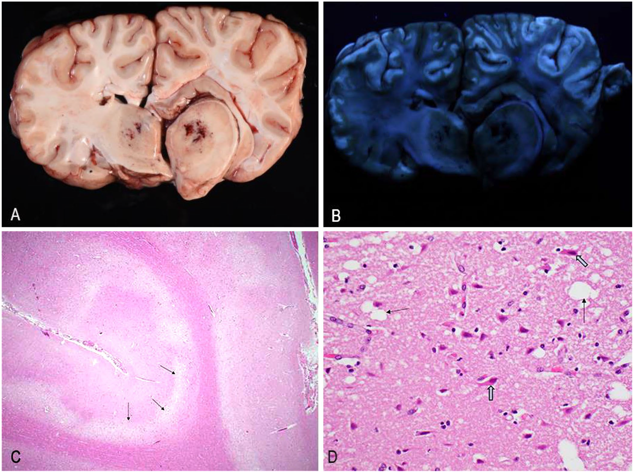
Gross and histologic brain lesions observed in polioencephalomalacia in cows 1 and 2.
Histologically, the brain of cow 1 had mild-to-severe, acute deep laminar neuronal necrosis (Fig. 1C), perivascular and neuropil edema or spongiosis, thalamic perivascular hemorrhage, and minimal ventral neutrophilic meningitis. Similarly, the brain of cow 2 had mild-to-severe, cerebral mid-to-deep laminar neuronal necrosis (sulci and gyri) with regional and perivascular edema, and rare small vessel fibrinoid degeneration. These sections also had areas of prominent perivascular and periglial clear space throughout the white matter attributed to both edema and likely autolysis as brains were shipped fresh. Additionally, thalamic neuronal necrosis, neuropil rarefaction (Fig. 1D), and perivascular hemorrhage were present. Glial changes were limited to rare swollen nuclei (presumptive astrocytes) with marginated chromatin.
To discern the cause of PEM and to rule out causes of acute death, ocular ammonia, liver lead, and brain sodium were tested (Table 1). All results were within normal limits. Sulfur concentration analysis of the TMR was performed at Iowa State’s Veterinary Diagnostic Laboratory (Ames, IA, USA; 5800 inductively coupled plasma optical emission spectrometer, Agilent). The total concentration of sulfur in the TMR was ~0.70%, which is above the maximum tolerable dietary concentration of sulfur (0.30–0.50%). To ascertain which commodity was contributing the sulfur, the ration was manually separated into gin trash and poultry litter. The soy hulls were not easily separated from the smaller particles of the other constituents, so a sample labeled “mids” was analyzed. This sample type was similar in makeup to the TMR as all constituents were present. The poultry litter contained 10,227 ppm (1.02%) sulfur, compared to 134 ppm (0.013%) in the gin trash and 3,376 ppm (0.33%) in the “mids.” Thus, the poultry litter contributed nearly all of the sulfur to the TMR, which was measured at 6,593 ppm (0.659%). Analyzing water sources for sulfur concentration is also necessary to estimate the total dietary intake by the animal. Water was not submitted for analysis in this case, but any sulfur in the water would only be additive to the TMR which already exceeded tolerable dietary concentrations. The offending diet had been suspended immediately from feeding, and no additional PEM cases had developed.
Results of the laboratory tests run on the provided samples for cows 1 and 2, and the 3 other affected cows.

ND = not done.
As drought continues to limit more conventional feed sources, producers need to know about the risk of sulfur-induced PEM associated with poultry litter, especially given its attractive nature as a cheaper source of protein. Although it is not uncommon for by- and co-products from other industries to be used as sources of feed for animals, producers should be aware that these products can be extremely variable due to no formal regulation regarding their use.5,7 To eliminate the risk of PEM from excess sulfur in the diet, nutrient analysis of TMRs should be utilized before using poultry litter as a feed source.