
Abstract
A 5-y-old, Piedmontese cow had a 4-mo history of ongoing development of skin masses. This was the only cow affected in a herd of 20 cows. Up to 12, hairless, red-to-black, raised nodules-to-plaques were distributed along the dorsum and tail head. Biopsies were taken for histopathology and ancillary testing. An ulcerated skin section contained dermal infiltrates of eosinophils, plasma cells, neutrophils, macrophages, lymphocytes, and multinucleate giant cells, and pyogranulomas. Fungal hyphae were seen within the dermis, multinucleate giant cells, and pyogranulomas. In pyogranulomas, fungi were surrounded by a Splendore–Hoeppli reaction. Dematiaceous (pigmented) hyphae were rarely observed with H&E-stained and unstained (cleared and mounted) sections, but stained well with a Fontana–Masson stain. Exserohilum mcginnisii was identified by fungal culture, followed by PCR assay and sequencing. Exserohilum is a dematiaceous fungus that causes disease in humans and rarely in animals. The use of unstained sections and Fontana–Masson stain are important to demonstrate pigment because dematiaceous fungi have little melanin and appear as hyaline hyphae histologically. PCR assay and sequencing aid in the differentiation and classification of fungal species. To our knowledge, E. mcginnisii dermal granulomas have not been reported previously in cattle.
Exserohilum (Pleosporales, Pleosporaceae) is a dematiaceous (pigmented), filamentous, environmental fungus that causes a spectrum of diseases in humans and rarely in animals. 6 Exserohilum spp. fungi are common in grass and soil, less common in marine environments, and are considered cosmopolitan pathogens, occurring mainly in warm tropical and subtropical regions. 6 Infection with this fungus attained significance in the human medical field when Exserohilum rostratum was involved as the cause of the largest reported outbreak of infections associated with contaminated steroid injections in humans that resulted in 63 deaths and 749 confirmed drug-related infections in 2012–2013 across 20 states in the United States.2,15 Three of the ~35 species of Exserohilum have been reported as causes of human disease: E. rostratum, E. longirostratum, and E. mcginnisii.2,6 In a review of 48 cases of natural infection with Exserohilum before the 2012 U.S. outbreak, most infections were caused by E. rostratum (60.4%), followed by E. longirostratum (6.3%), and E. mcginnisii (2%); in 31.3% of the cases, the species was unidentified. 6 Human cases are observed as systemic, cutaneous, corneal, and subcutaneous infections. 6 An unidentified species of Exserohilum has been reported as the cause of dermal granulomas in cattle, 16 and E. rostratum as the cause of rhinitis in horses 10 and goats. 3 We report here the clinical, histopathologic, and microbiologic findings of dermal granulomas caused by E. mcginnisii in a cow.
A 5-y-old, multiparous, pregnant, Piedmontese cow had a 4-mo history of ongoing development of raised skin masses. This was the only cow affected in a herd of 20 cows. At clinical examination, the animal had a 6 of 9 body condition score (9-point scale) and was bright, alert, and responsive. The palpable lymph nodes were within normal limits. Up to 12, hairless, red-to-black, raised nodules-to-plaques were distributed along the dorsum and tail head (Fig. 1). The lesions were painful and fluctuant but firm when palpated. Clinical differential diagnoses considered by the referring veterinarian included melanoma, squamous cell carcinoma, warbles, and papilloma. An incisional biopsy from one of the nodules was taken by the referring veterinarian, placed in 10% neutral-buffered formalin, and submitted to the Tifton Veterinary Diagnostic and Investigational Laboratory, College of Veterinary Medicine, University of Georgia (Tifton, GA, USA). The formalin-fixed skin biopsy received was ~1.5 × 1.4 × 0.4 cm. Sections were prepared and stained with H&E, periodic acid–Schiff (PAS), Gomori methenamine silver (GMS), and Fontana–Masson (FM) stains. In addition, unstained (cleared and mounted) sections were made.
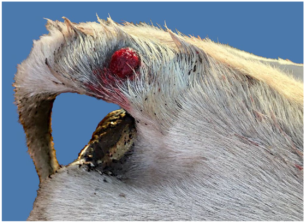
Exserohilum mcginnisii dermatitis in a cow. An ulcerated, red, round, and raised skin lesion on the tail head.
Histologically, an ulcerated skin section contained dermal infiltrates of eosinophils, plasma cells, neutrophils, macrophages, lymphocytes, and multinucleate giant cells, and pyogranulomas (Fig. 2A). Fungi were seen within the dermis, pyogranulomas (Fig. 2A, 2B), and multinucleate giant cells (Fig. 2C). In pyogranulomas, fungi were surrounded by a Splendore–Hoeppli reaction (Fig. 2B). Hyphae were septate, 2–6-µm wide, branched, with thick-walled, up to 25-µm vesicular swellings that resembled yeast-like structures, and were present singly or in chains and occasionally appeared to be budding. Dematiaceous hyphae were rarely observed in H&E-stained sections (Fig. 2D). Fungi stained well with GMS stain (Fig. 3A) and PAS reaction (Fig. 3B). Hyphae stained well with FM stain, demonstrating melanin pigment (Fig. 3C). A few pigmented hyphae were visible in unstained (cleared and mounted) sections (Fig. 3D). We diagnosed marked pyogranulomatous eosinophilic mycotic dermatitis. After the biopsy results were obtained, fresh tissue was submitted for fungal culture and PCR.
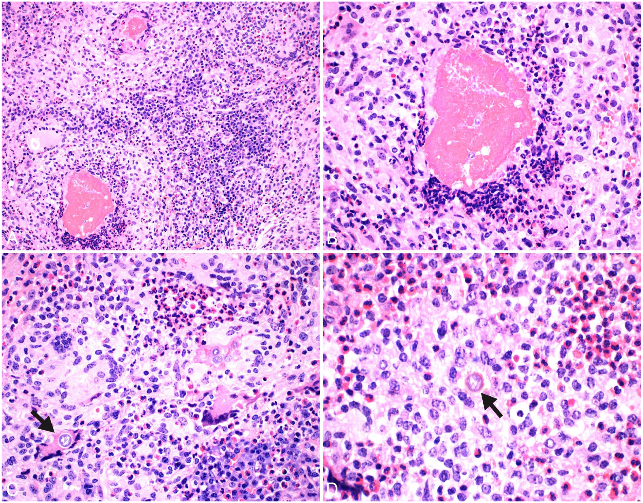
Histopathology of Exserohilum mcginnisii dermatitis in a cow.
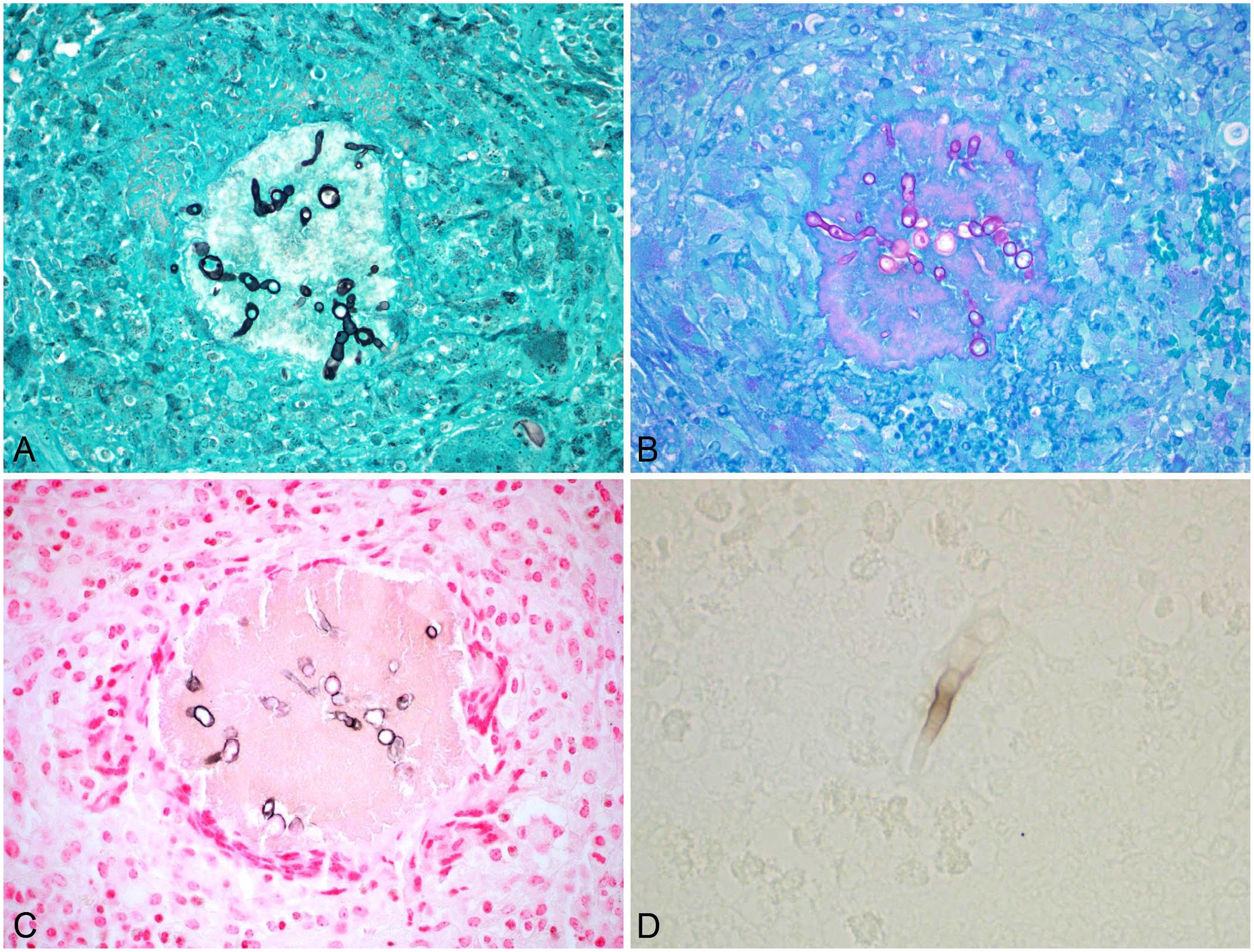
Hyphal morphology in Exserohilum mcginnisii dermatitis in a cow.
Fresh tissue samples were inoculated in Sabouraud dextrose agar (SDA) and mycobiotic agar (MA). After 2 wk, 2 dark-gray fungal colonies were observed on the SDA plate, but there was no growth on MA. A sample of a fungal colony from the SDA plate was used for a panfungal PCR assay targeting the internal transcribed spacer (ITS) gene, using the ITS1 (5′-TCCGTAGGTGAACCTGCG-3′) and ITS4 (5′-TCCTCCGCTTATTGATATGC-3′) primers, as described previously. 4 The PCR product was sent for sequencing (Eurofins Genomics, Louisville, KY, USA). The organism was identified using BLAST as Exserohilum mcginnisii, with 99.5% identity.
In a follow-up communication with the referring veterinarian 74 d after the initial diagnosis of mycotic dermatitis, the cow was reported as still having skin lesions despite treatment. The cow had been treated weekly for ~2 mo with systemic sodium iodine and a topical fungicide spray containing benzalkonium chloride as the active ingredient. The lesions had not grown or spread further and appeared less inflamed. At 100 d, the cow aborted; an autopsy of the fetus was not performed. The abortion may have been iatrogenic from treatment with systemic iodine. Shortly after the abortion, an experimental treatment of 0.45 kg of melted petroleum jelly and 17 g of Captan fungicide (N-trichloromethylthio-4-cyclohexane-1,2-dicarboximide; Southern Agricultural Insecticides) was used topically twice a week for 1 mo. Following this treatment, the lesions stopped expanding and decreased in size. At 5 mo, the cow still had lesions, but hair regrowth had started, and no discomfort could be elicited when pressure was applied to the skin lesions.
E. mcginnisii was first described in 1986 from a human patient with sinusitis. 11 In 2013, E. mcginnisii was reported as causing mycotic keratitis. 14 In a review of 48 cases of natural infection with Exserohilum before the 2012 U.S. outbreak, E. mcginnisii was responsible for only 2% of the infections in human cases. 6 Thus, in contrast to E. rostratum, E. mcginnisii is not a common cause of human infection. 6
Some isolates from human and animal cases of phaeohyphomycosis originally reported to be Drechslera and Helminthosporium spp. may be cases in which the etiologic agent was misidentified or classified with obsolete taxonomy. 8 These etiologic agents were most likely species within the genera Exserohilum or Bipolaris. 8 Several reports of Drechslera spp. causing mycotic bovine nasal granulomas have been reported, including cases caused by Drechslera sp.1,12 and D. rostrata. 9 D. rostrata has also been reported as the cause of eumycotic mycetoma in a cow affecting the nasal cavity, skin, and lymph nodes. 13 D. rostrata is considered an obsolete name for E. rostratum. 8
The first isolation of E. mcginnisii 11 coincided with the proposed reclassification of fungal organisms causing phaeohyphomycosis in humans and animals in the genera Bipolaris and Exserohilum. 8 No obsolete names were used in the previous literature for E. mcginnisii. 8 Although mycotic granuloma in cattle associated with Exserohilum spp. has been reported, the fungal species was not identified in one report 16 and in another report D. rostrata (now considered to be E. rostratum) was identified. 13 To our knowledge, E. mcginnisii dermal granulomas have not been reported previously in cattle. We retrieved no cases of E. mcginnisii infection in cattle in a search of Google, PubMed, Web of Science, CAB Direct, and SciELO, using the following search terms “Exserohilum mcginnisii cattle,” “Exserohilum mcginnisii cow,” “Exserohilum mcginnisii bovine,” suggesting that this fungal species has not been reported in cattle.
Human cases of Exserohilum infection occur mainly in warm, tropical, and subtropical areas, with most cases documented in the southern United States, India, and Israel. 6 Our case was from Georgia, southern USA. Many cases of Exserohilum spp. and Drechslera spp. in animals have been reported from similar climates. Exserohilum infections in animals have been documented in a cow from Texas, 16 a horse from Florida, and a goat from Brazil. 3 Mycotic bovine nasal granulomas and dermal mycetoma caused by Drechslera spp. have been reported in Brazil, 1 South Africa, 12 and Australia. 13
The most frequent underlying conditions associated with natural cases of Exserohilum spp. infection in humans include immunosuppression, trauma, and atopy. 6 Keratitis is mainly associated with corneal trauma from contaminated plants; trauma and atopy are the main predisposing factors in immunocompetent individuals. 6 In our case, a predisposing factor was not identified. In the report of dermal granuloma in cattle caused by an unidentified species of Exserohilum, the clinical presentation was similar to our case, with lesions distributed on the dorsal aspect of the shoulders, back, and hindquarters, and no other signs of illness. 16 In the case of D. rostrata (E. rostratum) infection in a cow, cutaneous lesions were also distributed over the rump and thighs as well as in the nasal cavity and lymph nodes. 13
Morphologic expressions of dematiaceous fungal infections of skin and soft tissue can be classified as mycetoma, chromoblastomycosis, and phaeohyphomycosis. 5 Mycetoma is characterized by the production of grains that histologically are interwoven mycelial aggregates covered by intense eosinophilic material (Splendore–Hoeppli phenomenon). 5 Phaeohyphomycosis can be confused with eumycotic mycetomas caused by dematiaceous fungi. The fungi in mycetomas form discrete organized nodules, whereas fungi of phaeohyphomycosis appear as individual hyphae and small aggregates scattered throughout the lesion. 7 The aggregates of hyphae in phaeohyphomycosis may be surrounded by Splendore–Hoeppli material, but this does not constitute a granule. In phaeohyphomycosis, fungi are usually within epithelioid macro-phages and multinucleate giant cells; granules of mycetomas are nearly always extracellular. 7 Therefore, even though some of the fungi in our case were surrounded by Splendore–Hoeppli material, ours is a case of phaeohyphomycosis rather than mycetoma. Similar histopathologic findings were observed in other cases of Exserohilum spp. and Drechslera spp. infection in cattle, including the presence of a Splendore–Hoeppli9,13,16 reaction and hyphae within giant cells, macrophage, or free in tissues.1,3,9,12,13,16 Eosinophils have also been observed in lesions associated with these fungi in cattle.9,12,13,16
Although a few fungal elements were pigmented in the H&E sections that we examined, the use of unstained sections and FM stain were very useful in demonstrating pigment. Dematiaceous fungi have pigmented hyphae; however, the degree of pigmentation can vary. Some dematiaceous fungi may have very little melanin and may appear as hyaline hyphae, 5 as in our case. In cases in which pigmentation is not evident with H&E stain, FM stain is needed to demonstrate the pigment. 5 However, FM staining must be interpreted with caution because other fungi, such as Aspergillus spp., some Mucorales genera, and Trichosporon, can stain positively. 5 The use of unstained sections aids in demonstrating the fungal pigment.
The different dematiaceous fungi cannot be distinguished from one another by histology. 5 Fungal culture followed by PCR and sequencing aids in the differentiation and classification of fungal species. Immunohistochemistry was more sensitive in detecting Exserohilum in paraffin-fixed tissues and/or fresh tissues than PCR for fungal detection in one study. 15 The lower sensitivity of the PCR assay was attributed in part to the few intact fungi in tissues and the difficulty in breaking down fungal cell walls during DNA extraction. 15
In the report of an unidentified species of Exserohilum sp. causing dermal granuloma in cattle, the lesions gradually resolved over 8–10 wk leaving only scars. 16 In another report of D. rostrata (E. rostratum) in a cow, skin lesions improved over 7 mo and appeared quiescent; 4 mo later there was recrudescence of skin lesions with dissemination of infection to the nasal cavity and lymph nodes. 13 In our case, the lesions had begun to resolve 5 mo after treatment.
Footnotes
Acknowledgements
We thank the histology, microbiology, and molecular sections at the Tifton Diagnostic Veterinary and Investigational Laboratory for their work on this case.
Declaration of conflicting interests
The authors declared no potential conflicts of interest with respect to the research, authorship, and/or publication of this article.
Funding
Publication costs for this manuscript are covered by the Tifton Veterinary Diagnostic and Investigational Laboratory.