
Abstract
Domestic rabbits (Oryctolagus cuniculus) are commonly kept as pets or bred for laboratory investigation, meat, fur production, or a combination of these purposes. We conducted a retrospective study to assess the prevalence of diseases in domestic rabbits according to purpose. We retrieved results of autopsies, biopsies, and cytologies from 2,583 cases received at 4 diagnostic laboratories in California from 2013–2022. Rabbits were classified as pets (2,241; 86.8%), laboratory animals (92; 3.6%), meat-production animals (60; 2.3%), or multipurpose animals (190; 7.4%). A final diagnosis was reached in 2,360 (91.4%) cases and was classified by system, etiology, and type of process. Pet rabbits had the highest median age (5.9 y; vs. 3 y, 0.67 y, and 0.25 y in meat, multipurpose, and laboratory rabbits, respectively), and most of the neoplasms were diagnosed in this group (872 of the total 896 neoplasms in the study; 97.3%), with tumors of the skin, female reproductive tract, and hematolymphoid system being the most common. Laboratory rabbits had a high prevalence of infectious enterotyphlocolitis (40 of 92; 43.4%), and ~45% of those cases were due to opportunistic colibacillosis. Infectious and parasitic pneumonias were common in meat rabbits (18 of 60; 30%); pneumonic pasteurellosis accounted for >60% of those cases. Infectious cholangiohepatitides were common in multipurpose rabbits (61 of 110; 55.5%), with rabbit hemorrhagic disease representing the most common etiology (82.4% of those cases). Our results demonstrate that purpose of use can predict prevalence of disease in rabbits submitted to diagnostic laboratories.
European domestic rabbits (Oryctolagus cuniculus) are frequently kept under human care worldwide for different purposes. In the United States, domestic rabbits are popular pets, but they are also kept for meat or fur production, or as backyard animals with a combined farming and pet purpose. Rabbits are also important for research; they are among the most common animal models used to study human disease. Rabbit carcasses and biopsies are commonly submitted to veterinary diagnostic laboratories in the United States for diagnostic purposes. Many rabbit diseases are well characterized, and their pathogeneses are well understood. Examples of these are myxomatosis 5 and rabbit hemorrhagic disease (RHD), 16 which have a devastating impact on wild and domestic rabbit populations. Other diseases of rabbits are of public health interest due to their zoonotic nature and include, among others, tularemia 14 and encephalitozoonosis. 21 However, despite the great body of knowledge on pathogenesis of rabbit diseases and medicine, the overall prevalence of diseases in this species is seldom described in the literature and could greatly inform diagnosticians of possible differential diagnoses, particularly when stratified by purpose of use.
Diseases affecting rabbits can have economic, animal welfare, and zoonotic impacts,10,15,16,21,24,26 and it is therefore important to be aware of the ongoing disease processes in different populations around the world. It is also very useful for diagnosticians to know the current prevalence of diseases to help establish differential diagnoses. Few studies analyze disease prevalence in rabbits, and those available are mostly focused on a particular body system3,6,17,25,32 or a rabbit type or purpose of use (e.g., pet rabbits).3,6,8,29,32 Differences in breed, management, housing, and overall purpose of use may result in differences in physiology, behavior, and, consequently, disease presentation. 31
We collected pathology records of rabbits submitted to several diagnostic laboratories in California for diagnostic purposes, for a 10-y period. We identified the prevalence of the main processes affecting pet, meat, laboratory, and multipurpose rabbits.
Materials and methods
Animals and institutions
We gathered archived diagnostic data from rabbits submitted to 4 laboratories in California, USA, between 2013 and 2022 for our study. The laboratories included were Zoo Exotic Pathology Services (Carmichael, CA; 2013.01.01–2018.04.01), California Animal Health & Food Safety Lab System (University of California, Davis, San Bernardino, Tulare, and Turlock branches; 2013.01.01–2022.11.01), William R. Pritchard Veterinary Medical Teaching Hospital (University of California, Davis; 2013.01.01–2022.11.01), and the Comparative Pathology Laboratory (University of California, Davis; 2013.01.01–2022.11.01). The animals were classified as pets, laboratory, meat production, or multipurpose (rabbits that are kept for a combination of 2 or more, as a pet and/or for meat- and fur-production purposes).
Diagnostic workup
Main morphologic diagnoses and etiology were established after considering macroscopic and/or microscopic lesions and, when available, ancillary tests. We included autopsy, biopsy, and cytology examinations in our study. Diagnoses were classified according to the system involved and the etiology.
Statistical analyses
Descriptive statistics were performed (Excel for Office 365 MSO 16.0; Microsoft). Prevalence of disease was calculated based on the total number of cases with final conclusive diagnoses per group (e.g., pet rabbits). System-specific prevalence of disease was calculated based on the total number of system-specific diagnoses (e.g., respiratory cases). Chi-square tests were used to compare prevalence between groups (GraphPad QuickCalcs, https://www.graphpad.com/quickcalcs/contingency1/). For all statistical analyses, p ≤ 0.05 was considered statistically significant.
Results
We included 2,583 cases in our study: 2,217 autopsy, 238 biopsy, and 128 cytology examinations. Of those, 2,241 cases were pet rabbits, 92 laboratory rabbits, 60 meat-production rabbits, and 190 multipurpose rabbits (Table 1). Median ages were higher for pet rabbits compared to other groups (t-test, p < 0.0001; 5.9 y for pets [range: 7 d to 15 y], 3 y for meat rabbits [35 d to 8 y], 0.67 y for multipurpose rabbits [7 d to 11 y], and 0.25 y for laboratory rabbits [1 d to 7 y]). Sex distribution was 1,289 females (49.9%) and 1,171 males (45.3%); sex was not reported for 123 (4.8%) rabbits. There was no difference in sex distribution within pet, laboratory, and multipurpose rabbits (p > 0.05). Among meat rabbits, females were overrepresented (p = 0.012; Table 2).
Number of diagnostic cases included in our study of disease in 2,583 domestic rabbits from 4 diagnostic laboratories, categorized by purpose and by sample submission type.
Number of cases by age and sex included in a retrospective study of disease in 2,583 domestic rabbits from 4 diagnostic laboratories, categorized by purpose.
A final diagnosis was reached in 2,360 cases (91.4%), which included 2,128 autopsies, 184 biopsies, and 48 cytologies. The cases in which a final diagnosis was reached included 2,040 pet (Figs. 1–18), 81 laboratory (Figs. 19–23), 58 meat, and 181 multipurpose (Figs. 24–29) rabbits. Sex distribution was 1,182 females (50.1%) and 1,074 males (45.5%); sex was not reported for 104 (4.4%) rabbits. The most frequently affected system was integumentary (554; 23.5%), and most of the cases involving this system were from pet rabbits. The next most frequently affected systems were hepatobiliary (487; 20.6%), respiratory (344; 14.6%), urinary (336; 14.2%), alimentary (290; 12.3%), cardiovascular (259; 11.0%), and female reproductive (259; 11.0%). The most common process across all groups was neoplasia (Table 3) with 896 cases (38.0%; 775 autopsies, 78 biopsies, 39 cytologies). After neoplasia, pneumonia (307; 13.0%), hepatitis (260; 11.0%), and nephritis (259; 11.0%) were the most frequently diagnosed processes; for each of those processes, an infectious etiology was identified in 36.0%, 18.2%, and 17.3% of the cases, respectively. Of the total animals received for autopsy with a final diagnosis, 1,058 (49.7%) cases were diagnosed with multiple conditions or illnesses.
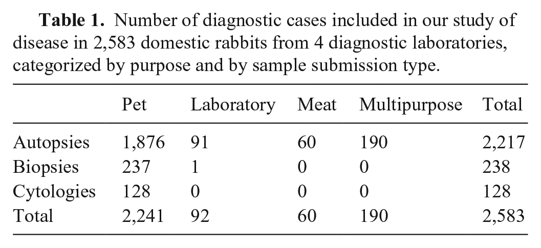
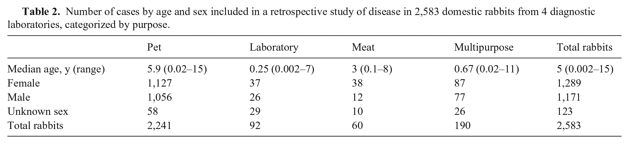
Gross lesions of diseases in pet rabbits.
Gross and microscopic lesions of diseases in pet rabbits.
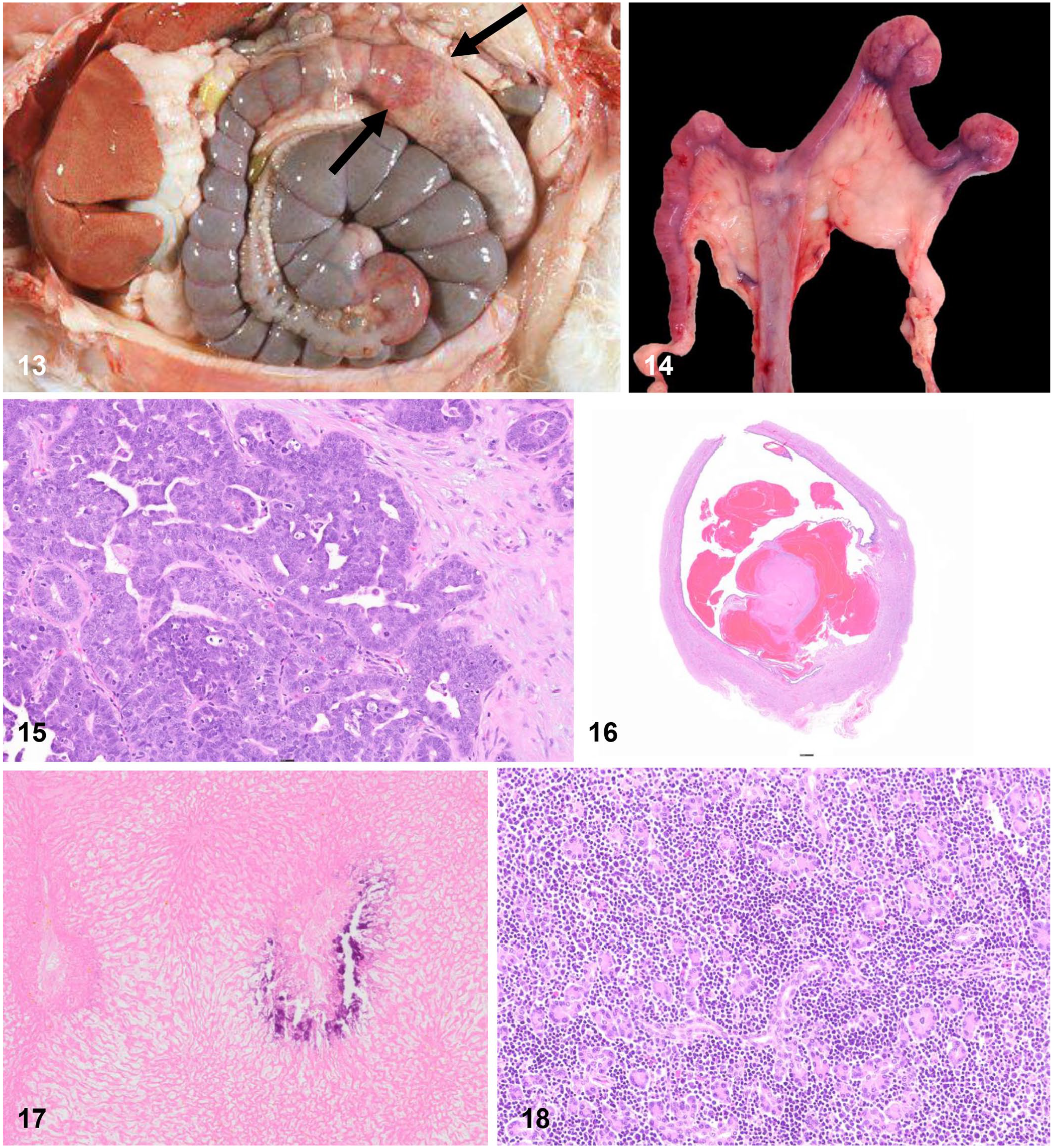
Gross and microscopic lesions of diseases in pet rabbits.
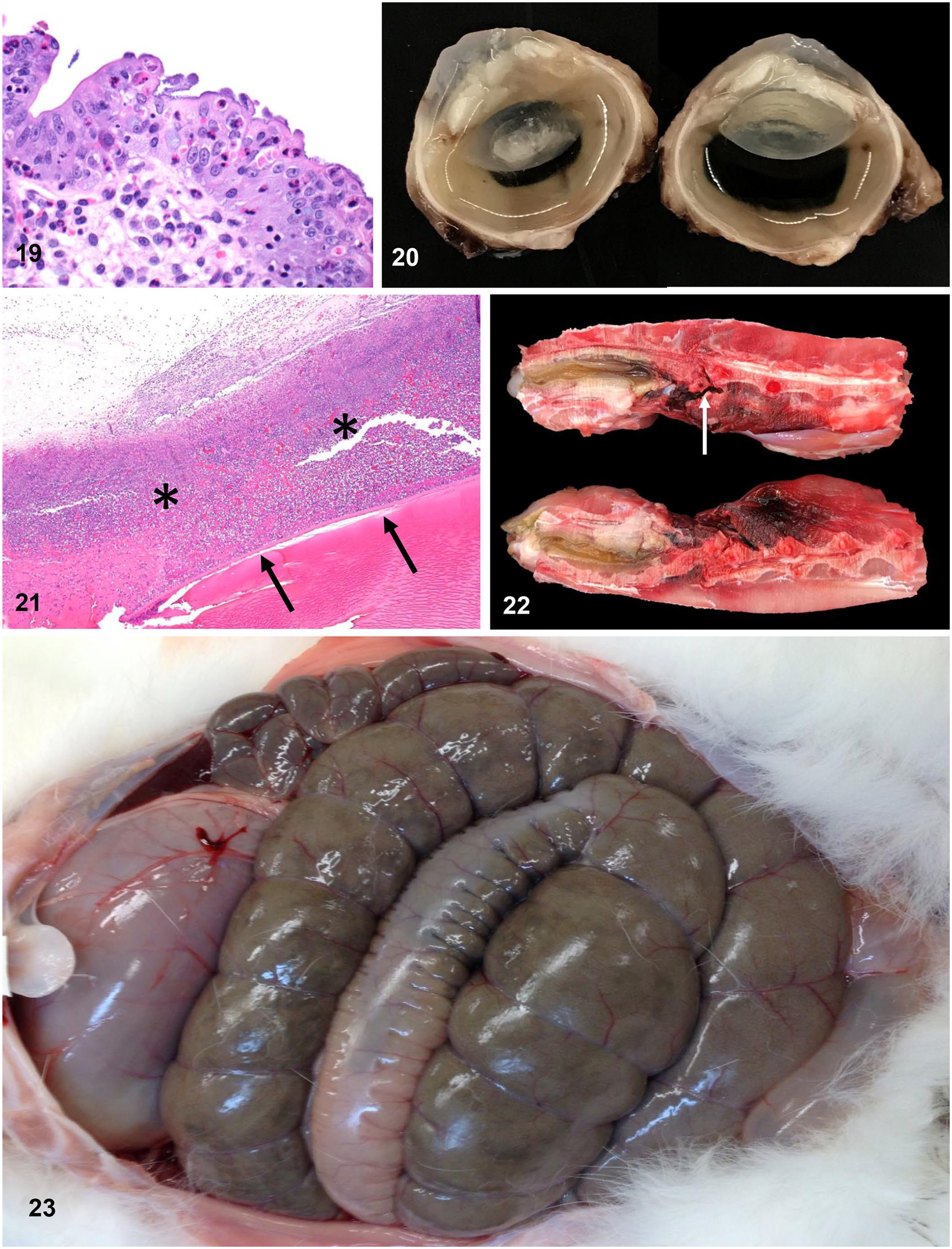
Gross and microscopic lesions of diseases in laboratory rabbits.
Gross and microscopic lesions of diseases in meat-production and multipurpose rabbits.
Most common neoplastic lesions found in domestic rabbits from 4 diagnostic laboratories, categorized by purpose. Percentage of the prevalence of each neoplasm within neoplastic lesions of its group are shown. Only neoplasms with a total prevalence higher than 20 are represented.
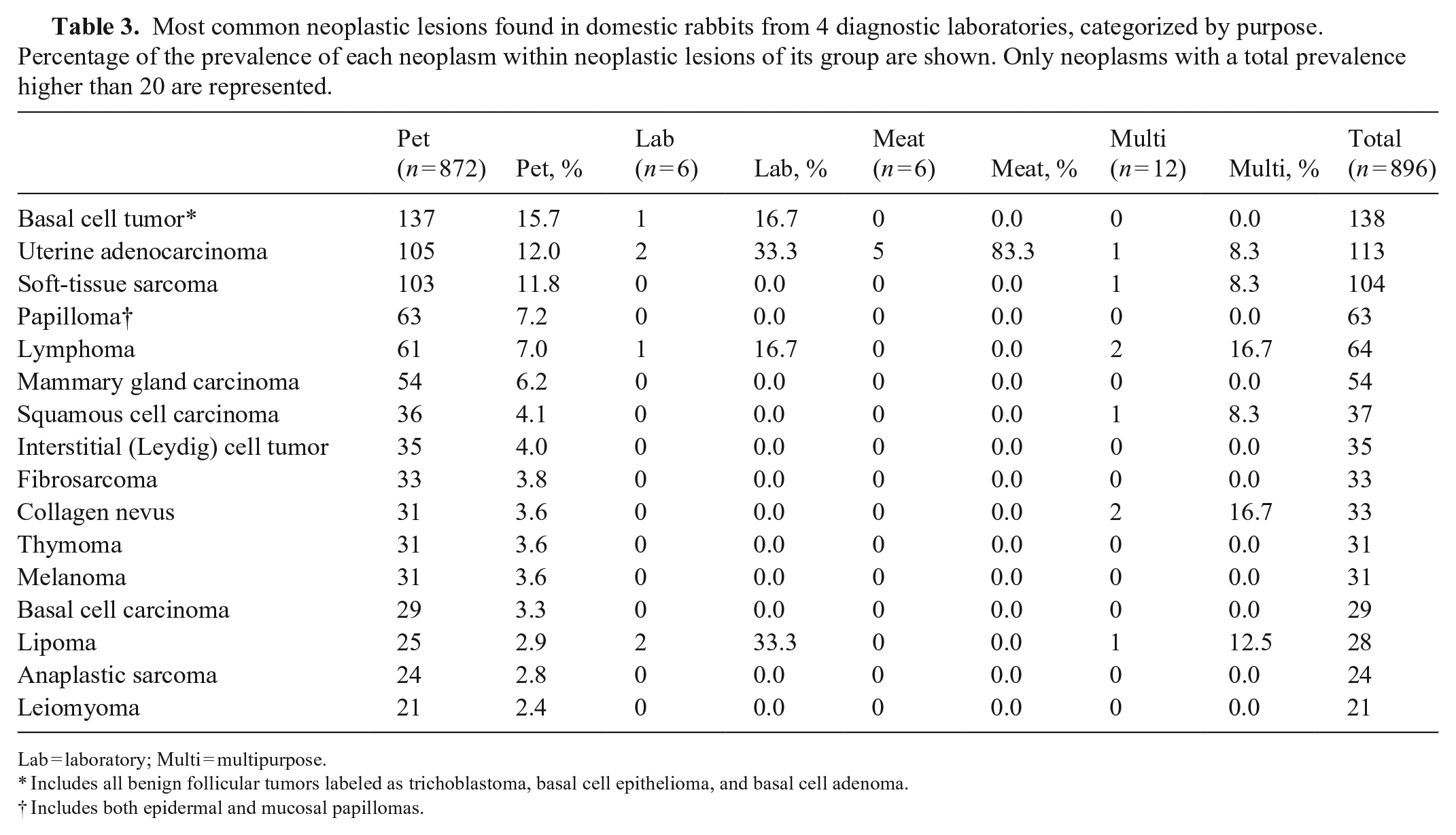
Lab = laboratory; Multi = multipurpose.
Includes all benign follicular tumors labeled as trichoblastoma, basal cell epithelioma, and basal cell adenoma.
Includes both epidermal and mucosal papillomas.
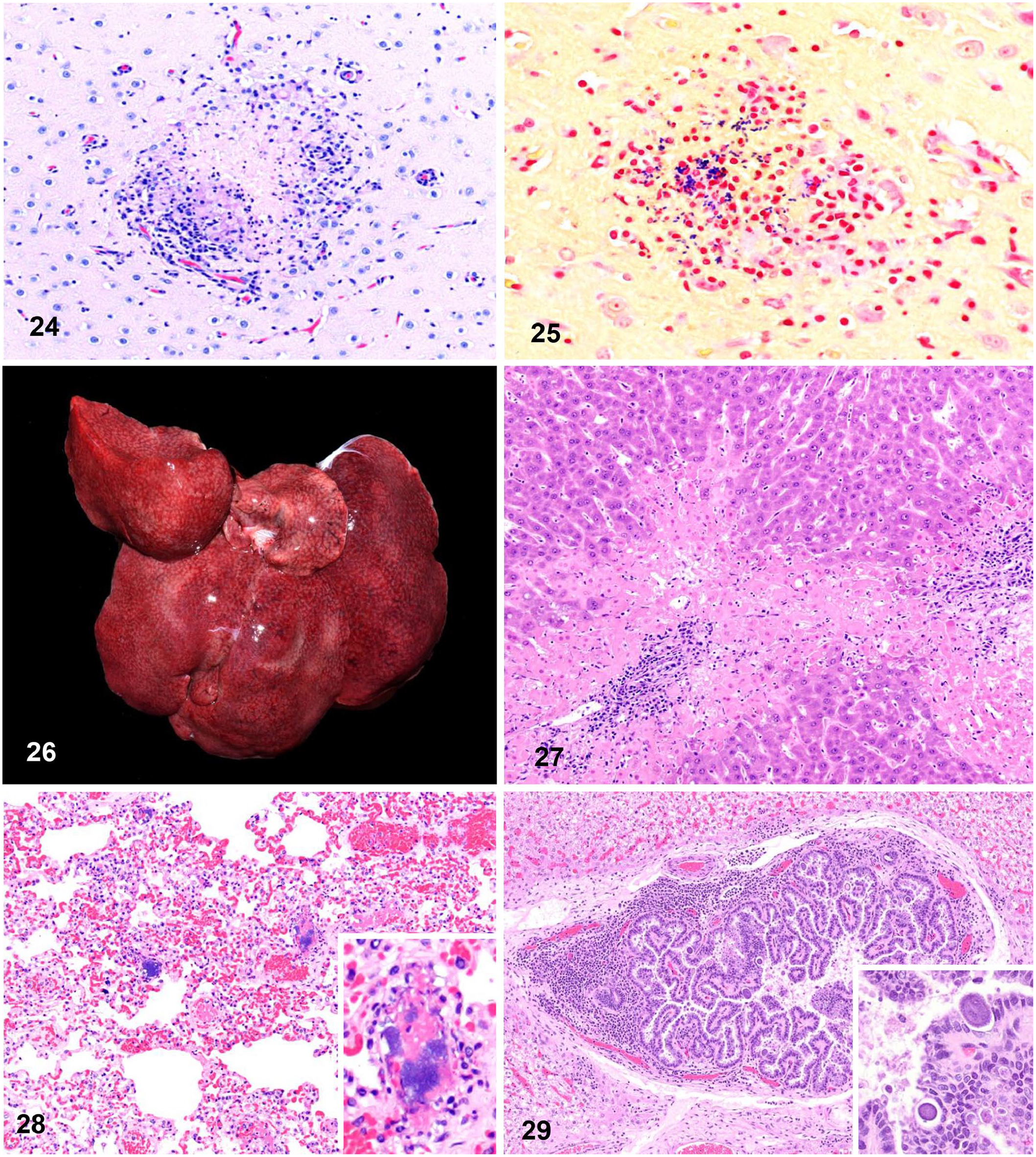
Overall, an infectious process was diagnosed in 1,379 cases (58.4%). The most common infectious agents detected were bacteria (255; 18.5% of infectious disease cases), followed by parasites (127; 9.2%), viruses (112; 8.1%), and fungi (107; 7.8%). The most common infectious etiologies across all groups were Pasteurella spp. (Fig. 3) and Encephalitozoon cuniculi (Figs. 11, 12, 24, 25). Among meat and multipurpose rabbits, Bordetella spp. was also frequently isolated. Meat rabbits had a significantly higher prevalence of myocarditis (11 of 60 vs. 75 of 2,523; p < 0.001) and encephalitis (8 of 58 vs. 101 of 2,302; p < 0.001) than the other groups combined.
Neoplasms
Of the 896 rabbits diagnosed with neoplasia (Table 3), 872 of 2,040 (42.7%) were pet rabbits. Neoplastic lesions were more frequent in pet rabbits than in the other groups (p < 0.0001). In the other groups, neoplasms were present in ≤10% of the total cases: 6 of 81 (7.4%) laboratory rabbits, 6 of 58 (10.3%) meat rabbits, and 12 of 181 (6.6%) multipurpose rabbits. The most common neoplasms in pet rabbits were basal cell tumors, uterine adenocarcinoma (Figs. 14, 15), and soft-tissue sarcoma. Other neoplasms worth noting in pet rabbits included lymphoma (Figs. 13, 18; 61 cases, 7.0%), mammary gland carcinoma (54 cases; 6.2%), thymoma (Fig. 2; 31 cases, 3.6%), melanoma (31 cases; 3.6%), biliary cystadenoma (16 cases; 1.8%), and giant cell sarcoma (14 cases; 1.6% of neoplasm cases in pet rabbits). Uterine adenocarcinoma was the most common neoplasm in meat rabbits (5 cases; 83.3% of neoplasia cases in meat rabbits).
Hepatobiliary system
Hepatobiliary lesions were diagnosed in 344 pet rabbits, 13 laboratory rabbits, 20 meat rabbits, and 110 multipurpose rabbits (Table 4). Of those, hepatobiliary lesions were responsible for the death of the animal in 14.3% of the cases. The most common hepatic lesion was hepatitis of undetermined etiology: 53.2% of liver lesions in pet rabbits, 53.9% in laboratory rabbits, 45.0% in meat rabbits, and 10.0% in multipurpose rabbits. The most common infectious agent causing hepatobiliary disease was RHD virus 2 (RHDV2; Figs. 26, 27), which was detected in 80 cases, 76.3% of which were multipurpose rabbits. Hepatic microsporidiosis due to E. cuniculi was present in 4 pet (1.2% of hepatobiliary lesions), 2 laboratory (15.4%), 3 meat (15.0%), and 2 multipurpose (1.8%) rabbit cases. Proliferative cholangitis due to coccidiosis (Fig. 29) was observed in 3 pet (0.9%) and 4 multipurpose (3.6%) rabbit cases. Another relevant process was liver lobe torsion (Figs. 1, 17), which was found in all groups, but with higher prevalence in pet rabbits (13.4% of the rabbits with hepatobiliary disease in this group had liver lobe torsion). Hepatic lipidosis (29 cases) was also commonly found in pet (6.7% of hepatobiliary cases) and multipurpose (7.7%) rabbits.
Hepatobiliary pathologic processes diagnosed in domestic rabbits from 4 diagnostic laboratories, categorized by purpose.
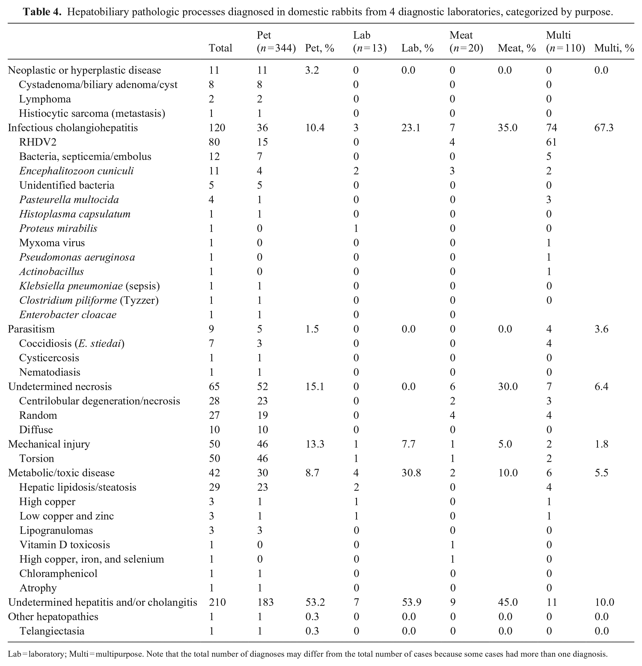
Lab = laboratory; Multi = multipurpose. Note that the total number of diagnoses may differ from the total number of cases because some cases had more than one diagnosis.
Respiratory system
Respiratory lesions were diagnosed in 213 pet rabbits, 22 laboratory rabbits, 28 meat rabbits, and 79 multipurpose rabbits (Table 5). The most common respiratory lesions were pneumonias of undetermined etiology: 43.7% of respiratory lesions in pet rabbits, 22.7% in laboratory rabbits, 10.7% in meat rabbits, and 12.7% in multipurpose rabbits. Bacterial pneumonias for which no isolate was obtained (either because bacteriology was not performed or because culture was negative) were also common, and they were 14.0% of the total respiratory lesions in all groups combined. The prevalence of bacterial pneumonia was particularly high in pet rabbits, encompassing 16.9% of respiratory lesions in the group. Pasteurella multocida was commonly isolated from pneumonic lungs with a total of 38 cases (11.1% of the total respiratory lesions in all groups combined); of those, 11 rabbits were septicemic (Figs. 3, 28). P. multocida pneumonia with or without septicemia was particularly common in multipurpose (24.1% of respiratory lesions in the group) and meat (39.3%) rabbits. Aspiration pneumonia was also a common respiratory process, particularly in pet (14.6%) and laboratory (9.1%) rabbits. Primary upper airway disease, including tracheitis or bronchitis and rhinitis or sinusitis, was not as common as pulmonary lesions, and was only relevant in pet rabbits, in which 9.9% of respiratory lesions were in upper airways.
Respiratory pathologic processes diagnosed in domestic rabbits from 4 diagnostic laboratories, categorized by purpose.
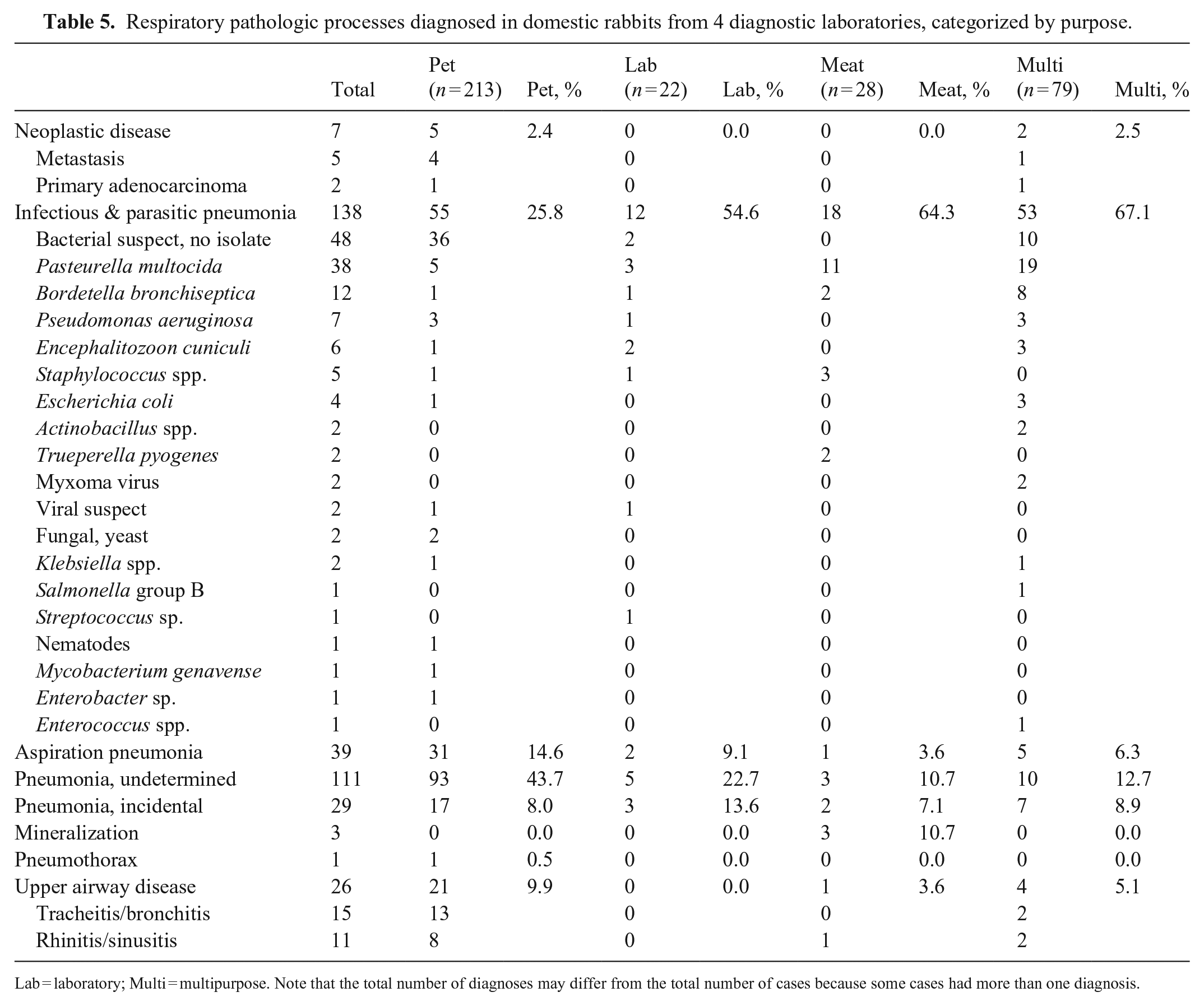
Lab = laboratory; Multi = multipurpose. Note that the total number of diagnoses may differ from the total number of cases because some cases had more than one diagnosis.
Alimentary system
Alimentary lesions were diagnosed in 174 pet rabbits, 48 laboratory rabbits, 12 meat rabbits, and 56 multipurpose rabbits (Table 6). In all types of rabbits, the most common alimentary process diagnosed was infectious enterotyphlocolitis (Figs. 19, 23), representing 16.1% of pet rabbits, 83.3% of laboratory rabbits, 41.7% of meat rabbits, and 53.6% of multipurpose rabbit alimentary cases. In multipurpose animals, coccidia were common parasites of the alimentary tract (28.6% of alimentary cases in the group); in laboratory animals, colibacillosis was the most common finding (37.5%). In pet rabbits, there was a high diversity of infectious agents, but most were identified in only one case. In pet rabbits, the prevalence of gastrointestinal stasis and/or obstructive syndrome (GSS 24 ; Figs. 7, 9, 10; 35 cases, 20.1%) and idiopathic ulcers or perforation (30 cases; 17.2%) closely followed that of the infectious processes. Dental disease, including malocclusion, incisor or molar root elongation, tooth root abscess (Figs. 4, 8), and periodontitis, was present at low frequency in pet (85 cases; 4.2%) and multipurpose (2 cases; 1.1%) rabbits and was not seen in laboratory or meat rabbit submissions during this time period. The rest of the alimentary processes identified were incidental in all groups.
Alimentary tract pathologic processes diagnosed in domestic rabbits from 4 diagnostic laboratories, categorized by purpose.
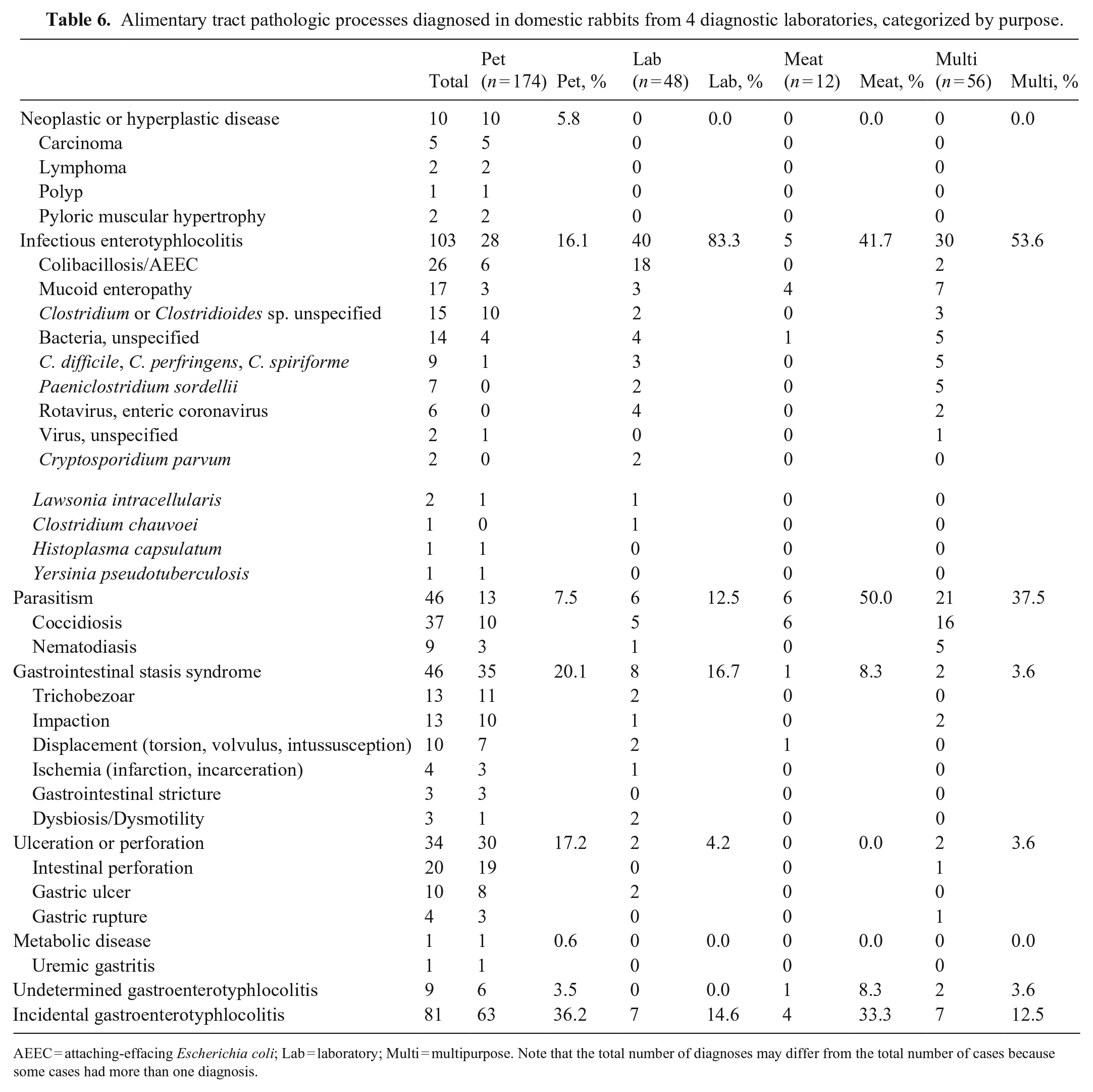
AEEC = attaching-effacing Escherichia coli; Lab = laboratory; Multi = multipurpose. Note that the total number of diagnoses may differ from the total number of cases because some cases had more than one diagnosis.
Urinary system
Urinary tract lesions were diagnosed in 262 pet rabbits, 6 laboratory rabbits, 28 meat rabbits, and 40 multipurpose rabbits (Table 7). The most common urinary processes were nephritis of unknown origin: 179 cases (68.3%) in pet rabbits, 2 cases (33.3%) in laboratory rabbits, 22 cases (78.6%) in meat rabbits, and 20 cases (50.0%) in multipurpose rabbits. Chronic renal disease, including infarcts (Fig. 5), was observed in pet (53 cases; 20.2%), meat (4 cases; 14.3%), and multipurpose (7 cases; 17.5%) rabbits, but not in laboratory rabbits. Cystitis was observed mostly in pet rabbits, particularly sedimentary cystitis (Fig. 6), which was a pet rabbit–only disease (5 cases; 1.9%) during the study period.
Urinary pathologic processes diagnosed in domestic rabbits from 4 diagnostic laboratories, categorized by purpose.
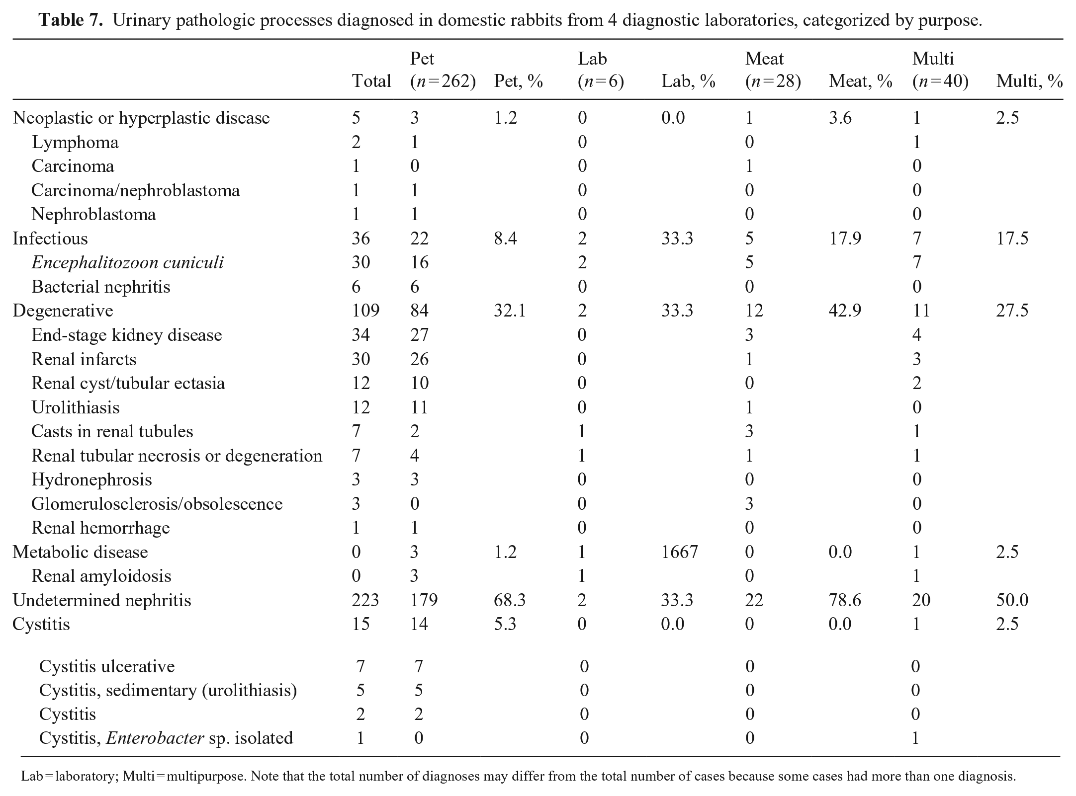
Lab = laboratory; Multi = multipurpose. Note that the total number of diagnoses may differ from the total number of cases because some cases had more than one diagnosis.
Cardiovascular system
Cardiovascular lesions were diagnosed in 197 pet rabbits, 8 laboratory rabbit, 32 meat rabbits, and 29 multipurpose rabbits (Table 8). The most common cardiovascular lesions were degenerative lesions: 50.8% of cardiovascular lesions in pet rabbits, 37.5% in laboratory rabbits, 56.3% in meat rabbits, and 48.3% in multipurpose rabbits. They were followed closely by inflammation of unknown origin: 42.6% of lesions in pet rabbits, 37.5% in laboratory rabbits, 37.5% in meat rabbits, and 17.2% in multipurpose rabbits. Most of these cardiac lesions were incidental or not part of the main pathologic process and were generally mild. Degenerative lesions were commonly correlated with hypertrophic and dilated cardiomyopathies. Infectious agents were uncommon and mostly consequences of septicemia.
Cardiovascular pathologic processes diagnosed in domestic rabbits from 4 diagnostic laboratories, categorized by purpose.
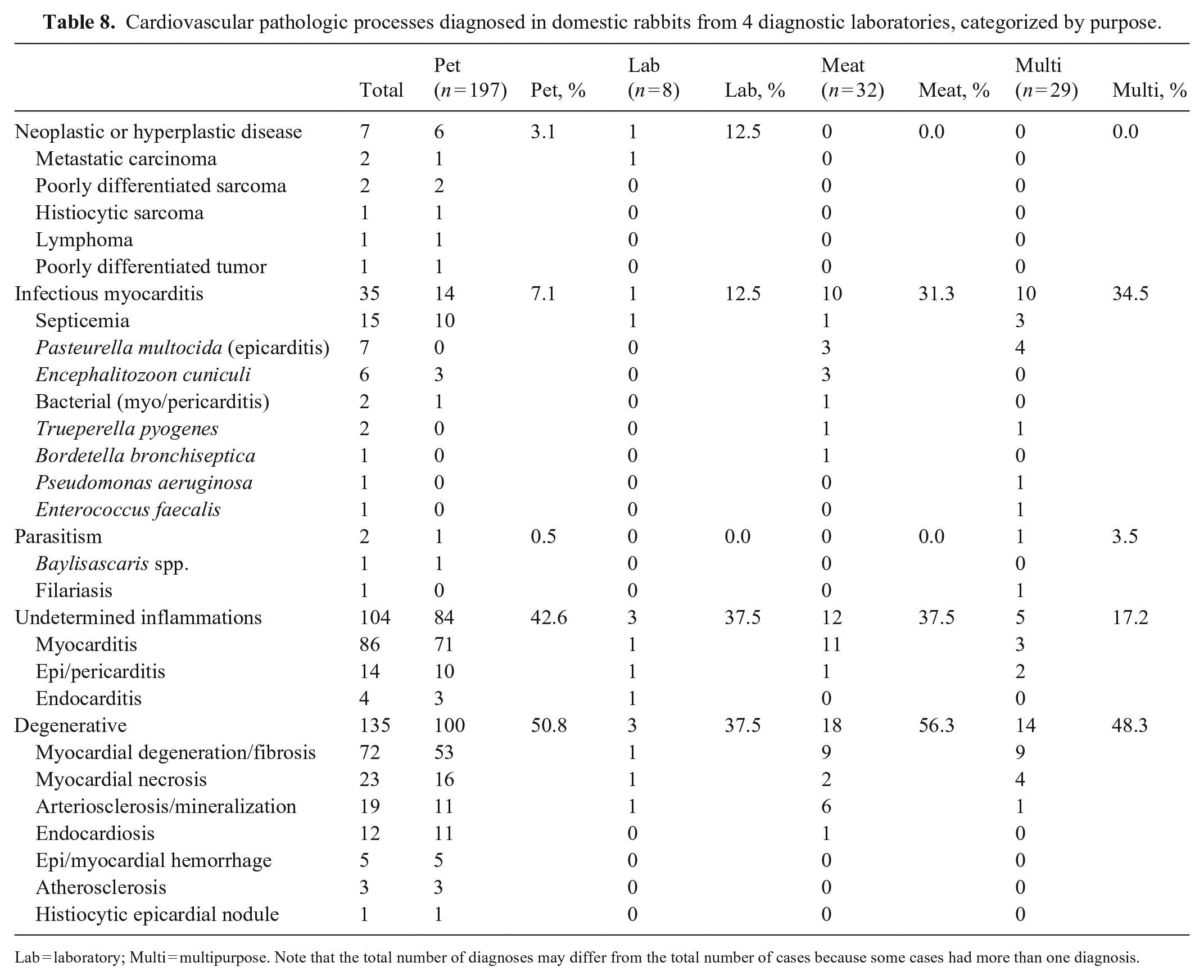
Lab = laboratory; Multi = multipurpose. Note that the total number of diagnoses may differ from the total number of cases because some cases had more than one diagnosis.
Female reproductive system
Female reproductive tract lesions were diagnosed in 296 pet rabbits, 3 laboratory rabbits, 10 meat rabbits, and 4 multipurpose rabbits (Table 9). Most of these lesions were in pet rabbits and were proliferative (hyperplasia or neoplasia) or degenerative diseases of the uterus. Uterine adenocarcinoma alone represented 43.4% of the reproductive tract disorders in pet rabbits and the next 3 most common uterine neoplasms (adenoma, leiomyoma, leiomyosarcoma) accounted for 25.2%. Neoplastic disorders were commonly co-diagnosed with endometrial hyperplasia. Endometrial venous aneurysm (Fig. 16) was observed only in pet rabbits (30 cases; 10.1%). In the rest of the groups, reproductive tract disorders were not common, but uterine adenocarcinomas were 83.3% of all neoplasms in meat rabbits (Table 3).
Female reproductive pathologic processes diagnosed in domestic rabbits from 4 diagnostic laboratories, categorized by purpose.
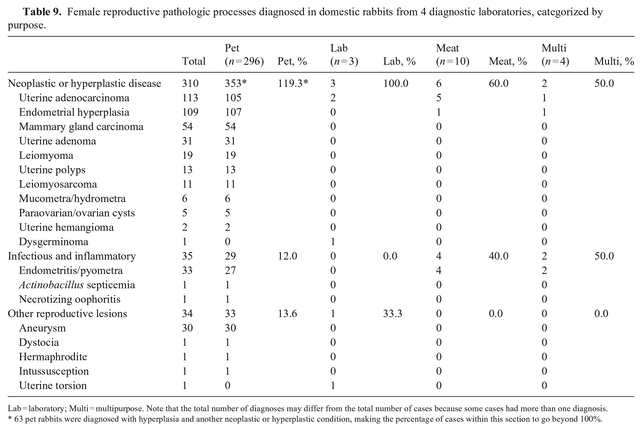
Lab = laboratory; Multi = multipurpose. Note that the total number of diagnoses may differ from the total number of cases because some cases had more than one diagnosis.
63 pet rabbits were diagnosed with hyperplasia and another neoplastic or hyperplastic condition, making the percentage of cases within this section to go beyond 100%.
Nervous system
Neurologic lesions were diagnosed in 74 pet rabbits, 2 laboratory rabbits, 25 meat rabbits, and 28 multipurpose rabbits (Table 10). As seen in the hepatobiliary and urinary systems, inflammation of unknown cause was the most common finding in all groups: 44.6% of neurologic lesions in pet rabbits, 50.0% in laboratory rabbits, 32.0% in meat rabbits, and 42.9% in multipurpose rabbits. The encephalitides were usually lymphoplasmacytic (32 of 54 cases; 59.3%) or lymphohistiocytic (18 cases; 33.3%). Among the known etiologies of encephalopathies, E. cuniculi was the only consistent agent causing disease in rabbits (Figs. 24, 25): 32 cases (43.2%) in pet rabbits, 1 case (50%) in a laboratory rabbit, 15 cases (60.0%) in meat rabbits, and 12 cases (42.9%) in multipurpose rabbits. Parasites, bacteria, and viruses caused only sporadic disease.
Neurologic pathologic processes diagnosed in domestic rabbits from 4 diagnostic laboratories, categorized by purpose.
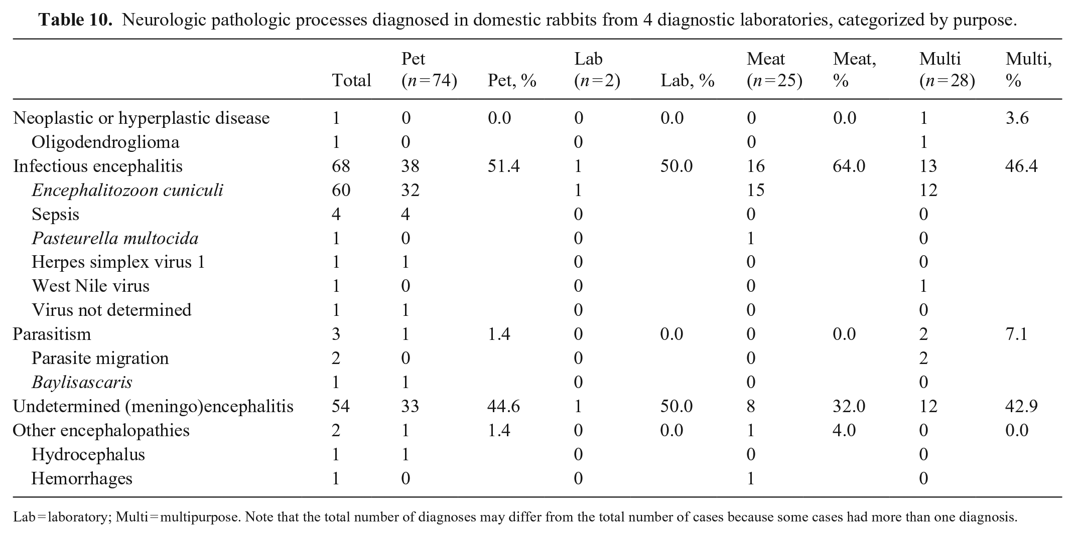
Lab = laboratory; Multi = multipurpose. Note that the total number of diagnoses may differ from the total number of cases because some cases had more than one diagnosis.
Integumentary system
Integumentary lesions were diagnosed in 596 pet rabbits, 7 laboratory rabbits, 4 meat rabbits, and 20 multipurpose rabbits (Table 11). Neoplastic lesions (66.9%) were reported most frequently in pet rabbits. The prevalence of infectious dermatitis was most frequent in multipurpose rabbits (70.0%) and was predominantly due to myxoma virus infection (50% of all skin diagnoses in multipurpose rabbits). Abscesses or pyogranulomas and bacterial dermatitis were most common in pet rabbits (58 of 85 [68.2%] skin diagnoses in pet rabbits). Splendore–Hoeppli material was observed in 10 of 42 abscesses or pyogranulomas (23.8%) in pet rabbits, although a causal agent was not identified. Noninfectious dermatitis and pododermatitis (sebaceous adenitis, pododermatitis, moist/ulcerative dermatitis, urine scald) was present in all groups, representing 18.8% of integumentary lesions in pet rabbits, 57.1% in laboratory rabbits, 75.0% in meat rabbits, and 10.0% in multipurpose rabbits.
Integumentary pathologic processes diagnosed in domestic rabbits from 4 diagnostic laboratories, categorized by purpose.
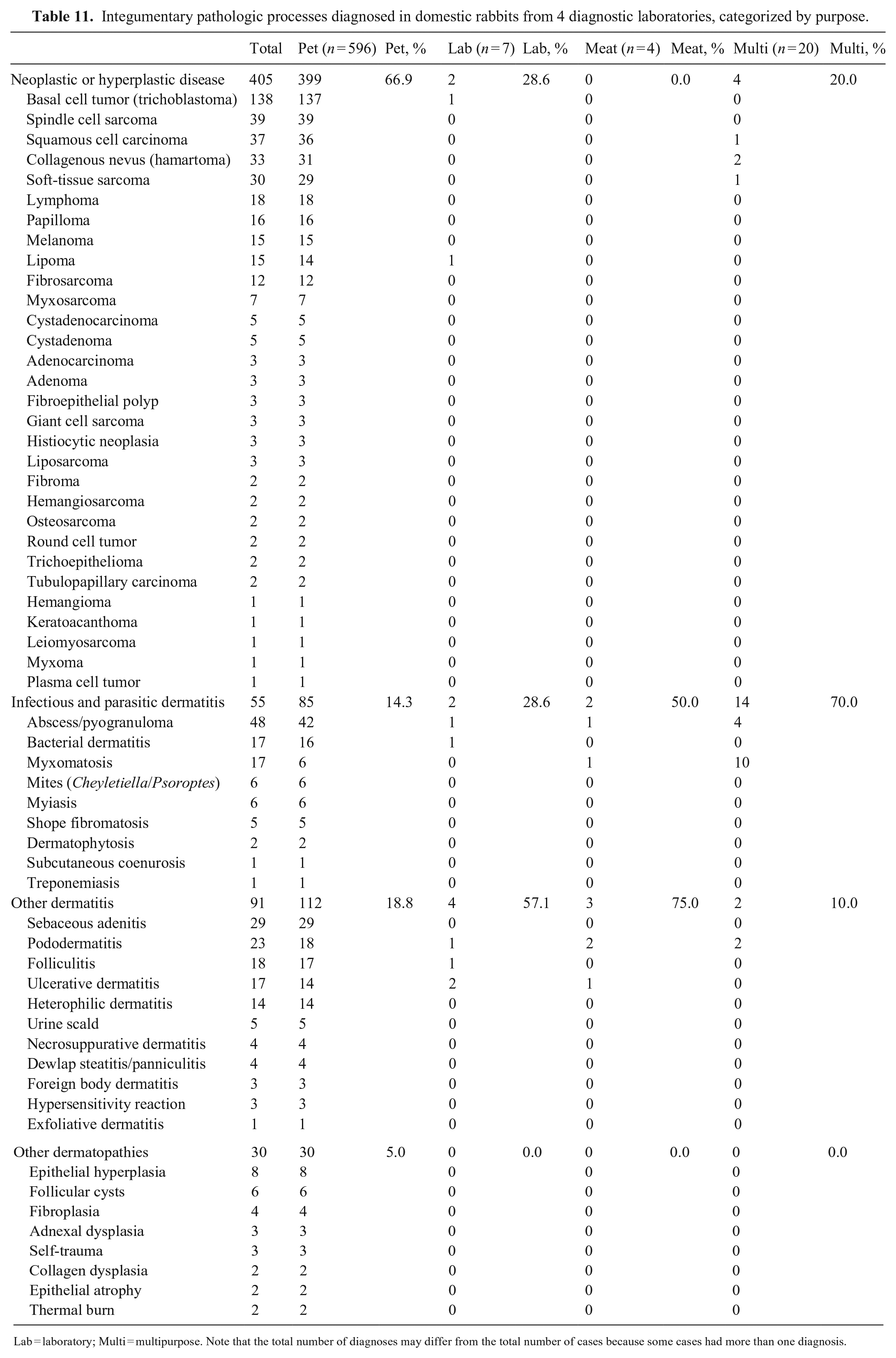
Lab = laboratory; Multi = multipurpose. Note that the total number of diagnoses may differ from the total number of cases because some cases had more than one diagnosis.
Discussion
Most of the data in our study came from autopsies (85.8%). All of the cytology examinations and all but one of the biopsies came from pet rabbits, which is a limitation of our study. This disparity of results may be due to the fact that, in practice, it is uncommon to perform such in vivo techniques for non-pet rabbits (laboratory, meat, and multipurpose in our study), and carcasses are often only submitted to assess the overall health of the group. Lethal processes can be overrepresented compared to milder diseases that do not commonly result in death. Geographic differences are not accounted for and, as the diagnostic laboratories in this study are located within California, the sample population is biased towards the West Coast of the United States. Another limitation is that in some cases, ancillary testing was not pursued to determine the specific cause of the lesion(s). Despite this, a conclusive diagnosis was reached in 91.4% of the cases, which is significantly higher than that achieved in previous studies. 13
Our results clearly indicate that the prevalence of certain rabbit diseases varies based on purpose of use. In a study performed in Spain to assess the prevalence of several diseases, 13 26 different conditions were identified, as opposed to the 169 different categories used to classify the diagnoses of the cases in our study. In that study, 13 the most frequent conditions were parasitosis but, in our case, parasitoses represented a small percentage, except in meat and multipurpose rabbits. This is probably due to the high percentage of farm and wild rabbits included in the aforementioned study. 13 In California, multipurpose animals are frequently backyard animals, making them more exposed to the environment than indoor rabbits (pets) or rabbits under controlled conditions (laboratory). 15 Parasites can have complex lifecycles involving multiple species, or they may have wildlife and other free-roaming animals as reservoirs. Multipurpose or backyard rabbits are therefore more easily infested due to their contact with other species. Lastly, pet rabbits tend to live longer than other rabbits, making it easier for them to have higher prevalence of certain processes, such as degenerative and some neoplastic diseases.
We identified 42 different neoplasms in our study. The greatest diversity of neoplasms was seen in pet rabbits; only 7 types of tumors were found in the other 3 groups combined. Neoplasms are common in pet rabbits, and they can represent over half of the biopsy and cytology diagnoses. 28 In our cases, neoplasms represented 872 of 2,040 (42.7%) pet rabbit cases. The higher prevalence of neoplasia in pet rabbits compared to other groups of our study could be attributed to their higher average age; there is an association between age and development of cancer in rabbits.6,8,28 The most common neoplasms were uterine adenocarcinoma, basal cell tumor, soft-tissue sarcoma, lymphoma, papilloma, and mammary gland carcinoma. Our results are mostly in accord with previous studies, in which high prevalence of uterine adenocarcinoma, soft-tissue sarcoma, mammary gland adenocarcinomas, and trichoblastomas (included in our study under basal cell tumors) are commonly reported.8,13,28 However, other neoplasms regarded as common in previous studies had a very low prevalence in our study, such as thymomas8,19 (3.6% of pet rabbit neoplasms in our study), giant cell sarcoma 7 (1.6% of pet rabbit neoplasms), and gall bladder tumors 2 (1.8% of pet rabbit neoplasms).
In pet rabbits, aside from integument, the 2 organ systems with lesions diagnosed more commonly were the hepatobiliary and the urinary systems. However, lesions of the liver and kidneys were not always associated with a specific disease process and were of little value to determine the cause of death of the rabbit. Most were interpreted to be inflammatory conditions related to systemic processes and not primary lesions of these organs because they were commonly found in conjunction with lesions of higher severity in other organs. Even in some cases in which there were severe hepatic or renal lesions, determining the cause was challenging. In cases with acute necrotic lesions of undetermined etiology, diagnosticians suspected toxic exposure (endotoxemia), septicemias, or, when the distribution was focally extensive or affected an entire lobe, resolved liver torsions. Pet rabbits also had a high number of alimentary tract lesions, particularly those attributed to infectious diseases and several types of motility problems, such as GSS. 23 Previous retrospective studies 28 have found a high prevalence of reproductive disorders in pet rabbits, but as the diagnoses were based on biopsy and cytology information, those findings were likely biased towards non–life-threatening processes that can be surgically resolved, such as reproductive tract masses. Other autopsy-based studies 13 also did not report a high prevalence of enteropathies in pet rabbits, but their sample population was considerably smaller than ours (~1.5%). In the skin, sebaceous adenitis was a disease solely observed in the pet rabbit group. Treponemiasis 2 is likely under-represented; 1 case was confirmed but at least 5 other cases were suspected to be due to Treponema paraluiscuniculi. Unfortunately, identification of intralesional spirochetes by histochemical staining is unlikely in many cases due to prior antimicrobial therapy (D. Reavill, pers. comm., 2020 Oct 31).
In laboratory rabbits, studies have reported spontaneous pulmonary 11 and renal 9 lesions in aged individuals (>2-y-old). This contrasts with our study population that was markedly younger, with a median age of 0.25 y. In fact, there is a gap in the knowledge of diseases affecting young laboratory rabbits. A 2011 study discussed the incidence of ocular lesions, 18 but we had only 2 cases in our study. A high percentage (40 of 92; 43.5%) of the laboratory rabbits in our study were diagnosed with an infectious enterotyphlocolitis, of which 18 of 40 (45.0%) were attributed to Escherichia coli. Rather than being a primary infectious cause, these cases of colibacillosis were likely opportunistic cases secondary to rabbit GSS. Laboratory rabbits are known carriers of enteropathogenic E. coli, 30 which can cause sporadic outbreaks of disease 29 ; but in our study, the cases were distributed along the time period, thus we interpret them as sporadic disease rather than large outbreaks. Gastrointestinal stasis, bacterial/aspiration pneumonia, and vertebral fractures were common life-limiting diagnoses in laboratory rabbits and often attributed to handling, compound administration, anesthesia, or husbandry practices that result in insufficient roughage, sedentary behavior, or stress.2,15
Rabbit meat consumption is common in some European countries and in China. 12 Despite not being a common meat source in the United States, meat rabbit production has acquired importance in an increasing number of states.4,20 The small number of meat animals in our study makes it difficult to draw relevant conclusions, but the prevalence of myocarditis (11 of 60; 18.3%) was significantly higher than that found in the other groups (75 of 2,523; 2.9%). Myocarditis in meat rabbits was also more common than in pet rabbits in reported studies. 25 The high prevalence of myocarditis in meat rabbits could be attributed to undiagnosed E. cuniculi infestations, which have been demonstrated to cause myocarditis. 21 The prevalence of nephritis and encephalitis in meat rabbits were also higher than in the rest of the groups, and may also be related to E. cuniculi, although the cause was not always confirmed. Meat rabbits also had a significantly high prevalence of uterine adenocarcinoma (8.6%), which was also the most common neoplasm. Our population of meat rabbits included a high number of intact aged female rabbits, which could explain these numbers.
Multipurpose rabbits had a combined purpose of pet and meat or fur production, and this was the second largest group. These animals are usually kept outdoors and/or in backyards, occasionally with limited sanitary control, as discussed before, and movement of rabbits and related products among different premises is believed to be constant. This background was reflected by the fact that multipurpose rabbits presented the highest prevalence of RHDV2, a virus that has circulated in California since at least 2020. 1 The liver is the main target organ of RHDV2, but lesions can also occur in other organs21,22; however, for the purpose of our retrospective study, the RHDV2 cases were counted within the hepatobiliary system only, and the array of lesions in each system will be described in a future study. Lesions associated with infectious etiologies were more common in the multipurpose group, in general, accounting for 75.0% of the alimentary lesions, 65.1% of the respiratory lesions, 50.0% of the neurologic lesions, and 40.4% of the hepatic lesions. Infections by E. cuniculi were also common in this group, with 12 cases of cerebral encephalitozoonosis (6.6% of all multipurpose rabbits) and 7 of renal encephalitozoonosis (3.9%). Coccidiosis in the intestines (28.6%) and liver (2.2%) was a common parasitic process.
In the literature, when lesions of the CNS are discussed, a causative agent is commonly identified. 17 That was not the case in our study, and 54 (44.6%) of the CNS lesions remained as encephalitis of unknown origin. Some meningoencephalitis and encephalitis cases without a confirmed etiology, especially those with granulomatous or histiocytic inflammation, were suspected to be caused by E. cuniculi, but the agent could not be identified. It is possible that the lack of ancillary testing precluded reaching a definitive diagnostics in these cases. 27 For the purposes of our studies, E. cuniculi was considered responsible for granulomatous or histiocytic encephalitis.
In pet rabbits, the expected diseases encountered by a diagnostic pathologist will be age-related and neoplastic, with tumors of the skin, female reproductive tract, and hematolymphoid system being the most common. In laboratory rabbits, the expected diseases (GSS, enteric colibacillosis, vertebral fractures, aspiration pneumonia) will be due to stress, management, handling or procedure-related, or a combination of the aforementioned. Meat and mixed-use rabbits that are housed outdoors are expected to have more infectious diseases, including pasteurellosis and encephalitozoonosis, and in these groups, uterine adenocarcinoma was observed with highest prevalence. In all groups, mild-to-moderate pneumonia, hepatitis, and nephritis of unknown origin were also common. Our survey emphasizes the importance of purpose-use, as an indirect reflection of, but not limited to, husbandry, potential stressors, and the relative risk of infectious, reproductive, and neoplastic disease.
Footnotes
Acknowledgements
Melea Hunrath and Briana Luna, necropsy technicians, and multiple generations of anatomic pathology and laboratory medicine residents from the UCD CPL, CAHFS, and VMTH are deeply acknowledged.
Declaration of conflicting interests
The authors declared no potential conflicts of interest with respect to the research, authorship, and/or publication of this article.
Funding
The authors received no financial support for the research, authorship, and/or publication of this article.