
Abstract
The European domestic rabbit (Oryctolagus cuniculus domesticus) is commonly kept as a pet, with increasing popularity among pet owners. Despite the increasing body of information on lagomorph medicine and pathology, comprehensive published compilations of causes of mortality in pet rabbits are limited. We analyzed health disorders, pathology findings, and most probable causes of death in 100 pet rabbits submitted to the Anatomopathological Diagnostic Service of the Veterinary School of the University of Las Palmas de Gran Canaria, Canary Islands, Spain, from 2011 to 2022. We reviewed clinical data and gross and histopathologic findings to classify the various disorders into infectious (n = 39), noninfectious (n = 45), and idiopathic conditions (n = 16). Within infectious disease processes, Encephalitozoon cuniculi infection (n = 14) and rabbit hemorrhagic viral disease (n = 14) had the highest prevalence. Regarding the noninfectious conditions, perioperative mortality (n = 10), gastrointestinal syndrome (n = 7), and trauma (n = 6) were the most common clinicopathologic entities observed.
The European domestic rabbit (Oryctolagus cuniculus domesticus) is a non-traditional companion animal, becoming more popular in many countries such as Spain. Pet rabbits have become regular patients in primary-care small animal clinics and in exotic animal referral centers. 7 According to a census estimation by Veterindustria and ANFAAC (Asociación Nacional de Fabricantes de Alimentos para Animales de Compañía), the ownership rate for pet rabbits in Spain increased from 1.9% in 2017 to 2.5% of households in 2021. 1 Furthermore, the pet rabbit population rose from 718,364 individuals to 1,122,060, an increase of 70%. 1
Despite domestic rabbits being a common pet species and the number of clinical reports in scientific literature steadily increasing, comprehensive clinical studies, including those evaluating the causes of mortality, are needed to inform the rabbit and exotic pet veterinary professional.16,21 We analyzed health disorders, pathology findings, and the most probable causes of death of pet rabbits in the Canary Islands, Spain.
Materials and methods
Animals
We included in our study domestic rabbits submitted for postmortem examination to the Anatomopathological Diagnostic Service of the Veterinary School of the University of Las Palmas de Gran Canaria, Canary Islands, Spain, from January 2011 to December 2022. Submissions came from different veterinarians and pet rabbit owners from the Canary Islands, Spain. Ethics approval was not required for this research according to European Union Directive 2010/63.
Whenever possible, referring owners or veterinarians provided a brief clinical history (sex, age, clinical signs, date of death) for the submitted rabbit. The 100 rabbits included in our study were in various stages of decomposition (codes 1–3). 15 Eight cases were recently dead rabbits (code 1), 74 were fresh or slightly decomposed (code 2), and 18 were moderately decomposed (code 3). Rabbits in advanced decomposition conditions were not included in our study. In addition, we classified cases into 3 age groups: young rabbit (<12-mo-old), adult (12-mo to 5-y-old), and geriatric (>5-y-old). 19
Autopsy, histologic evaluation, and clinicopathologic entity assignment
Our autopsy procedure followed a published protocol. 5 Representative samples from tongue, esophagus, trachea, thyroid gland, thymus, heart, lung, stomach, small intestine, cecum, colon, liver, spleen, kidney, adrenal glands, pancreas, urinary bladder, brain, skeletal muscle, sciatic nerve, bone marrow, and either uterus or testis had been collected systematically. Tissue samples were fixed in 10% neutral-buffered formalin at room temperature for 24 h, processed routinely, and 5-µm sections were stained with H&E for histologic examination. Periodic acid–Schiff, Gram, Giemsa, Ziehl–Neelsen, Congo red, Masson trichrome, and Prussian blue stains were performed on selected tissue sections when necessary.
Clinical histories, autopsy findings, and histology slides were reviewed and re-analyzed by a team consisting of a European Specialist in Veterinary Pathology (O. Quesada-Canales), a European Specialist in Wildlife Population Health (M. Arbelo), and a clinical veterinarian specializing in rabbits and exotic animals (J. Espinosa García-San Román). After histology slide review and considering the anamnesis and clinical history, cases were assigned to the following clinicopathologic entities: bacterial infection, fungal infection, viral infection, gastrointestinal syndrome, trauma, neoplasia, metabolic-nutritional disease, heat stroke, dental disorder, perioperative mortality, and undetermined-idiopathic causes of death. These 14 categories of clinicopathologic entities were developed based on the following guiding principles: clinical interest, previous use in similar studies,6,16,23 International Classification of Diseases (ICD-11) nomenclature, 25 frequency of cases (entities were formed when >2 cases shared a characteristic; otherwise, they were placed in a miscellaneous category). Diseases known to occur in the species but not detected as possible causes of death in this study were excluded.
Statistical analysis
Data were analyzed using SPSS Statistic (v.26; IBM). A one-sample Kolmogorov–Smirnov test tested the normality of the distribution of variables. Continuous variables with normal distribution were presented as x̄, SD, and 95% CI; non-normal variables were reported as median (interquartile range [IQR]). Frequency values and 95% CIs for each clinicopathologic entity were presented, in addition to individual values for different sexes and age groups. The chi-square test was used to compare the distribution of clinical entities between sexes and age groups. When 20% of the cells had expected frequencies <5, the Fisher exact test was used. The Mann–Whitney U test was used to compare sexes and the variables of age and body weight. p ≤ 0.05 was considered significant.
Results
Submission analysis
We studied 100 rabbits submitted for postmortem examination. Forty-nine rabbits were male, 43 were female, and the sex could not be stated in 8 cases due to the lack of development of the kit or the preservation status of the rabbit. In the submissions analyzed, 27 were young rabbits, 58 were adults, and 15 were geriatric rabbits. Age ranged from 1 mo to 11 y, with a median of 1.8 y (IQR = 0.58–4 y). Females had a median age of 2 y (IQR = 0.83–3.50); the median age of males was 1.8 y (IQR = 0.73–4.75). Rabbits weighed 0.30–3.20 kg, with a median of 1.50 kg (IQR = 0.94–1.94). Weight medians by sex were 1.80 kg (IQR = 1.20–2.00) for females and 1.40 (IQR = 1.00–1.65) for males. Weight medians by age group were 0.90 kg for young rabbits (IQR = 0.46–1.45), 1.58 kg for adults (IQR = 1.20–2.00), and 1.43 kg for geriatric rabbits (IQR = 1.22–1.94). No statistically significant differences between sexes and the variables age (p = 0.619) and weight (p = 0.061) were observed. Significant differences in body weight were observed comparing young rabbits to adult (p = 0.001) and geriatric (p = 0.031) rabbits.
Mortality and clinicopathologic entities
A morphologic diagnosis was established in 95 cases and presumptive cause of disease in 82 cases. Considering both, we were able to classify 84 cases in one of the established clinicopathologic entities (Table 1). Idiopathic cases were statistically significantly more prevalent in young rabbits (8 of 27; p = 0.024). No statistically significant differences were observed in clinicopathologic entities between sexes (Table 1).
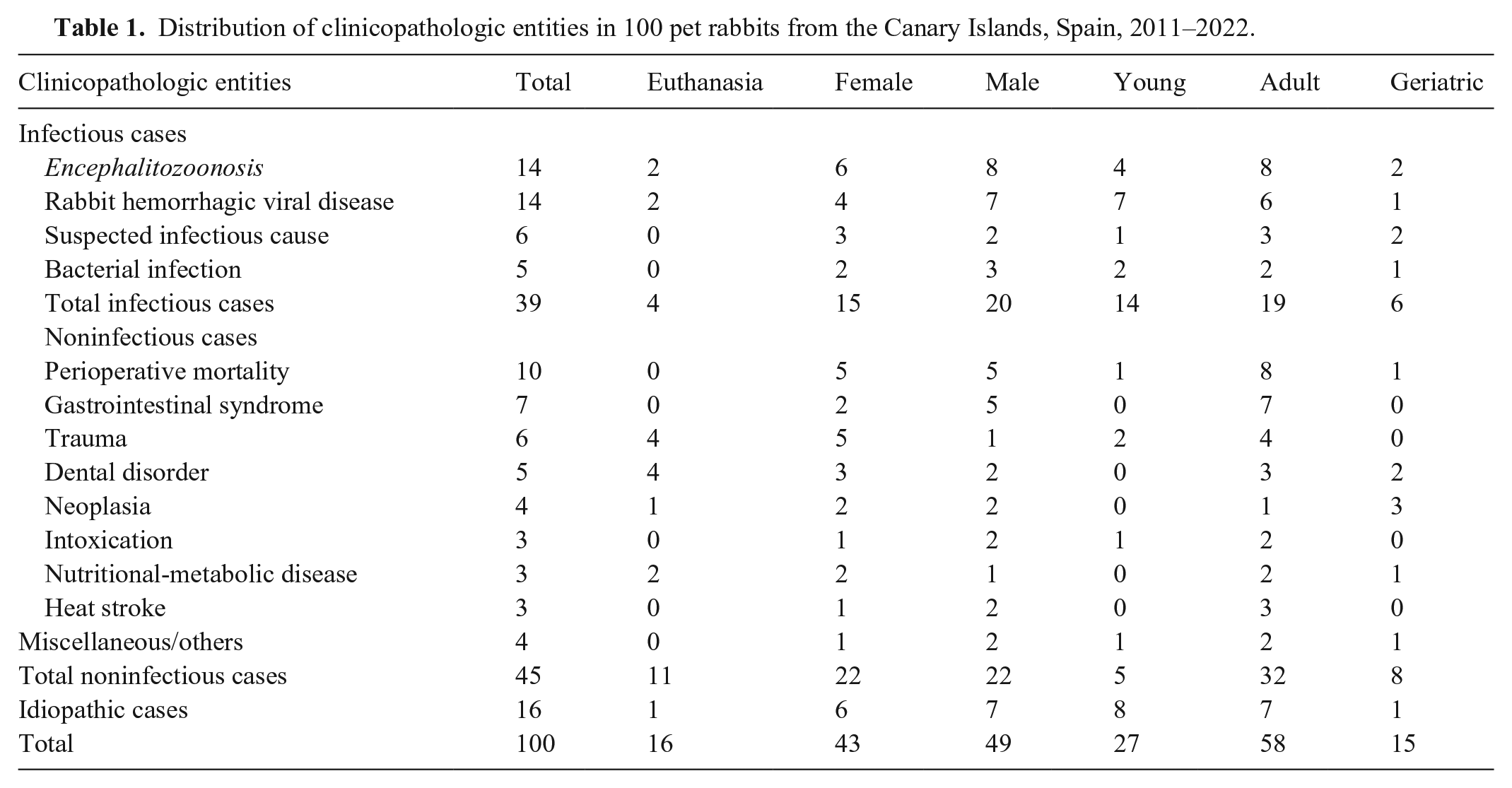
Distribution of clinicopathologic entities in 100 pet rabbits from the Canary Islands, Spain, 2011–2022.
Infectious processes
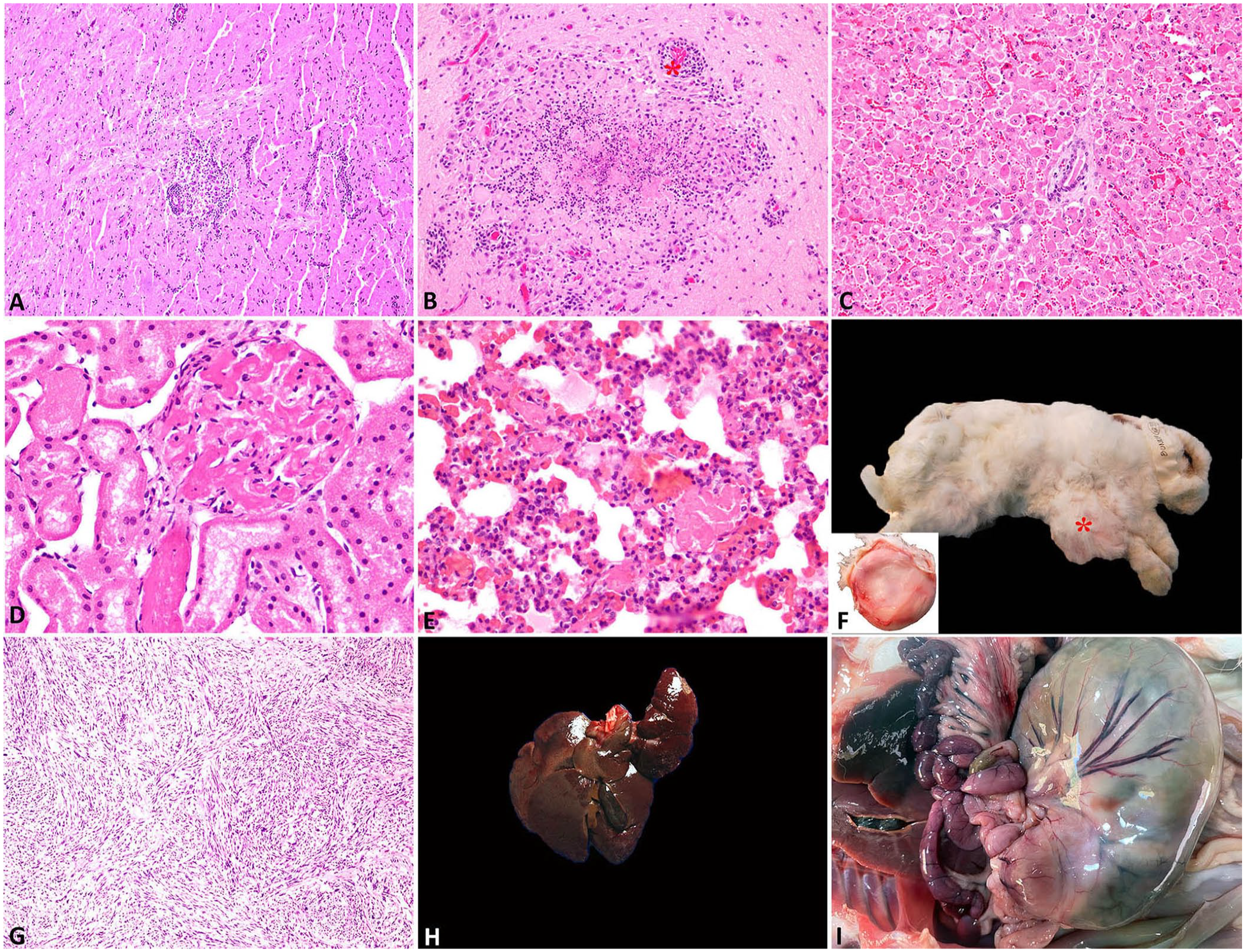
There were no statistically significant differences between the frequency of infectious disease and age ranges (Table 1). Lesions consistent with Encephalitozoon cuniculi infection were found in 23 rabbits and contributed to the death of 14. Histologic lesions included granulomatous meningoencephalomyelitis with astrogliosis and perivascular lymphocytic cuffs (Fig. 1A, 1B), and granulomatous interstitial nephritis. Renal lesions were observed in 14 of 23.
Lesions found in autopsies of pet rabbits from the Canary Islands, Spain.
Lesions compatible with rabbit hemorrhagic disease (RHD) were necrotizing hepatitis or hepatic necrosis (14 of 14; Fig. 1C). Additionally, there was renal, pulmonary, intestinal, or generalized thrombosis (11 of 14; Fig. 1D, 1E), diffuse severe splenic necrosis (4 of 14), and interstitial pneumonia (2 of 14).
Noninfectious causes of death were statistically significantly greater in adults (32 of 58; p = 0.025), and significantly lower for young rabbits (5 of 27, p = 0.001; Table 1).
Perioperative mortality
In 10 cases, the cause of death was perioperative mortality, based mainly on the clinical history and anamnesis, either during pre-anesthesia, induction, surgery, or in the 48–72 h after surgery. No specific or common lesions were found in the rabbits included in this entity.
Gastrointestinal syndrome
Seven cases were included in the gastrointestinal syndrome. All cases were concentrated in adults (7 of 7; p = 0.020). Gross lesions were abdominal dilation, distended stomach, and accumulation of gas, food, or hairs in the stomach and intestine, occasionally becoming impaction or obstruction processes. In 3 of 7 cases, the gastric mucosa had multifocal-to-coalescing, circular, erosions, ulcers, or necrotic areas. Duodenal perforation had occurred in one case.
Trauma
Four of the 6 trauma cases were euthanized because of irreversible neurologic damage; 3 of the 6 trauma cases were caused by falling from a height. Two of 6 cases had lumbar fractures; the other 4 cases included hemothorax with lung contusion, an open transverse tibial fracture, a laceration with a necrotizing skin infection in the dorsum, and bilateral hindlimb muscle atrophy secondary to neurologic injury.
Dental disorders
Five rabbits died of circumstances arising from dental problems. Four were euthanized due to the severity and irreversibility of the condition or because the owners declined to continue treatment. The fifth case died during hospitalization with hyporexia and hypercreatinemia. Gross lesions observed included incisor or molar overgrowths, exophthalmos, weight loss, abscesses, and osteomyelitis.
Neoplasia
In 4 cases, neoplasia was determined as the cause of death, including a uterine adenocarcinoma with lung metastasis, an intestinal adenocarcinoma, a disseminated undifferentiated sarcoma (Fig. 1F, 1G), and a case of multiorgan metastasis most likely of endometrial origin. A fifth case had died of a liver torsion, but a uterine adenocarcinoma had been removed a week before. The frequency of neoplasia was also statistically significantly greater in geriatric rabbits than in young and adult rabbits combined (3 of 4; p = 0.01).
Intoxication
In 3 cases, intoxication was determined as the cause of death based mainly on the clinical history and anamnesis. Specific or common lesions were not found in the rabbits included in this entity. In 2 cases, death was associated with the use of fipronil to treat ectoparasites, following recommendations in specialized stores. In the third case, the owner accidentally overdosed the rabbit with gabapentin, which was being used to treat chronic neuropathic pain.
Nutritional and metabolic disorders
Three deaths were attributed to metabolic or nutritional disorders, 2 to hepatic lipidosis, and 1 to nephrolithiasis. This latter case had multifocal interstitial nephritis with ureteral calculi.
Heat stroke
Three casualties were attributed to heat stroke, based on the anamnesis, clinical history, and multiorgan congestion and hemorrhage.
Miscellaneous
Four cases were grouped in the miscellaneous category, including 1 case of hepatic torsion, 1 intestinal torsion, and 2 cases of sudden death associated with handling-induced stress. In the hepatic torsion, the caudate lobe had areas of hemorrhage and congestion, with hepatocellular degeneration and necrosis (Fig. 1H). In the intestinal torsion case, necrosis and hemorrhage with venous infarction were observed in the segment involved (Fig. 1I). One of the cases associated with handling stress had moderate-to-severe multifocal and monophasic acute muscle fiber necrosis and degeneration. These lesions are compatible with capture myopathy. In the other case, the rabbit died while being manipulated for diagnostic tests and had acute multiorgan congestion.
Discussion
Encephalitozoonosis and rabbit hemorrhagic disease were the leading causes of death diagnosed in our case series, with 14 cases each, which constituted 28 of the 39 infectious diseases, 28 of the 100 total deaths, and 28 of the 84 deaths with determined cause. All individuals diagnosed with potential E. cuniculi had brain lesions; compatible renal lesions were less frequent. E. cuniculi has been implicated as an opportunistic zoonotic agent in immunocompromised humans.11,14 Disease surveillance and transmission reduction are crucial in the One Health framework. Primary transmission occurs through ingestion or inhalation of E. cuniculi spores in the urine or feces of an infected individual.11,22
Half of the animals (7 of 14) that presumably died from RHD virus (RHDV; Lagovirus europaeus) were <7-mo-old, and all died in 2016 during the emergence of the new variant (RHDV2) in Spain, indicating a high likelihood that they were affected by this new variant. 4 High mortality associated with RHD is remarkable in urban environments and among pet rabbits. 18 However, RHD would be included as a differential diagnosis in rabbits with nonspecific clinical signs and sudden death, especially if they are unvaccinated, with histopathology proving valuable in confirming or refuting the diagnosis and uncovering other potential causes of death, as gross evidence of the disease is often lacking. 9 Vaccination, including for the new RHDV variant, is strongly recommended for all rabbits, even indoor pets.
As evidenced by the number of perioperative deaths observed and the reported greater incidence of peri-anesthetic mortality in rabbits compared to dogs and cats, 12 protocol enhancement may be necessary. Gastrointestinal syndrome, which is commonly encountered in rabbit medicine, has multifactorial and synergistic components, including electrolyte disorders, dehydration, hypoglycemia, hepatic lipidosis, enterotoxemia, or gastrointestinal perforation. Given this complexity, we included the gastrointestinal syndrome as a separate clinicopathologic entity. 13 In the same way, dental problems may not be a direct cause of mortality, but unfortunately, many rabbits with chronic or recurrent dental disorders become hyporexic or anorexic, have gastric hypomotility or stasis, or develop abscesses or osteomyelitis, which lead to spontaneous death or euthanasia. Few studies explain how much dental disease contributes to euthanasia or death. 10 The other primary cause of euthanasia was trauma and its related surgical complications such as severe infections, vascular compromise, non-union fractures or non-healing open fractures, as well as irreversible neurologic damage.
Lack of knowledge about the feeding, management, and treatments of rabbits could be a determining factor in certain lesions and deaths. Cases attributed to intoxication were unintentional poisoning by owners, by fipronil and gabapentin overdose. 24 Urolithiasis is prevalent in pet rabbits due to their calcium metabolism and can be reduced with proper nutrition. 20 Additionally, any cause of anorexia can rapidly lead to hepatic lipidosis in the rabbit if it is not addressed promptly. 24 Although we observed a low prevalence of deaths attributable to heat stroke, rabbits are highly susceptible to this condition, potentially exacerbated by climate change, and it is something of which owners should be aware.8,17
Neoplasia was diagnosed in only 5 cases, and 4 of them were considered the cause of death. However, although our study has few cases, it is consistent with other studies in which uterine adenocarcinoma was the most frequent tumor found.2,3 The high number of young animals in our study could explain the low prevalence of neoplasia.
The median age of rabbits in our study contrasts with those of other studies; a high proportion of young rabbits lowered our average age. The elevated mortality rate noted among young rabbits could be linked to ownership patterns influenced by factors such as insufficient rabbit care knowledge together with an increased demand suggesting a lack of experience or maturity in the Spanish rabbit pet market.
The main limitations of our study are that the data were from a single institution, which may not be fully representative, as well as its retrospective nature. Additionally, economic constraints, technical, and legal limitations hindered the performance of ancillary tests such as bacterial culture, molecular assays, or immunohistochemical analysis to confirm the etiologic agent in infectious diseases. Moreover, incomplete clinical information provided by clinicians posed a significant limitation, underscoring the importance of accurate and comprehensive clinical histories for more complete diagnoses. Our results may contribute to the knowledge of diseases and health issues affecting pet rabbits and improve preventive health care measures and clinical decision-making.
Footnotes
Acknowledgements
We thank all of the veterinary practitioners who submitted samples to the Anatomopathological Diagnostic Service. We gratefully acknowledge the technical team from the Department of Morphology, University of Las Palmas de Gran Canaria (ULPGC), for their excellent technical work.
Declaration of conflicting interests
The authors declared no potential conflicts of interest with respect to the research, authorship, and/or publication of this article.
Funding
The authors received no specific financial support for the research, authorship, and/or publication of this article.