
Abstract
Fish maintained in managed care may have longer lifespans as a result of advances in veterinary medicine and husbandry and reduced risk of predation. Neoplasia is of increasing interest in managed aquarium populations. However, few studies have systematically evaluated neoplasia in managed fish populations. Our objective in this retrospective study was to review and describe neoplasia diagnosed in fish at a large public display aquarium between 2005 and 2021. Any fish diagnosed with neoplasia on either antemortem or postmortem evaluation during the study period was included, and all medical records, biopsy, and autopsy reports were reviewed. Sixty-two fish met the inclusion criteria; 37 species were included in the study population, most of which were tropical freshwater fish (n = 34 fish). Thirty-two types of neoplasia were identified. Ten fish had benign neoplasms, and 53 fish had malignant neoplasms. The most common neoplasms were of epithelial and neuroectodermal origin. The most common site of tumor origin was the skin. Our data suggest that mesenchymal neoplasms may be more common in cold saltwater fish than in tropical freshwater and saltwater fish. Malignant neoplasms were most commonly diagnosed in the study population and should be a top differential when neoplasms are identified in fish managed under human care. Our study contributes to the overall knowledge of the health of aquarium fish and may aid clinicians in characterizing neoplasia that may be present in fish under human care.
Fish maintained in managed care may have longer lifespans as a result of advances in veterinary medicine and husbandry and reduced risk of predation. Early reports of fish neoplasms were generally limited to the laboratory use of fish as models of carcinogenesis.2,8,17,18,23,35,36,46 A 2021 study identified 76 neoplasms within a managed population of 41 Atlantic bumper fish (Chloroscombrus chrysurus). 47 However, few studies have systematically described neoplasia in different fish species.
Neoplasms in fish have been reported as spontaneous individual cases, idiopathically in epizootic proportions, and following chemical induction in laboratory fish. Specific neoplasms reported in fish include squamous cell carcinoma,13,24,31,39,42,51 infiltrative tubular carcinoma, 40 adenocarcinoma, 25 exocrine pancreatic carcinoma, 25 intestinal carcinoma, 25 soft-tissue sarcoma,36,47 hemangiosarcoma,20,47 rhabdomyosarcoma,25,47 lepidosarcoma, 47 fibrosarcoma, 47 neu-ronal embryonal tumors,23,46 lymphoma,3,5,10,11,18,19,25,26,45,47,48 malignant ovarian germ cell tumor,21,22,28 malignant nerve sheath tumor,2,47 melanoma,35,46 and a variety of benign tumors.12,16,30,32,38,44,46,47,50 There are well-documented causes for fish neoplasia, such as a retrovirus causing walleye dermal sarcoma.36,41 However, the causes of most fish neoplasms are unknown.
Our objective was to retrospectively review and describe neoplasia in fish at a large public display aquarium between 2005 and 2021, and specifically to 1) describe neoplastic lesions, and 2) identify the major categories of neoplasia diagnosed in the study population.
Materials and methods
We conducted a retrospective review of medical records and postmortem reports of fish from the Vancouver Aquarium (Vancouver, British Columbia, Canada) from July 2005 to October 2021. Inclusion criteria included any fish that was diagnosed with neoplasia on either antemortem or postmortem evaluation during the study period.
Tissue samples were collected as surgical biopsies or during postmortem examinations. Tissues were fixed in 10% neutral-buffered formalin and were submitted to the Animal Health Centre–British Columbia Ministry of Agriculture (Abbotsford, British Columbia, Canada) for routine histologic evaluation. Over the study period, 5 different ACVP board–certified veterinary pathologists, all with experience in fish pathology, contributed to the review of the H&E-stained slides. Each case was reviewed by ≥ 2 pathologists to ensure that a consensus diagnosis was achieved for each case.
Immunohistochemical staining was not attempted in the study population. The classification of tumors as benign versus malignant was based on standard histologic criteria as used in mammalian counterparts (i.e., local or vascular invasion; cellular and nuclear atypia; mitotic activity, necrosis).
Data collected during review of medical records and postmortem reports included species, date of postmortem examination, physical examination findings, disposition, primary cause of death (if applicable), postmortem findings (if applicable), and histopathology findings.
Results
In total, 62 fish met the inclusion criteria (60 teleost fish and 2 elasmobranchs). Thirty-seven species were included in the study population, including 18 species of tropical freshwater fish (n = 34), 9 species of tropical saltwater fish (n = 11), and 10 species of cold saltwater fish (n = 17). Data reviewed included 14 biopsy reports from 11 fish and 54 postmortem reports. Three fish had both biopsy and postmortem reports available for review. Postmortem examinations were completed for 30 fish that were euthanized and 24 that died. Eight of the fish had no final disposition reported as only the biopsy reports were available for review. The 62 fish (1 fish had 2 tumors) diagnosed with neoplasia were tracked over time, and the highest number occurred in 2017 (Fig. 1). Thirty-two types of neoplasia were identified (Table 1). There were 10 benign neoplasms and 53 malignant neoplasms reported (Table 1). Twenty tumors were of neuroectodermal or neural crest cell origin, 3 tumors were of germ cell origin, 24 tumors were of epithelial origin, 12 tumors were of mesenchymal origin, and 4 tumors were lymphoma (Table 1). One fish had both an epithelial tumor (basal cell carcinoma) and a neuroectodermal or neural crest cell tumor (iridophoroma). Neuronal embryonal tumors (n = 7), papillomas (n = 7), thyroid carcinomas (n = 6), peripheral nerve sheath tumors (n = 6), lymphomas (n = 4), and iridophoromas (n = 4) were the most common neoplasms reported in our study population. No viral inclusions were seen in our cases.
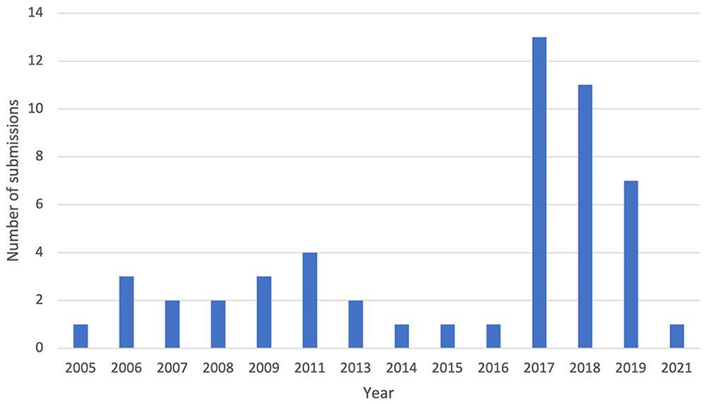
Number of fish diagnosed with neoplasia from 2005–2021 at a large, display aquarium.
Species, number of fish, tumor type, and tissue location for neoplasia in fish at a large display aquarium, 2005–2021.
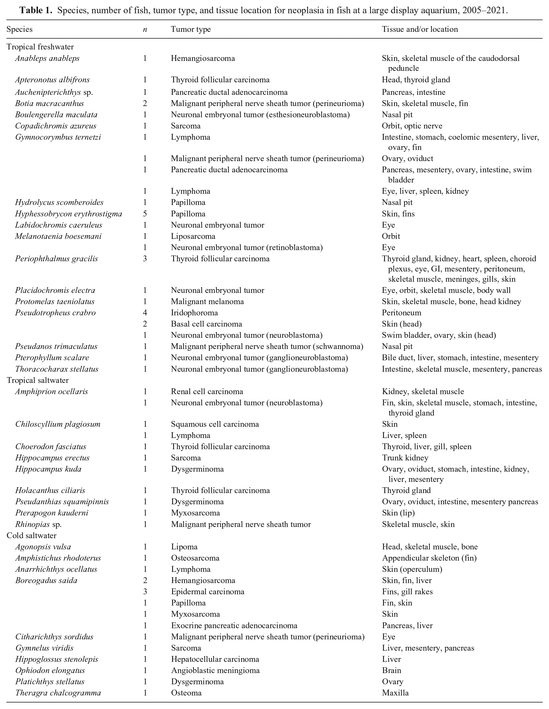
Thirty-five tumors were diagnosed in the tropical freshwater fish population. Of these tumors, 16 were of neuroectodermal or neural crest cell origin (Fig. 2), 14 were of epithelial origin, 3 were of mesenchymal origin, and 2 were lymphoma. There were no germ cell tumors in this population. The most common tumor site of origin was the skin (n = 11). Most tumors were malignant (n = 29).
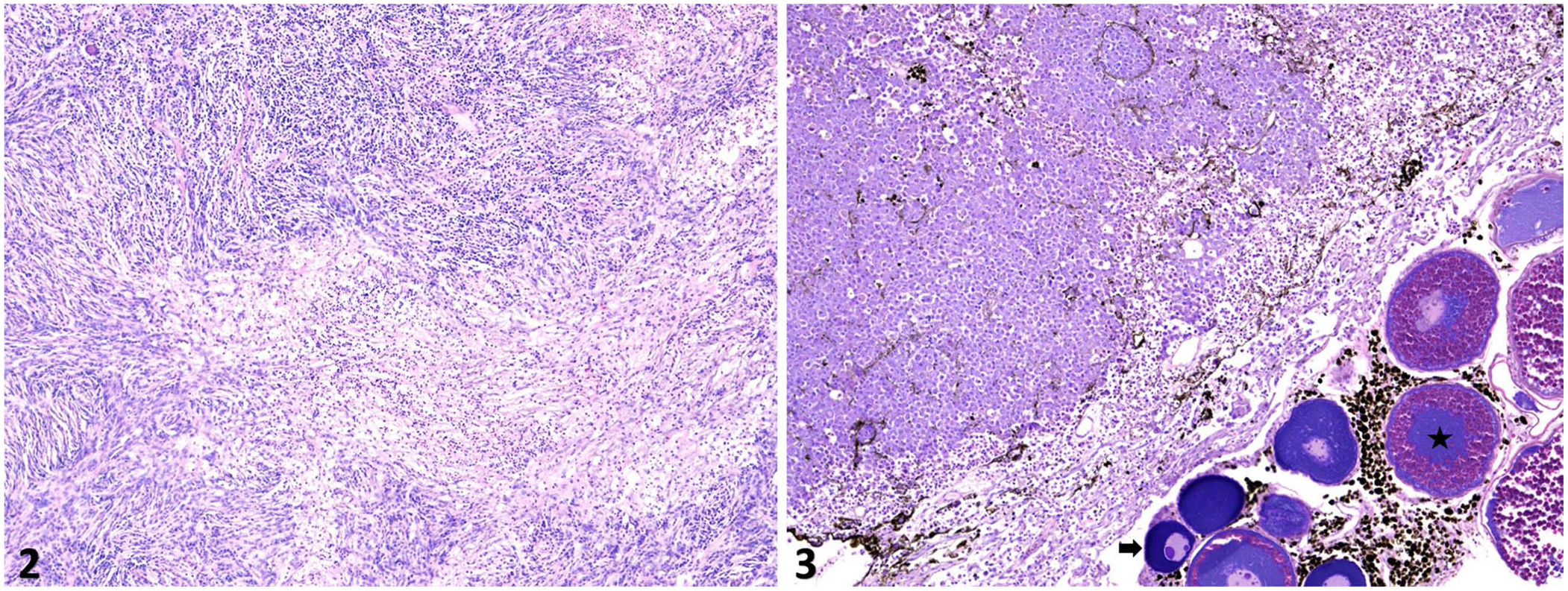
Ganglioneuroblastoma arising from the peripheral nervous system of a silver angelfish (Pterophyllum scalare). A well-differentiated region within the neoplasm consists of multiple tissue and cell types including glial cells, scattered neurons, and Schwannian stroma. Areas of necrosis are largely confined to the Schwannian stroma. H&E.
Eleven tumors were diagnosed in the tropical saltwater fish population. Of these tumors, 4 were of epithelial origin, 2 were of neuroectodermal or neural crest cell origin, 2 were of germ cell origin, 2 were of mesenchymal origin, and 1 was lymphoma. All tumors in this population were malignant. The most common site of tumor origin was the skin (n = 4). Seventeen tumors were diagnosed in the cold saltwater fish population. Of these tumors, 7 were of mesenchymal origin, 6 were of epithelial origin, 2 were of neuroectodermal or neural crest cell origin, 1 was of germ cell origin (Fig. 3), and 1 was lymphoma. The most common site of tumor origin was the skin (n = 8). Most tumors were malignant (n = 13).
Discussion
We believe that the reason for the increased number of neoplasia cases seen in 2017 was due to more specimens submitted for histopathology rather than an increased prevalence of neoplasia compared to other years with a lower number of submissions.
Lymphoma was one of the more common neoplasms diagnosed in our study population. Lymphoma has been described in various fish species.3,5,10,11,18,19,25,26,30,45,47,48 In our study, half of the lymphoma cases were diagnosed in tropical freshwater fish, which is in contrast to most of the literature in which lymphoma was diagnosed in cold saltwater fish.
Iridophoroma was one of the more common neoplasms diagnosed in our study population. Iridophoroma has been reported in teleosts.29,37,43 All of the iridophoroma cases were reported in bumblebee cichlids (Pseudotropheus crabro) in our study population. We retrieved no cases of iridophoroma in bumblebee cichlids in a search of PubMed and Google Scholar, using search terms “iridophoroma,” “bumblebee cichlid,” and “Pseudotropheus crabro,” suggesting that iridophoroma has not been reported previously in bumblebee cichlids.
Two elasmobranchs were diagnosed with neoplasia in our study population. A whitespotted bamboo shark (Chiloscyllium plagiosum) was diagnosed with lymphoma of the spleen and liver. 48 Another conspecific in our study population had a diagnosis of squamous cell carcinoma of its rostrum. 9 Neoplasia is relatively uncommon in elasmobranchs, with only a few reports in the primary literature.15,26,34 One of these reports was a 16-y retrospective study that reported neoplasia in only 6 of 1,546 elasmobranchs (0.4%), further illustrating the relative scarcity of neoplastic conditions. 15
There were several neuronal embryonal tumors diagnosed in our study population, including esthesioneuroblastoma, retinoblastoma, ganglioneuroblastoma, and neuroblastoma. Neuronal embryonal tumors are relatively well documented in teleost fish.12,16,23,47,49 In our study population, all of these tumors occurred in tropical freshwater fish. Most fish had nasal or ocular lesions, which is consistent with the current teleost literature. A neuronal embryonal tumor should be considered a top differential in a fish with a nasal or ocular mass.
Germ cell tumors, 2 of which were diagnosed in our study population, have been reported in teleosts and elasmobranchs, but are rare.21,22
Several slender mudskippers (Periophthalmus gracilis) had proliferative thyroid masses identified during the study period. Similar masses have been reported in other teleost fish.1,4,30,32 Most reports of proliferative lesions in the thyroid are hyperplastic as opposed to neoplastic.7,27,33 In our study period, 3 of 4 P. gracilis were diagnosed with thyroid follicular carcinoma and were included in the study population. One P. gracilis was diagnosed with thyroid hyperplasia and was excluded from the study population. Proposed factors that may cause thyroid tissue proliferation in teleost fish include iodine deficiency and other nutritional imbalances, seasonal factors, genetic susceptibility, water quality, and exposure to goitrogenic substances.6,14
Some limitations of our study include variability in medical record data and lack of proven immunohistochemical stains or other diagnostic stains. Immunohistochemistry (IHC) using mammalian antibodies is often of limited utility as an aid to determining tumor diagnoses in fish and requires the use of both mammalian and fish internal positive controls for valid interpretation. Physical antemortem and postmortem examinations were performed by various clinicians and pathologists throughout the study period. Additionally, not all fish were submitted for histopathology (e.g., if marked autolysis was noted, the fish were discarded as opposed to being submitted). Future research should focus on comparing neoplasms across institutions, comparing cases in free-ranging versus managed populations, and investigating IHC stains and therapeutic options for neoplasms in managed fish populations.
Footnotes
Acknowledgements
We thank Kristi Heffron, Michael Manalang, and Drs. Gary Marty, Doran Kirkbright, Laura Baseler, and Brock Chappell for their contributions to this study. We also thank the aquarists and Animal Health team for maintaining a focus on quality of life for each animal in their care.
Declaration of conflicting interests
Heindrich Snyman is a guest editor of the JVDI special section on “Aquatic animal health and disease” but was not involved in the editorial evaluation or decision to accept this article for publication
Funding
The authors received no financial support for the research, authorship, and/or publication of this article.