
Abstract
A 13-year-old female Boer goat with a history of chronic arthritis and recurrent episodes of recumbency, increased serum gamma-glutamyl transferase, and complete inability to stand was submitted for necropsy. Gross changes included the presence of a white, firm, smooth, 6 cm × 4 cm × 3 cm mass that diffusely expanded and partially effaced the gall bladder and infiltrated the adjacent hepatic parenchyma. On cut surface, the mass was pale yellow and had small, irregular, dark yellow areas. Scattered through the pancreas were 2 gray, well-demarcated, soft, homogeneous, 1 cm in diameter nodules that bulged out from the capsular surface. The right femoral head had a locally extensive area of cartilage erosion. Histological and immunohistochemical evaluation of the gall bladder and pancreatic nodules revealed a primary cholecystic adenocarcinoma and multifocal insulinomas, respectively. A metastatic focus from the gall bladder neoplasm was observed infiltrating the right adrenal gland medulla. The goat also had mild pulmonary infestation by Muellerius capillaris. Primary hepatobiliary and pancreatic neoplasia is rare in goats, and to the authors’ knowledge, neither cholecystic adenocarcinoma nor pancreatic insulinomas have been previously reported in this species.
Keywords
Primary hepatobiliary neoplasia is relatively common in domestic animals. 9 Benign and malignant hepatocellular tumors, biliary tumors, neuroendocrine tumors, and mesenchymal tumors are included in this group and have all been well described in the veterinary medical literature. 9 Biliary neoplasia can originate from intra- or extrahepatic biliary ducts or from the gall bladder, 9 and although primary gall bladder neoplasia is fairly common in human medicine, it has been rarely described in veterinary medicine.8,9 Descriptions of primary gall bladder (cholecystic) papillomas, adenomas, and adenocarcinomas are restricted to a few cases in cattle,2,3 cats,10,21 dogs,4,22 pigs,1,19 lions, 25 bearded dragons, 16 fat sand rats, 27 and mice. 28 Although a few reports of hepatic cholangiocarcinomas have been described in goats,15,23,24 to the authors’ knowledge, there have been no reports of primary cholecystic neoplasia in this species.
Pancreatic islet cell neoplasia is fairly common in dogs, 12 ferrets, 6 cattle, 17 and less often cats. 13 The most common islet cell tumors are either adenomas or carcinomas arising from insulin-secreting beta cells (insulinomas). These tumors may be functional and lead to clinical signs associated with hypoglycemia.6,12,13,17 To the authors’ knowledge, no previous reports of islet cell tumors affecting goats have been reported in the veterinary literature.
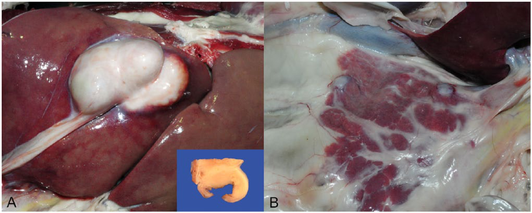
The current study describes a case of cholecystic adenocarcinoma and multiple pancreatic insulinomas in a 13-year-old female Boer goat with a history of chronic arthritis and recurrent episodes of recumbency. The clinical signs worsened 2 days prior to referral, and the goat presented with lateral recumbency and complete inability to stand. Blood work revealed increased serum gamma-glutamyl transferase (230 IU/l; reference range: 27 ± 3 IU/l). 20 Because of the progressive nature of the clinical signs and the possibility of rabies, the goat was subjected to euthanasia and necropsy. The main gross findings were restricted to the liver and pancreas (Fig. 1). A white, firm, smooth, 6 cm × 4 cm × 3 cm mass was observed infiltrating and partially effacing the gall bladder and extending to the adjacent hepatic parenchyma (Fig. 1A). The cut surface was pale yellow, with small, irregular dark yellow areas. The pancreas had 2 gray, well-demarcated, soft, homogeneous, 1 cm in diameter nodules that bulged out from the capsular surface (Fig. 1B). A locally extensive area of cartilage erosion was present on the right femoral head (chronic arthritis). Fluorescent antibody testing for rabies and Listeria spp. was performed on fresh samples of brain, and results were negative. No gross findings were observed within the ear canal or internal ear and in the cranial nerves. Multiple tissues were fixed in 10% formalin, trimmed, routinely processed for histology, sectioned at 4-µm thickness, and stained with hematoxylin and eosin. Histological examination revealed 2 different neoplasms affecting the gall bladder and pancreas (Fig. 2). The gall bladder architecture was almost completely effaced by an extensive, transmural, and infiltrative mass composed of neoplastic cells that were densely arranged in numerous acini and tubules containing luminal basophilic secretory material and supported by a dense fibrovascular and collagenous stroma (desmoplasia). Neoplastic cells had distinct cell borders and moderate amounts of eosinophilic, granular cytoplasm. Nuclei were oval and had finely stippled to vesicular chromatin with 1–3 nucleoli. Mitoses averaged from 0 to 1 per each high power field at 400×. Anisocytosis and anisokaryosis were moderate. Necrotic neoplastic cells were frequent and often located superficially toward the lumen of the gall bladder where they were admixed with small mineralized foci and basophilic, flocculent secretory material. The tumor multifocally infiltrated and compressed the immediate adjacent hepatic parenchyma. Hepatocytes in these areas were dissociated and admixed with hemorrhage and small numbers of neutrophils, lymphocytes, and plasma cells. No histological changes were observed throughout the remaining hepatic parenchyma. A well-demarcated, nodular, 1 mm in diameter metastatic focus from the gall bladder tumor was observed infiltrating the right adrenal medulla. Neoplastic cells were morphologically similar to those observed in the gall bladder neoplasm and were supported by abundant fibrous stroma.
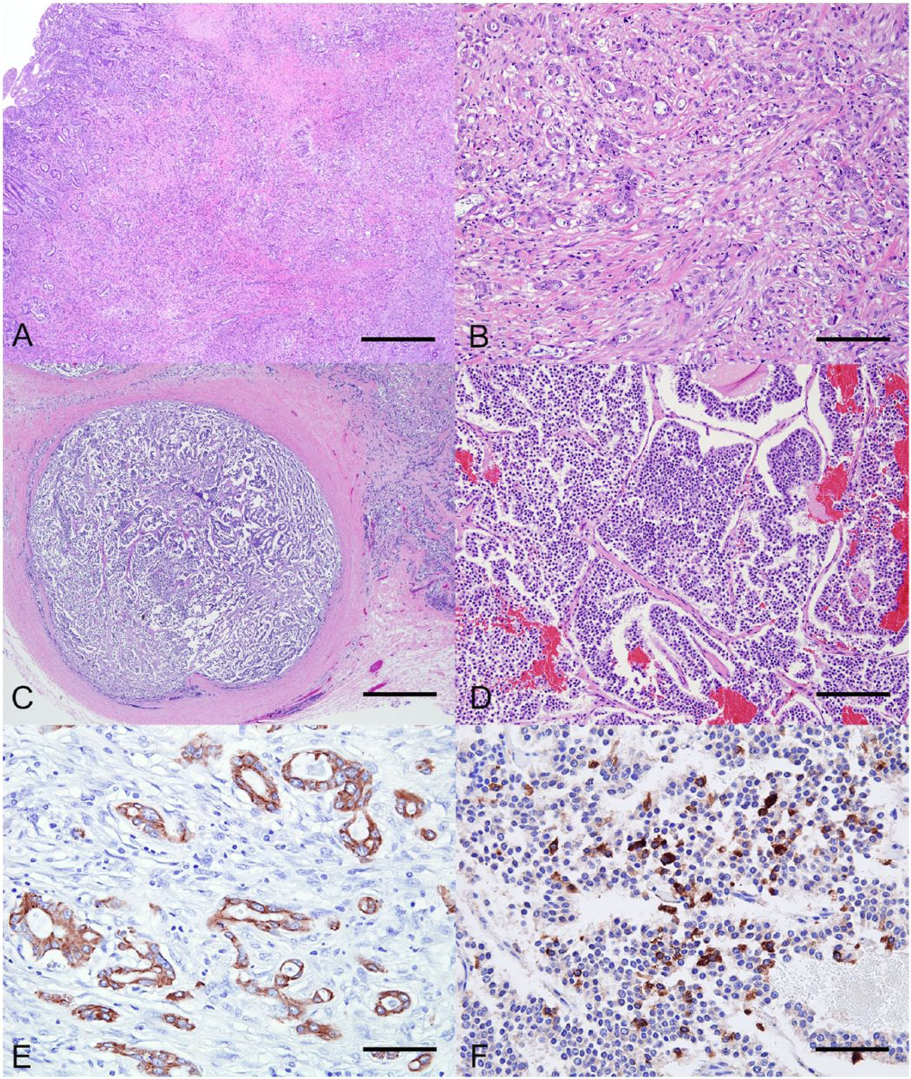
The pancreatic nodules were composed of well-demarcated, fully encapsulated accumulations of neoplastic cells arranged in small nests, packets, or cords supported by a fine fibrovascular stroma. Neoplastic cells were polygonal and had indistinct cell borders, with small amounts of eosinophilic, granular cytoplasm. Nuclei were oval and had finely stippled chromatin with 1 basophilic nucleolus. Mitoses were rare. Rare areas of hemorrhage were present within the neoplastic nodules.
In addition, affecting 10% of the pulmonary parenchyma were multifocal areas of alveolar septal fibrosis associated with clusters of foamy and epithelioid macrophages that were admixed with unembryonated and embryonated nematode eggs with a thin, eosinophilic shell. The eggs were filled with multiple, 5-µm-diameter blastomeres. There were also a few adult nematodes measuring up to 60 µm in diameter that were covered by a thin, smooth, eosinophilic cuticle with polymyarian–coelomyarian musculature and a pseudocoelom containing an intestine lined by multinucleate cells. The nematodes and nematode eggs were morphologically consistent with Muellerius capillaris. 11
Immunohistochemical staining (IHC) for pancytokeratin (Lu-5) and insulin was performed in sections of gall bladder and pancreas, respectively. The primary antibody used for pancytokeratin IHC was a mouse monoclonal pancytokeratin a at a dilution of 1:100 with an incubation period of 90 min. Haired skin and adjacent normal biliary epithelium were used as control and internal control, respectively. Insulin primary antibody was monoclonal guinea pig anti-insulin b at a dilution of 1:800 and an incubation time of 30 min. Adjacent pancreatic tissue was used as internal control. Approximately 70% of neoplastic cells in the gall bladder and adjacent hepatic parenchyma expressed strong cytoplasmic immunostaining for pancytokeratin, and approximately 90% of neoplastic cells in the pancreatic nodules showed moderate to strong cytoplasmic immunostaining for insulin.
The gross, histological, and immunohistochemical findings in this goat are consistent with a primary cholecystic adenocarcinoma (with metastasis to the adrenal medulla) and multifocal pancreatic insulinomas. The pulmonary infestation by M. capillaris was mild and considered incidental in the present case. Cholecystic adenocarcinomas may be locally invasive and destructive, and therefore their differentiation from disseminated cholangiocarcinomas may be difficult, especially when they diffusely affect the hepatic parenchyma and their primary site of origin can no longer be determined. 25 In the current case, the presence of a single gall bladder mass with invasion only into the immediate adjacent hepatic parenchyma supported the diagnosis of a primary gall bladder tumor. Although IHC for pancytokeratin was performed to confirm and illustrate the cholecystic tumor in the present case, biliary adenocarcinomas usually have characteristic morphological features that would not require such ancillary tests for confirmation in the diagnostic routine. For this reason, no IHC was run on sections of the adrenal gland containing the metastatic focus. Cholecystic adenocarcinomas are relatively common in human beings and have been associated with chronic irritation and inflammation or infection caused by gallstones in the majority of cases. 8 In contrast, no information regarding potential etiology, risk factors, or breed, age, and sex predilection have been described for these tumors in veterinary medicine because only a few single case reports have been published. 9 Cholecystic adenomas have been described as one of the most common hepatic neoplasms occurring in slaughtered cattle1,2 and have been less often reported in dogs and cats,9,26 but cholecystic adenocarcinomas are rare in all veterinary species 9 and have never been described in goats. In fact, 2 recent retrospective studies performed on caprine cases submitted to the Department of Pathology and Athens Veterinary Diagnostic Laboratory at the University of Georgia (Athens, Georgia) 14 and to the Veterinary Diagnostic Laboratory at the Oregon State University (Corvallis, Oregon) 18 revealed a prevalence of neoplastic lesions in approximately 10% of the diagnosed cases, but no primary hepatic neoplasia was observed in these studies. The most common neoplasms in these studies included lymphoma, cutaneous squamous cell carcinoma, thymoma, and pheochromocytoma.14,18
The multiple pancreatic tumors in the current case were confirmed as islet cell tumors of beta cell origin (insulinomas) by IHC. Beta cell tumors may occur as single or multiple nodules affecting the same or different pancreatic lobes. 5 While beta cell adenomas are encapsulated from the remaining parenchyma, carcinomas tend to be larger, locally invasive, and can also metastasize to regional lymph nodes and liver. 5 However, it may be difficult to distinguish benign from malignant islet cell tumors based on morphological features. The presence of well-demarcated and encapsulated nodules with no invasion of surrounding tissues supported the diagnosis of benign pancreatic neoplasia in this case.6,12,13,17 Because of the lack of a complete biochemical profile that could have measured blood glucose levels, it was not possible to determine whether the insulinomas were functional in this case. Although the periodic episodes of recumbency could be attributed to severe hypoglycemia,6,12,13,17 such signs are nonspecific and could also be secondary to the chronic arthritis.
Muellerius capillaris is the most common pulmonary nematode of small ruminants. Although heavy infestations may produce respiratory clinical signs, infestation in the goat in the present study was mild, and no evidence of clinical significance was noted. 7
In summary, a case of primary cholecystic adenocarcinoma with adrenal gland metastasis in a goat that also had multifocal insulinomas is described. The diagnosis was made based on histology and confirmed by IHC for pancytokeratin and insulin. The pulmonary parasitism was mild and considered as an incidental finding.
Footnotes
Acknowledgements
The authors thank Patricia Rowe from the Histology Laboratory at the Athens Veterinary Diagnostic Laboratory (University of Georgia) for her amazing technical assistance with the immunohistochemistry.
a.
Pan-cytokeratin Lu-5, Dako North America Inc., Carpinteria, CA.
b.
Insulin, Dako North America Inc., Carpinteria, CA.
Declaration of conflicting interests
The author(s) declared no potential conflicts of interest with respect to the research, authorship, and/or publication of this article.
Funding
The author(s) received no financial support for the research, authorship, and/or publication of this article.