
Abstract
A flock of 48 sheep in Argentina grazing on a pasture of hybrid Urochloa (formerly Brachiaria) Mulato II (Urochloa ruziziensis × Urochloa decumbens × Urochloa brizantha) developed facial dermatitis, severe jaundice, and weakness after brief physical activity. Blood biochemistry of 3 animals revealed azotemia, elevated aspartate aminotransferase activity, and increased direct, indirect, and total bilirubin concentrations. The urine was markedly turbid and contained large concentrations of bile pigments and protein. At autopsy of 2 animals, there was severe jaundice and subcutaneous submandibular edema. The livers were enlarged, intensely yellow, and had a marked acinar pattern. Gallbladders were distended, and the kidneys were diffusely dark in one animal and yellow-green in the other. Microscopically, there was lymphoplasmacytic and histiocytic cholangiohepatitis with abundant crystals in the lumen of bile ducts and in the cytoplasm of macrophages. The proximal and distal convoluted renal tubules had protein casts in their lumens, and crystals were observed in the lumen and epithelial cells. Lectin histochemistry showed strong affinity for Arachis hypogaea agglutinin in hepatic macrophages. In the one sheep that was tested for heavy metals, copper concentrations in the liver and kidney were within the RIs. Despite the immediate change of pasture, morbidity and mortality were 100% within 3 mo. The association between the consumption of this pasture, and the clinical, biochemical, pathology, and lectin histochemistry findings confirmed intoxication with Urochloa hybrid Mulato II. To our knowledge, intoxication by this hybrid of Urochloa has not been reported previously.
Plants of the genus Urochloa (formerly Brachiaria), Panicum, and Tribulus have been described as the cause of hepatogenous photosensitization associated with lithogenic saponins.15,23,29,32,34 Spontaneous photosensitization associated with crystal hepatopathy and nephropathy has been described in sheep grazing on Tribulus terrestris pastures.20,36 The condition has been reproduced experimentally in sheep 2 and goats. 3 Similar lesions were described in cattle, 27 sheep, 1 goats, 35 and buffaloes 16 grazing Panicum miliaceum, Urochloa decumbens, Urochloa brizantha, Urochloa humidicola, or Urochloa ruziziensis. In goats 12 and sheep, 30 similar lesions were described in animals spontaneously intoxicated with Urochloa hybrid Mulato I (U. ruziziensis × U. brizantha).
Urochloa hybrid Mulato II is the result of 3 generations of crossbreeding of U. ruziziensis, U. decumbens, and U. brizantha conducted by the International Center for Tropical Agriculture (CIAT) in Colombia. It is a grass of high quality and forage production adapted to humid and sub-humid tropical regions. 37
Published information about the forage value and potential toxicity of this plant is scant. In the only experimental study feeding adult sheep with U. hybrid Mulatto II, no clinical, hematologic, or pathologic changes were observed. 30 After searching PubMed, CAB, and Google Scholar, we could not find reports of any toxic event associated with the consumption of Urochloa hybrid Mulato II. We describe here an outbreak of intoxication in sheep grazing Urochloa hybrid Mulato II, with special emphasis on the hepatic and renal lesions.
One of the authors of our report (R. Marin) visited a smallholding producer in the El Carmen Department, Jujuy Province, northwest Argentina. The producer had a flock of 48 sheep of different ages, including 36 native creole and 12 Corriedale animals. The sheep had recently been introduced to the property and, at the time of the visit, had been grazing a pasture of Urochloa Mulato II for 30 d, without access to other feed. The pasture planting had been carried out following the instructions of the company that supplied the Urochloa Mulato II seeds, which certified their identification. In addition, no other Urochloa species had ever been planted on the farm or neighboring premises, which most likely eliminates the possibility of contamination by other Urochloa species in the pasture.
At the time of the visit, 22 animals, including 10 creole and all of the Corriedale animals, had anorexia, weight loss, depression, and facial dermatopathy, with a clinical course of 5–7 d. The affected sheep were examined clinically. Eyelid edema, photophobia, conjunctivitis, dermatoblepharitis, nasal dermatitis, submandibular edema, jaundice, and weakness after brief physical activity were observed. The onset of clinical disease was observed after 23–25 d of grazing the Urochloa Mulato II pasture. At the time of the field visit, morbidity and mortality were 50% and 8.3%, respectively, but both reached 100% over 3 mo, despite the immediate change of diet at the time of the visit. The new diet consisted of alfalfa hay and corn, but the anorexia in the flock was very severe and consumption was poor.
Whole EDTA blood and serum were collected from 3 clinically affected animals and used for CBC and biochemistry. A urine sample was obtained from a sick animal for urinalysis. The blood biochemistry revealed elevated activity of aspartate aminotransferase (AST), and increased concentrations of urea, creatinine, and direct, indirect, and total bilirubin in the 3 animals studied (Table 1). RBC count, packed cell volume, and hemoglobin concentration were within RIs in all animals evaluated. The urine had marked turbidity and high concentrations of bile pigments and proteins; hemoglobin was not detected.
Blood chemistry results in sheep intoxicated with Urochloa hybrid Mulato II.
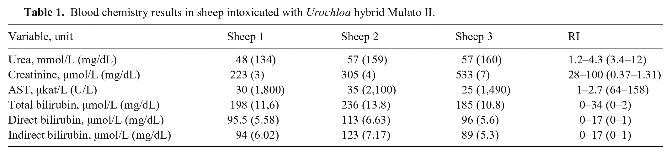
An autopsy of 2 adult sheep that died spontaneously was performed. Sheep 1 had clinical signs for 5 d; sheep 2 had been clinically sick for 12 d. In both cases, the autopsy was performed ~6 h after death. Both carcasses were in a mild-to-moderate state of postmortem decomposition. Sheep 1 was in good nutritional condition, with adequate fat reserves and muscle. Sheep 2 was in poor nutritional condition, with no fat reserves, serous atrophy of fat, and generalized and severe muscle atrophy. Gross lesions were similar in both animals. The skin of the periocular region had symmetrical lesions consisting of severe crusting and abundant serohemorrhagic exudate, which was most marked in the area surrounding the ocular medial commissure (Fig. 1); the skin surrounding the nares had moderate crusty lesions. Intense yellow discoloration was observed in the sclera (Fig. 1), oral and vulvar mucosae, articular cartilage, and subcutaneous and visceral fat. Submandibular subcutaneous edema was severe. The liver was enlarged, had rounded edges, was bright yellow, and had a marked acinar pattern (Fig. 2). The gallbladder was distended by a large amount of thick bile. The kidneys of sheep 1 were diffusely dark-brown to black (Fig. 3), and the urine was dark-brown. The kidneys of sheep 2 were diffusely green-yellow (Fig. 4); no urine was present in the urinary bladder of this animal.
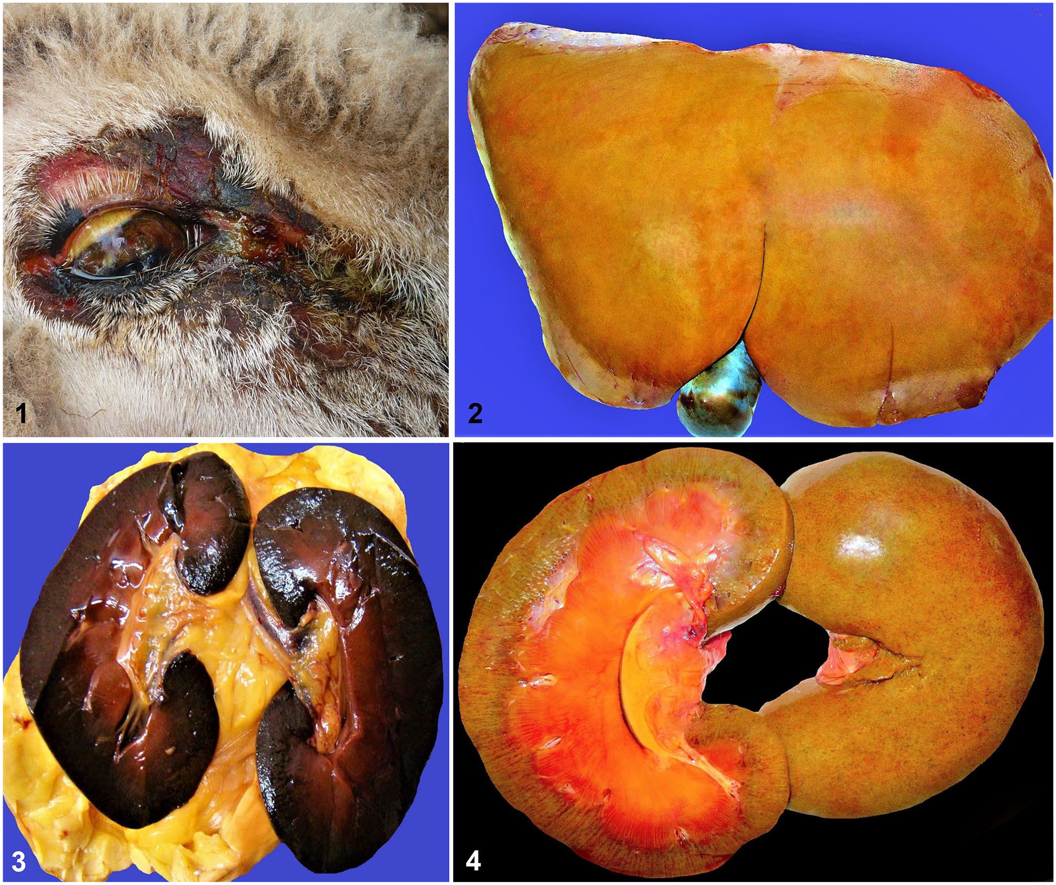
Gross findings in sheep intoxicated with Urochloa hybrid Mulato II.
Samples of liver and kidneys were collected and fixed in 10% neutral-buffered formalin, pH 7.2, for 24 h. Tissue samples were routinely processed for the production of 4-μm sections that were stained with H&E and Hall for bile. Microscopically, lesions were similar in both animals. There was disorganization of the hepatic cords, and mildly distended sinusoids with canalicular bile stasis (Fig. 5). Most hepatocytes had fine intracytoplasmic microvacuolation or a single large intracytoplasmic vacuole (Fig. 6). Randomly, individual hepatocytes had cytoplasmic swelling and acidophilia, karyorrhexis, or karyolysis. Multifocally, there was a moderate number of foamy macrophages, single or in clusters, many of them with ≥2 nuclei (Fig. 6). Frequently, these cells contained intracytoplasmic spiky birefringent crystals. Similar crystals were also observed in the lumen of a few bile ducts. Many bile canaliculi were distended by bile (Figs. 5, 6). In addition, mild periportal proliferation of bile ducts and lymphoplasmacytic infiltration were observed. In the kidneys, there were intratubular protein casts and many crystals, similar to those described in the liver, in the lumen of proximal convoluted tubules, admixed with eosinophilic homogeneous material (Figs. 7, 8). The epithelium of these tubules was diffusely degenerate or necrotic, and many of the epithelial cells had sloughed into the lumen. Occasionally, large cytoplasmic vacuoles were observed in the epithelium of the proximal and distal convoluted tubules. Intracytoplasmic brown pigment in the epithelium of proximal convoluted renal tubules and admixed with the crystals was diffusely observed.
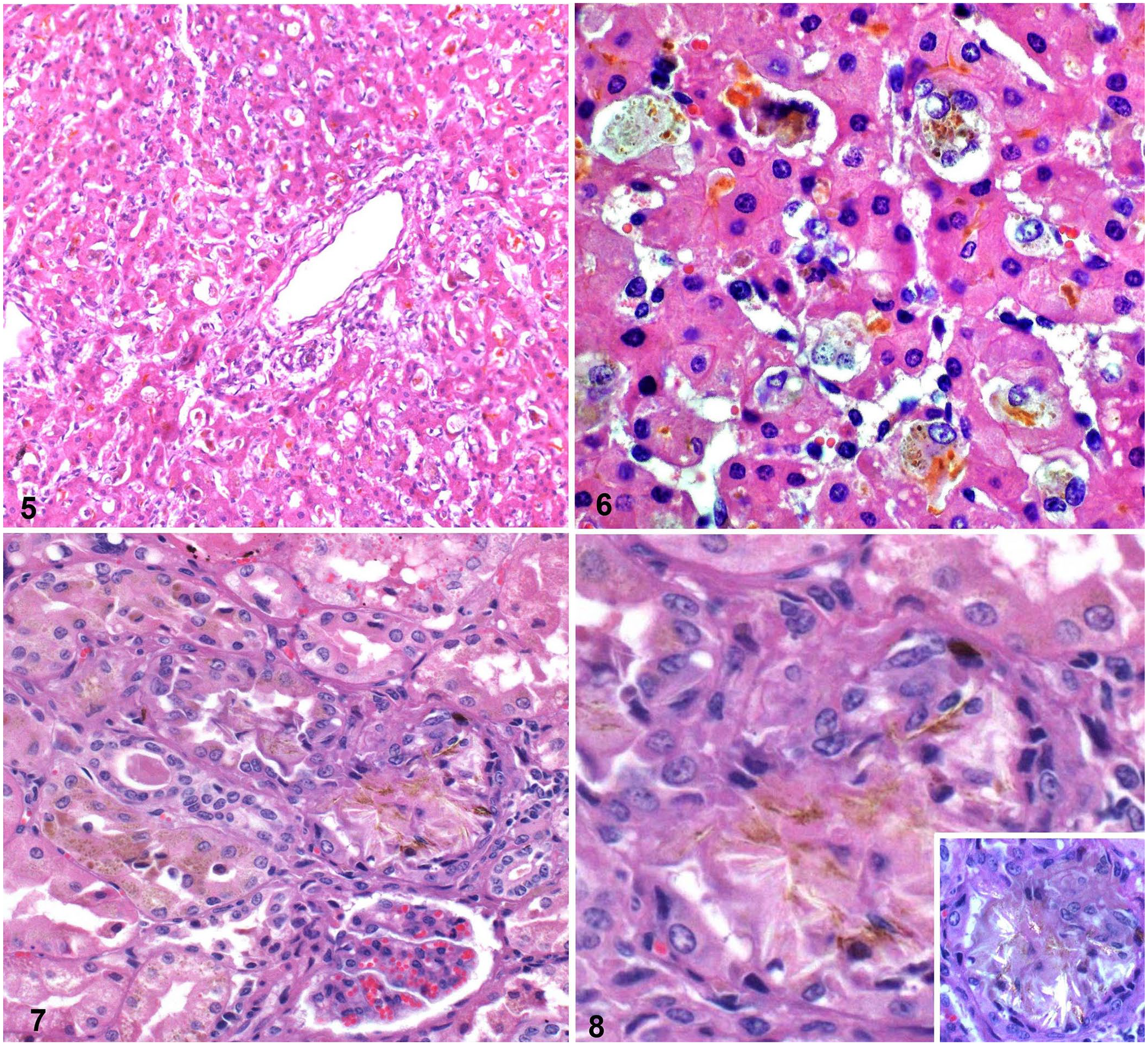
Microscopic hepatic and renal findings in sheep intoxicated with Urochloa hybrid Mulato II.
Paraffin sections of liver were processed for lectin histochemistry using biotinylated lectin Arachis hypogaea agglutinin (PNA; Vector) as described previously. 12 Cells with foamy cytoplasm had strong labeling by PNA lectin.
Formalin-fixed liver and kidney of sheep 1 were processed for heavy metals by atomic absorption spectrophotometry per California Animal Health and Food Safety Laboratory (Davis, CA, USA) standard operating procedures. This included copper, lead, cadmium, cobalt, arsenic, and zinc. All heavy metal concentrations were within RIs.
The clinical signs, gross and microscopic lesions, results of lectin histochemistry and blood biochemistry, in association with the history of exclusive consumption of Urochloa hybrid Mulato II for the previous 30 d, confirmed a diagnosis of intoxication by this plant. Other plants present in Argentina that could cause photosensitization and crystal-associated cholangiohepatitis and nephrosis in sheep include U. brizantha, U. humidicola, U. Mulato hybrid, T. terrestris, and Panicum spp. Intoxication by these plants was ruled out because the animals had not had access to them. Pithomyces chartarum, 13 another common cause of photosensitization, was ruled out based on microscopic hepatic and renal changes. A possible association with chronic copper intoxication in sheep 1 was ruled out based on normal hepatic and renal copper concentrations. The difference in color between the kidneys of sheep 1 and 2 may have been associated with the different clinical progression of the disease in the animals. The dark kidney of sheep 1 was compatible with hemoglobinuric nephrosis; however, hemoglobin was not detected in the urine of this animal, and the cause of the kidney discoloration in this animal could not be explained satisfactorily.
The most characteristic lesions of intoxication by Urochloa spp. are birefringent crystals in the bile ducts, macrophages, and hepatocytes, all of which were observed in our case. 14 Cells with foamy cytoplasm in the liver stained positively with PNA lectin, a finding that is compatible with macrophages. The presence of foamy macrophages in the liver and kidney is a characteristic of Urochloa spp. poisoning.14,21
In Argentina, U. brizantha and Mulato II, among other Urochloa species, are recommended as good forage resources for the sub-tropical northeastern region of the country. 9 In this region, 2 outbreaks of photosensitization were reported in cattle grazing U. brizantha 10 and U. decumbens, 17 respectively, contaminated by Pithomyces chartarum. However, the spore count of P. chartarum in the pasture was very low in one outbreak; no microscopic description of the hepatic lesions was provided for the second outbreak. In addition, no description of renal lesions was provided for either of these 2 outbreaks. It is possible that these 2 outbreaks were caused by intoxication with the Urochloa species and not by P. chartarum. In Brazil, poisoning by Urochloa spp. was misdiagnosed for many years as poisoning by P. chartarum. 33
The use of Urochloa spp. in northwestern Argentina seems to be more limited than in the northeastern region. However, an outbreak of severe dermatitis affecting 80% of 400 recently weaned Brangus red calves, which had been grazing Urochloa hybrid Mulato I and U. brizantha for 40 d, was described (authors’ unpublished observation). The animals had no previous exposure to Urochloa spp. and, after consumption of this plant, had anorexia, apathy, loss of condition, and swelling and scarring of the ears, the tips of which were necrotic.
In the area where our cases of intoxication by Mulato II occurred, anecdotal evidence suggests that intoxication by this plant has occurred frequently. However, because these farms are located in remote locations far away from veterinary assistance, it is likely that those cases went unreported. Intoxication by Urochloa may be underdiagnosed in Argentina.
Urochloa spp. is a highly productive grass that is widespread in several tropical and subtropical regions of the world and that adapts to a wide range of environments and soil types. However, a limiting factor for the use of this grass is its toxicity, which is associated with lithogenic steroidal saponins (dicotomin, protodioscin, saponin B) 28 that induce the formation of crystals in the biliary system. 33 Protodioscin is the main saponin found in Urochloa spp. 7 The most characteristic lesion of Urochloa spp. poisoning is the presence of birefringent crystals in the bile ducts, hepatic macrophages, hepatocytes, 18 and renal tubular epithelium. 33 These lesions were seen in our sheep cases. The gross and microscopic hepatic and renal changes observed in our cases were similar to, and indistinguishable from, those described previously in sheep intoxicated by other Urochloa spp. The elevated serum AST activity and total, direct, and indirect bilirubin concentrations, and the microscopic findings in our case demonstrate severe hepatic damage, and have been described in natural and experimental poisoning of cattle and sheep with U. brizantha, U. humidicola, and U. ruziziensis.27,33
In our case, we speculate that the skin lesions observed grossly were a consequence of the action of photodynamic substances accumulated because of liver failure. Skin was, unfortunately, not collected for histopathology during the autopsy of the animals, and we cannot draw definitive conclusions about the pathogenesis of the skin lesions. The gross lesions were, however, very characteristic of photosensitization, which, coupled with the clinical history and gross and microscopic lesions in liver and kidneys, is highly suggestive of this condition. The clinical, hematologic, histopathologic, and lectin histochemical findings associated with the consumption of Urochloa hybrid Mulato II were similar to cases described in intoxications by Urochloa spp.18,27,33 and led us to the final diagnosis of intoxication by Urochloa Mulato II.
Our findings differ from those obtained in the only report we could find of adult sheep fed Urochloa hybrid Mula-tto II experimentally, in which no clinical, hematologic, or histopathologic changes were observed. 30 The reason for this difference in outcome was not determined. The toxicity of Urochloa spp. varies depending on several factors, including the species and growth phase of the plant, and the animal species, age, susceptibility, and previous experience with the plant. U. decumbens is the most toxic of the Urochloa spp., and it contains the highest concentrations of protodioscin, followed in decreasing order of toxicity, by U. brizantha and U. ruziziensis, U. humidicola, and U. dictyoneura. Young leaves have higher concentrations of protodioscin than mature or old senescent leaves.4,5,25 Sheep are more susceptible than cattle and goats to intoxication by Urochloa spp., although, within the same species, there are differences in susceptibility between individual animals. Young animals of all species are more susceptible than adults to intoxication by this plant. Finally, naive animals with no previous contact with Urochloa spp. plants are more susceptible to poisoning than animals raised in areas where plants of this species are present.9,11,19,20,22,24 Some of these factors were considered in the outbreak reported here and were likely responsible for the 100% mortality observed. Sheep are the most susceptible animal species to intoxication by Urochloa spp.; in addition, the animals in our case were naïve to Urochloa spp. exposure. 19 The Urochloa hybrid Mulato II has a very high concentration of protodioscin, which is similar to that found in U. decumbens, 26 the most toxic Urochloa species known.6,8,25,33 Additionally, our cases did not have access to any other feed, the stocking rate was very high, and the pasture was sprouting, a stage at which there is a higher protodioscin concentration in the plants. Also, the sheep flock was continuously exposed to the sun with no shade available, a factor that exacerbates the toxicity of the plant. 31
In Brazil, U. decumbens was introduced in the 1950s, and in the early years caused numerous outbreaks of poisoning in livestock. The frequency of outbreaks, mainly in cattle, decreased over the years, mainly because there was natural selection: the susceptible animals died and the most resistant ones survived. 33 Another factor contributing to the decrease in the number of outbreaks is that U. decumbens was gradually substituted by less-toxic species, mainly U. brizantha. However, in sheep, Urochloa spp. still causes severe mortalities in Brazil, mainly in naive animals.
Our results and other reports of toxicity with U. brizantha and with hybrid Mulato I (U. brizantha × U. ruziziensis)4,12,30 suggest that it is risky to give naive livestock access to Urochloa pastures. The solution would be to select varieties producing lower protodioscin levels, which to date has not been considered in the genetic improvement of Urochloa spp. Other species of Urochloa with lower saponin content, such as U. humidicola or U. dictyoneura, should be considered as forage resources in tropical regions. Another option for the use of Urochloa spp. is to measure the concentration of protodioscin as part of the selection process for Urochloa spp. and their varieties.
Footnotes
Acknowledgements
We thank Ms. J. Beingesser and Mr. Jose Constante for excellent technical assistance.
Declaration of conflicting interests
The authors declared no potential conflicts of interest with respect to the research, authorship, or publication of this article.
Funding
This study was partly funded by the California Animal Health and Food Safety Laboratory, UCDavis.