
Abstract
Solanum glaucophyllum, a toxic plant known for its calcinogenic effects, causes enzootic calcinosis in ruminant and monogastric animals. We describe an outbreak of enzootic calcinosis that occurred in a herd of 110 horses grazing pastureland heavily contaminated with S. glaucophyllum in Buenos Aires province, Argentina. Ten horses developed clinical signs, and 6 horses died. Clinical signs included abnormal gait (stiff-legged action, short strides), stiffness, thoracolumbar kyphosis, reluctance to move, wide stance, chronic weight loss, weakness, recumbency, and difficulty standing. Autopsy of 2 horses revealed severe mineralization of the aorta, pulmonary arteries, heart, and lungs, consistent with enzootic calcinosis. Although horses usually have very selective grazing behavior, under food restriction conditions, they can ingest the toxic plants and can develop the disease. Enzootic calcinosis should be considered as a differential diagnosis in horses grazing S. glaucophyllum–invaded pasturelands with compatible clinical signs and lesions.
Enzootic calcinosis (EC) refers to a group of diseases characterized by extensive mineral deposition in soft tissues, induced by ingestion of calcinogenic compounds present in certain toxic plants, mostly within the Solanaceae family. These plants contain glycosides that are biologically active analogues of vitamin D3. The glycoside is hydrolyzed to release active 1,25-dihydroxycholecalciferol (vitamin D3; calcitriol). Ingestion of these plants causes increased absorption of calcium and phosphorus from the intestine, chronic hypercalcemia and hyperphosphatemia, and soft tissue mineralization.12,17 Of the calcinogenic plants known to induce EC, Solanum glaucophyllum (waxyleaf nightshade, duraznillo blanco; formerly S. malacoxylon), a perennial shrub native to South America, is one of the most extensively studied.2,19 EC induced by this plant, locally referred to as enteque seco, causes considerable economic losses to the livestock industry. 19
Other plants are well-recognized causes of EC worldwide, 17 including Nierembergia veitchii 16 and Nierembergia rivularis in South America, 8 Cestrum diurnum in North America,7,11 Trisetum flavescens in Europe,1,6,10,18 and Solanum torvum in Oceania. 13 All of these plants are within the Solanaceae family, except for T. flavescens, which is a grass in the family Poaceae. 6
S. glaucophyllum is widely distributed in Argentina, Paraguay, Uruguay, and southern Brazil, 12 at altitudes of 0–600 m above sea level (https://goo.gl/jEbRzT). It is considered a marsh plant that grows in low wet areas that occasionally flood. 14 Depending on environmental conditions, especially precipitation, temperature, and humidity, S. glaucophyllum grows from spring to autumn. It is deciduous and may lose its leaves in the winter or during drought. 14 S. glaucophyllum is not voluntarily ingested by livestock under normal circumstances. Because the toxic glycosides are contained mainly in the leaves, 5 most cases of intoxication result from accidental ingestion of dry leaves admixed with forage after dry summers, when livestock graze almost exclusively on lower grasslands.12,14 Hay contaminated with calcinogenic plants constitutes another potential source of poisoning for horses and cattle. 1
EC is more frequent in ruminants and rare in monogastric species; however, natural cases have been reported in pigs 3 and horses (Gimeno EJ, pers. comm., 2017). Given that vitamin D and its metabolites exist as glycosides in these plants, particularly as various fructoglucosides of variable molecular weight, ruminants are more susceptible to EC because glycosidases in ruminal fluid hydrolyze the glycosides and enhance vitamin D activity, presumably by increasing their bioavailability and absorption from the gastrointestinal tract. Consequently, monogastric species are generally less susceptible to intoxication, except for rabbits. 20 EC has been reproduced experimentally in ruminants, pigs, rabbits, rats, and guinea pigs.16,20
Prolonged ingestion of S. glaucophyllum causes chronic hypercalcemia as a result of the increased consumption of glycosides of vitamin D3.12,17 Based on the pleiotropic physiological role of vitamin D in classical target tissues, as well as evidence of the extraskeletal effects of calcitriol, 4 it is evident that the pathogenesis of EC is highly complex. Experimental studies performed in different animal models that administered ground leaves of S. glaucophyllum orally or intraruminally have shown changes in cell proliferation, differentiation, and apoptosis in the aorta and lungs, 15 skin, 9 thymus, lymph nodes, and spleen (Fontana PA, et al. Structural and functional changes in organs and cells of the immune system in Solanum glaucophyllum intoxicated heifers. Proc 7th Intern Symp Poisonous Plants; June 2005; Logan, UT. Available from: https://goo.gl/6uMxYL). 20 In horses, EC has been associated primarily with the ingestion of T. flavescens in Europe,1,10,18 and less frequently with C. diurnum in the United States. 11 EC has occurred in horses after grazing calcinogenic plants, 10 and also after ingestion of calcinogenic plants present in hay. 1 Clinical signs include weight loss, lameness, disturbances of movement, kyphosis, pain on palpation of the flexor tendons and suspensory ligaments, polyuria, and recurrent colic.1,10–12 Reports describing EC in horses are scarce in the literature, and none of them involve ingestion of S. glaucophyllum, to our knowledge.
We describe herein cases of EC in horses in Argentina, caused by the ingestion of S. glaucophyllum. The outbreak occurred during the late winter and early spring of 2009 at a ~300-hectare farm located in Lezama department, Buenos Aires province, Argentina (35° 52’21.30’’S, 57° 53’50.06’’W). The farm was in a plain area 14 m above sea level, and was divided into 30 hectares of lower natural pasturelands that were heavily populated by S. glaucophyllum, and 270 hectares of higher quality pasture. The 30 hectares of natural pastureland were continuously grazed exclusively by a herd of 110 mixed-breed adult horses year-round.
The first clinically affected animal was detected in August 2009. Subsequently, over a period of ~60 d, 9 more horses developed clinical disease (morbidity = 9%), 6 of which died (mortality = 5%). Clinical signs were slowly progressive and included abnormal gait (stiff-legged action, short strides), stiffness, thoracolumbar kyphosis, reluctance to move, wide stance, chronic weight loss, weakness, recumbency for extended periods of time, and difficulty standing without assistance. Veterinary pathologists visited the farm in October; 4 affected animals were still symptomatic. One of these horses, a 2-y-old mixed-breed gelding (case A) that was in poor body condition and had pale mucous membranes, was euthanized for postmortem examination. A second horse, a 4-y-old gelding (case B) that had died the day before, was also autopsied.
Autopsy findings were similar in both cases. Major arteries including the thoracic aorta and pulmonary arteries, had diffusely rigid walls with marked loss of elasticity, and were variably thickened and expanded by extensive, coalescing, irregular, variably sized, rough-surfaced, firm-to-hard, white, chalky, gritty (mineralized) intramural plaques and coalescing nodules that narrowed the vascular lumens, and were most notable from the luminal (endothelial) surface (Fig. 1). The aortic semilunar valves were moderately thickened and had lost their normal elasticity because of fibrosis, which may have resulted from valvular insufficiency secondary to the abovementioned aortic lesions. There were ~500 mL of clear amber fluid within the abdominal cavities. The caudodorsal aspects of the caudal lung lobes failed to collapse. No other remarkable lesions were seen at postmortem examination; however, the vertebral column and spinal cord were not examined. The parathyroid glands were undetectable grossly.
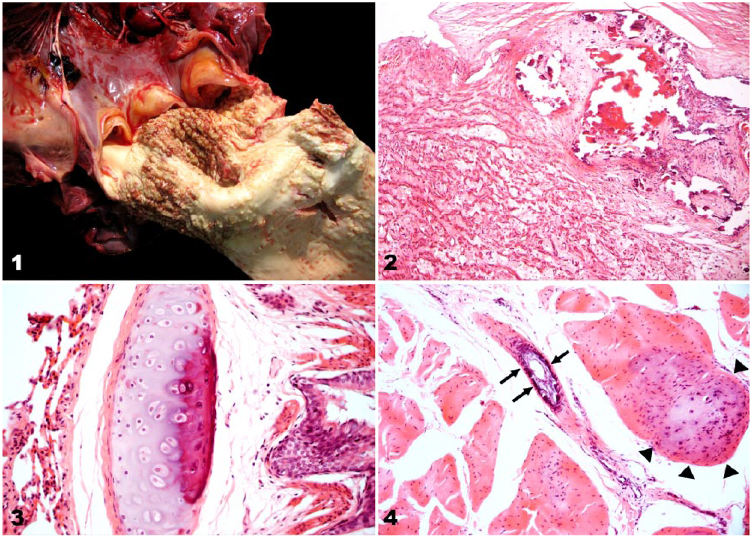
Lesions of enzootic calcinosis in horse A, caused by ingestion of Solanum glaucophyllum.
Several tissues, including aorta, heart, lung, liver, spleen, brain, pancreas, thyroid gland, kidney, superficial digital flexor tendon and skeletal muscle from case A, and heart/pulmonary artery from case B, were fixed in 10% neutral-buffered formalin, routinely processed and embedded in paraffin, sectioned at 4–5 µm, and stained with hematoxylin and eosin (H&E) for histologic examination. Selected sections of aorta/pulmonary artery and heart from both cases, and lung from case A were also stained with Von Kossa (VK) stain. Histologically, the heart of both horses had random multifocal cartilaginous metaplasia and mineralization in the myocardium; branches of the coronary arteries had hypertrophy and hyperplasia of medial smooth muscle cells, and intimal and medial mineralization. There were extensive mineral deposits in the tunica intima and media of the aorta and pulmonary arteries, with accompanying subendothelial intimal proliferation and fibroplasia (Fig. 2). Additionally, in case A there was mineralization of bronchial and bronchiolar cartilage (Fig. 3). In sections of tendon, there was multifocal cartilaginous metaplasia of tenocytes, and mineralization of the collagenous matrix, as well as multifocal subendothelial intimal proliferation and segmental medial mineralization of medium-sized arterioles (Fig. 4). There also was multifocal subendothelial mineralization in a branch of the splenic artery. There was inconspicuous multifocal mineralization in the renal cortical interstitium and tubular basement membranes. Mineral deposits were visible with both H&E and VK stains. No lesions were evident in the other tissues examined histologically.
EC induced by ingestion of S. glaucophyllum was diagnosed based on the history of exposure and ingestion of the plant over a period of several months, clinical signs, and gross and microscopic pathologic findings. The spring of 2008 and summer and autumn of 2009 had been unusually dry in this region. The accumulated rainfall in these periods was 27, 184, and 124 mm, respectively. The historical averages are 221, 300, and 186 mm, respectively (National Meteorological Service, Argentina). The low rainfall may have resulted in a reduction in available forage, thus, forcing the horses either to graze S. glaucophyllum, which is uncommon when horses have access to other feed sources, or favored accidental ingestion of defoliated S. glaucophyllum leaves with the underlying grasses. Clinical signs and lesions in these horses were comparable to those of T. flavescens intoxication in this species. 1
Footnotes
Acknowledgements
We thank Tania Lischinsky and Maria Kiki Poso for technical assistance with the histochemical stains.
Declaration of conflicting interests
The authors declared no potential conflicts of interest with respect to the research, authorship, and/or publication of this article.
Funding
The authors received financial support from the Project PNSA-1115054 of INTA for their research and/or authorship of this article.