
Abstract
Pneumonia is a significant disease of horses. Although pneumonia has traditionally been studied in racehorses, little information is available for non-racing horses. Non-racing horses that died with pulmonary lesions (n = 156) were available from cases submitted for autopsy from January 2015 to June 2020. Bronchopneumonia (35%), interstitial pneumonia (29%), embolic pneumonia (21%), granulomatous pneumonia (13%), and pleuritis (2%) were observed in the examined horses. Seventy-four horses died or were euthanized because of pulmonary diseases, and 82 horses died or were euthanized because of non-pulmonary causes but had lung lesions. Of the horses that died from pulmonary causes, the most common finding was bronchopneumonia, with abscesses and/or necrosis in the cranioventral aspect of the lung. Bacteria isolated from cases of bronchopneumonia were Streptococcus equi subsp. zooepidemicus (48.5%), Klebsiella pneumoniae (12.1%), and Actinobacillus equuli subsp. haemolyticus (9.1%). The most common extrapulmonary lesions responsible for death in horses that also had lesions in the lung were mainly in the gastrointestinal system (30%), multiple systems (septicemia and/or toxemia; 27%), and musculoskeletal system (12%). The main postmortem findings in cases of bronchopneumonia of non-racing horses were similar to those reported previously in racehorses. However, some non-racing horses also had interstitial and granulomatous pneumonia, patterns not described previously in racehorses in California, likely as a result of the inclusion of extended age categories for non-racing horses. We also found that the equine lung was frequently affected in cases of sepsis and gastrointestinal problems of infectious origin.
Diseases of the respiratory tract are common in all age groups and types of horses. 23 Several predisposing factors for respiratory diseases have been recognized, the most common are frequent or long travel, strenuous training, and little rest between travel and training. 9 Racehorses are affected commonly by respiratory disease, which causes a significant loss of training days. 7 With racing success being highly dependent on horse health, much research has been conducted on horses used for racing to understand how performance is impacted by various respiratory disease patterns.
Although non-racing horses may not be involved in high-performance exercise, the impact on their health is still significant given the large population of such horses in the United States. A 2015 study by the USDA found that 37.7% of equine operations in the western region of the United States (Arizona, California, Colorado, Montana, Oregon, Wyoming) primarily use their equids for pleasure. This same study found that from birth to 20-y-old, respiratory problems are the third leading cause of death among all equids. 26
The incidence of different respiratory diseases varies with age. Respiratory problems are a common cause of death in foals up to 12-mo-old and are an important cause of death in the general equine population.5,13,15,20 Neonatal foals are prone to develop pneumonia as a consequence of sepsis. 22 Infection may occur in utero (ascending infections of fetal membranes, or aspiration of contaminated fetal fluid), during parturition, or during the postnatal period. The respiratory system, gastrointestinal (GI) system, and umbilicus are common portals of pathogen entry.3,22 The prevalence of pneumonia in septic foals varies greatly, and ranges from 19% to 50%; Escherichia coli is the agent that is isolated most commonly. 25 In adult horses, bronchopneumonia, which is diagnosed frequently, can be either primary or secondary to another disease process. 16
We conducted our retrospective study to investigate the ways in which pneumonia impacts non-racing horses in California, to compare these results to racehorses, and to also understand the role that diseases in other organs play in the development of pulmonary lesions.
Materials and methods
Case selection
We conducted a retrospective study of non-racing horses submitted to the California Animal Health and Food Safety Laboratory System (CAHFS; Davis, Tulare, and San Bernardino branches) for autopsy from January 1, 2015 to June 30, 2020. For the purpose of our study, a non-racing horse was defined as a horse that was not participating in racing or training at a facility under the jurisdiction of the CHRB (California Horse Racing Board) at the moment of death and was submitted for autopsy by a private owner. Although some of these horses may have competed in the past, this information was not available to us in most of the cases. Contrarily, a racing horse (competition animal, not included in our study) was defined as a horse that died spontaneously or was euthanized in a facility under the jurisdiction of the CHRB and was submitted for autopsy by said board. We searched the CAHFS database for non-racing equine cases containing “pneumonia”, “pleuropneumonia”, “interstitial pneumonia”, and/or “pleuritis”. Information derived from autopsy reports was analyzed, including breed, age, sex, gross findings, organs affected, morphologic diagnosis, etiology, and cause of death.
Animals
Between January 1, 2015 and June 30, 2020, 2,024 equine cases (horses, mules, donkeys) were received for autopsy through the CAHFS laboratory system. Of those cases, the search terms “pneumonia”, “pleuropneumonia”, “interstitial pneumonia”, and/or “pleuritis” were included in the diagnosis field of 233 (11.5%) reports. All competition horses, fetuses, donkeys, mules, and animals with incomplete information (such as missing age, sex, clinical information), or submissions that included field autopsy samples, were excluded, leaving 156 horses for our study, which is 7% of all equine autopsies during this period. Animals were classified in age categories, which included group N (neonates; 0–14-d-old), group A (15-d to <1-y-old), group B (1–5-y-old), group C (6–14-y-old), and group D (≥15-y-old). Age categories were created to analyze the impact of different disease processes at different ages. The rationale for the creation of these groups is as follows: The neonatal period (0–14-d-old) is one of the most critical ages for infectious diseases, including but not limited to omphalitis, septicemias, and respiratory and enteric infections.11,13 Foals in group A are at higher risk of Rhodococcus spp. infections and idiopathic interstitial pneumonia, among others. Horses in group B include the ages of most of the racehorses submitted for autopsy in California, 9 which provides a matched case control for the non-competing animals included in our study. Group C are considered adult horses, and group D are classified as mature and aged horses. 18 By including age groups such as neonates and adults above racing age, our study can include animals that may go through stressors that are more prevalent with young and old age, poor immunity, or compounding disease processes specific to these age groups.
Of all horses included in our study, 72 (46%) were females, 54 (35%) were intact males, and 30 (19%) were geldings. Major breeds (with ≥5 cases) included 50 Quarter Horses (32%), 47 Thoroughbreds (31%), 10 Arabians (6%), 7 Warmbloods (5%), 6 Andalusians (4%), 5 Miniature Horses (3%), and 31 others (19%). Breeds with ≤4 cases included Mustang, Dutch Warmblood, Friesian, mixed breed, Oldenburg, Paint, Shetland Pony, Appaloosa, Holsteiner, Irish Draught, Morgan, Percheron, Peruvian Paso, Pony, Standardbred, and Tennessee Walker (Suppl. Table 1).
Cause of death
Final reports were reviewed, and information was extracted from each report. Cases were further classified into animals that died or were euthanized because of pulmonary causes (n = 74) and animals with non-pulmonary cause of death with lesions in the lungs (n = 82). Horses that died from non-pulmonary causes were placed in categories based on the primarily affected organ system that contributed to their death.
Pathologic findings and etiologies
Cases were classified based on morphologic diagnoses (bronchopneumonia, interstitial pneumonia, granulomatous pneumonia, embolic pneumonia, abscess, pleuritis only) and etiologies (bacterial, fungal, parasitic, viral, trauma, protozoa, undetermined). 9 In cases of bronchopneumonia, the presence and distribution of abscesses and/or areas of necrosis were assessed. For the 3 organ systems affected most frequently in the cases that died because of non-pulmonary causes, the findings in the lungs were additionally classified as: incidental (when the findings in the lung were unrelated to the findings in the system involved as the primary cause of death), terminal (findings in the lungs were considered agonal and/or perimortem, and were not directly associated with the system involved as the primary cause of death), and secondary (findings in the lungs were associated directly with the system involved as the primary cause of death).
Results
The most frequent pulmonary lesion was bronchopneumonia (n = 55; 35%), followed by interstitial pneumonia (n = 45; 29%), embolic pneumonia (n = 33; 21%), granulomatous pneumonia (n = 20; 13%), and pleuritis (n = 3, 2%; Suppl. Table 2). The most common etiologic agent class was bacteria, with the exception of interstitial pneumonia, for which viral infections and undetermined or unknown etiologies were the most frequent etiologic classes.
Pulmonary cause of death
The most frequent morphologic diagnosis in the 74 equids with a pulmonary cause of death was bronchopneumonia (n = 34, 46%; Table 1), and the most common cause was bacterial (n = 28; 82%), of which Streptococcus spp. (Fig. 1) were the agents isolated most frequently. Interstitial pneumonia was the second most frequent morphologic diagnosis (n = 22; 30%). Cause of interstitial pneumonia in animals <1-y-old could not be determined in 5 of 7 cases. A few cases of viral infections in cases of interstitial pneumonia were identified in animals <5-y-old, caused by equid herpesvirus 1 or 4 (EHV; Equid alphaherpesvirus 1 or 4) or influenza A(H3N8) viruses (Alphainfluenzavirus influenzae). In animals >5-y-old, the most frequent cause of interstitial pneumonia was EHV5 infection. Twelve cases (16%) of granulomatous pneumonia were identified. The most frequently detected agent in cases of granulomatous pneumonia was Rhodococcus equi, which was most commonly isolated from animals <1-y-old (n = 5; 7%). Embolic pneumonia was not associated with age group or specific causative agents. One case of pleuritis secondary to rupture of the esophagus was identified.
Classification of pneumonia and its etiology in horses that died or were euthanized as a result of pulmonary problems.
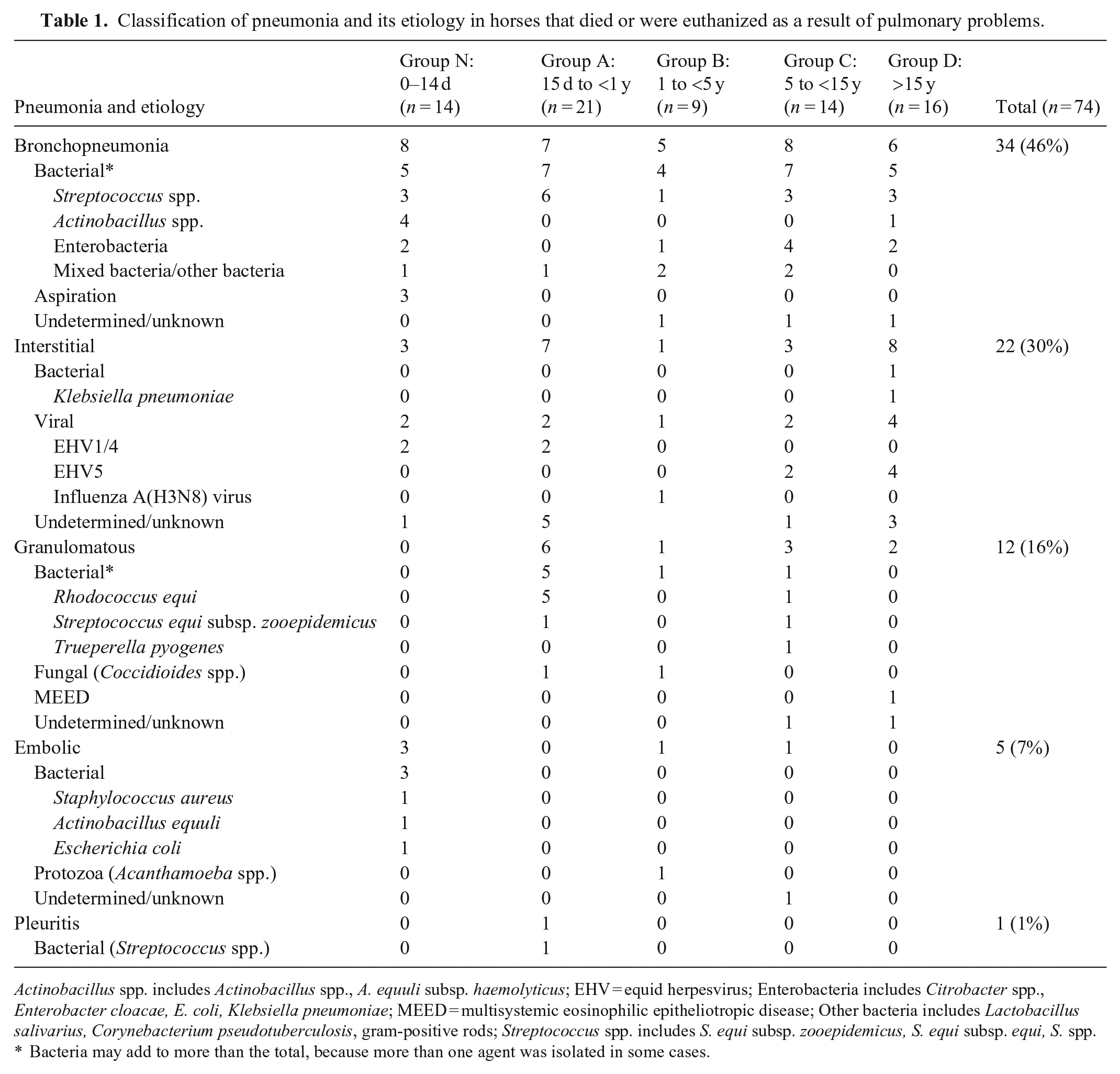
Actinobacillus spp. includes Actinobacillus spp., A. equuli subsp. haemolyticus; EHV = equid herpesvirus; Enterobacteria includes Citrobacter spp., Enterobacter cloacae, E. coli, Klebsiella pneumoniae; MEED = multisystemic eosinophilic epitheliotropic disease; Other bacteria includes Lactobacillus salivarius, Corynebacterium pseudotuberculosis, gram-positive rods; Streptococcus spp. includes S. equi subsp. zooepidemicus, S. equi subsp. equi, S. spp.
Bacteria may add to more than the total, because more than one agent was isolated in some cases.
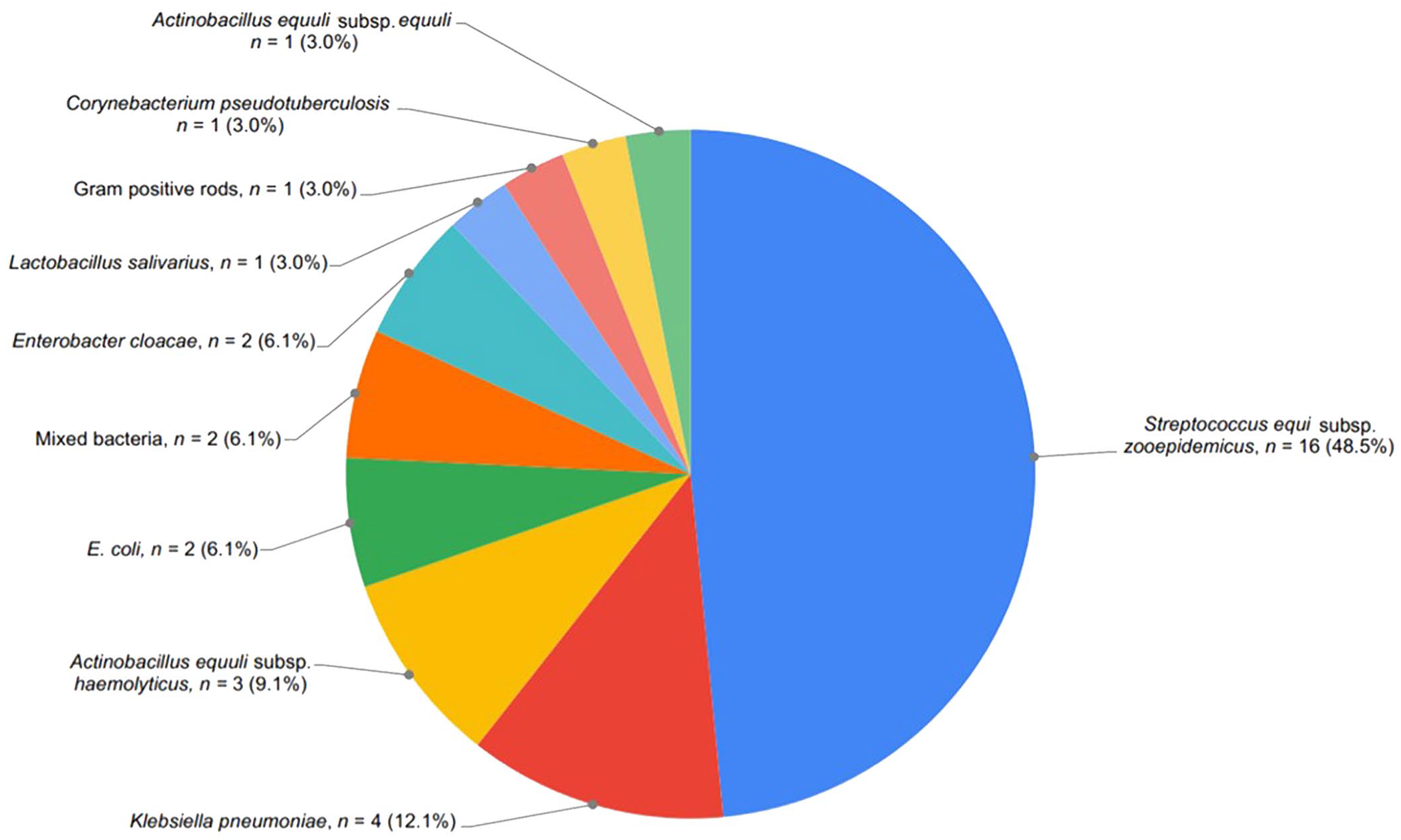
Bacteria isolated from cases of equine bronchopneumonia, in animals in which the lung was the organ affected most severely.
From the 28 cases of bacterial bronchopneumonia, 33 species of aerobic bacteria were isolated (5 cases had 2 isolates). The bacterial species isolated most frequently was Streptococcus equi subsp. zooepidemicus (n = 16; 48.5%), followed by Klebsiella pneumoniae (n = 4; 12.1%), and Actinobacillus equuli subsp. haemolyticus (n = 3, 9.1%; Fig. 1). S. equi subsp. zooepidemicus was isolated most frequently from animals <1-y-old (n = 9), with 1 isolate in animals >1-y-old to <5-y-old, and 3 isolates in animals >5-y-old. K. pneumoniae was isolated from 4 cases of animals >5-y-old old (data not shown).
The most frequent location for abscesses and/or areas of necrosis in the 28 cases of bacterial bronchopneumonia was in the cranioventral aspect of the right diaphragmatic lung lobe (n = 6; 21%), followed by bilateral presentation of bronchopneumonia in the same location (n = 5, 18%; data not shown).
Non-pulmonary cause of death with lesions in the lung
The most frequent extra-pulmonary cause of death in horses with pulmonary lesions (n = 82) was in the GI system (n = 25; 30%), followed by whole-body involvement (septicemia and/or toxemia; n = 22, 27%), musculoskeletal system (n = 10; 12%), nervous system (n = 7; 9%), cardiovascular system (n = 7; 9%), integumentary system (n = 5; 6%); lymphoid system (n = 3; 4%), and unknown/undetermined (n = 3, 4%; Suppl. Table 3).
The 3 most common non-pulmonary causes of death were additionally analyzed to understand the pathology of the lung in the deaths of these horses (Suppl. Table 4). Cases were subclassified as terminal, incidental, and secondary as defined previously. Only a few animals were recognized (5 of 74, 7%; Table 1) in which the most significant lesion was embolic pneumonia without a clear origin. Nevertheless, when all animals are considered, embolic pneumonia was identified in 33 of 156 (21.1%) cases (Suppl. Table 2), more frequently as a consequence of a lesion in another organ, particularly the GI system. For the 25 cases of horses that died or were euthanized because of a GI system problem (Fig. 2; Suppl. Table 4), lesions in the lungs were considered incidental in 8 cases, lesions were considered part of the terminal or agonal process in 5 cases, and the lung was affected secondarily in 12 cases. In 8 of those 12 cases, lesions were associated with embolic or interstitial lesions in the lung subsequent to inflammation of the small intestine, cecum, and/or colon.
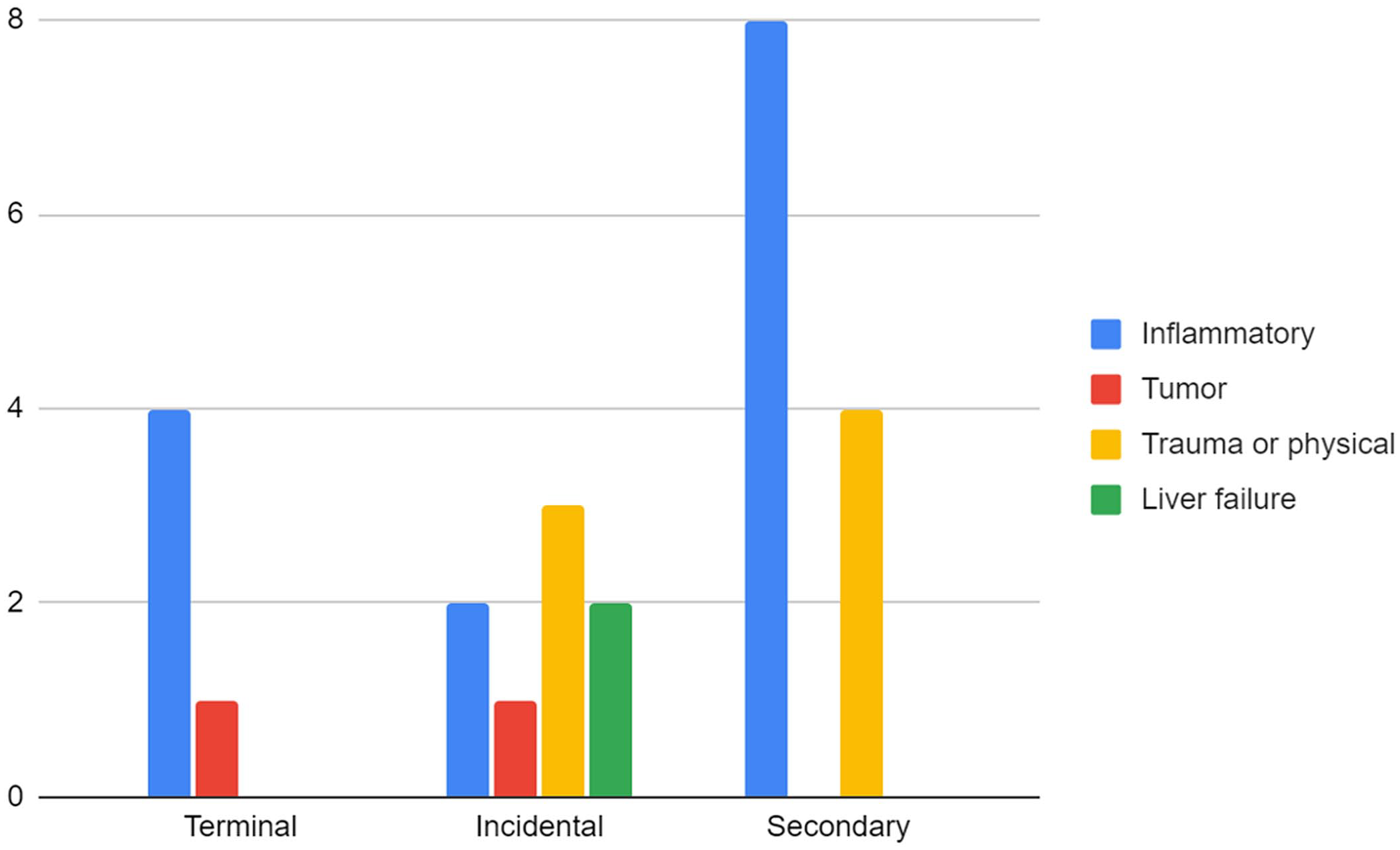
Pulmonary association (terminal, incidental, secondary) with the cause of death in animals that died or were euthanized with the gastrointestinal (GI) system as the most severely affected organ system. Bars represent the number of cases of the disease process identified in the GI system (inflammatory, tumor, trauma or physical, liver failure). For full information of each case, please refer to Suppl. Table 4. Incidental finding = finding in the lung is unrelated to the finding in GI system; Secondary finding = findings in the lung are directly associated with the lesion in the GI system; Terminal finding = finding in the lung is agonal or perimortem and is not directly associated with the GI system as the main affected system.
Of 22 cases of whole-body involvement (septicemia and/or endotoxemia), 21 cases had findings in the lungs that were classified as secondary, with the diagnosis of embolic (n = 14) or interstitial pneumonia (n = 6). In one case, bronchopneumonia was noted secondary to strangles. Findings in the lungs were considered a terminal event in only one case of septicemia and/or endotoxemia (Suppl. Table 5).
In the 10 cases in which lesions in the musculoskeletal system were considered the primary cause of death, findings in the lung were considered incidental in 5 cases, secondary in 4 cases, and terminal in 1 case. In 3 of the 4 secondary cases, lesions in the lung were the result of nutritional myopathy (vitamin E–selenium deficiency), with aspiration bronchopneumonia explained by skeletal muscle degeneration. No lesions in the myocardium were described in those cases (Suppl. Table 6).
Discussion
Bronchopneumonia was the most common pulmonary lesion observed in the horses examined, as described previously. 16 S. equi subsp. zooepidemicus was isolated from the majority of bronchopneumonia cases, similar to a report for racehorses in the same region. 9 S. equi subsp. zooepidemicus is a bacterial pathogen that is isolated frequently from cases of pneumonia of horses of any age, alone or in combination with other aerobic and anaerobic bacteria.2,4,5,9,21 S. equi subsp. zooepidemicus is a commensal bacterium found in the tonsils of many horses. 1 When conditions limit host immunity, the bacterium can overpower host defense mechanisms and lead to disease. 1 Although non-racing horses may be exposed to less physical stress compared to racehorses, they may still be exposed to travel, poor ventilation, emotional stressors, or inclement weather, especially in warmer weather in California.
Two other bacteria isolated commonly from cases of bronchopneumonia were K. pneumoniae and A. equuli subsp. haemolyticus. The age groups affected with K. pneumoniae were the C (6–14-y-old) and D (≥15-y-old) age groups (data not shown). K. pneumoniae is observed more frequently in animals that develop pneumonia after mechanical ventilation, strenuous exercise, or a history of prolonged travel. 12 It is possible that animals included in our study underwent similar scenarios and/or had debilitated immune status. In addition, it is common to encounter multidrug-resistant strains of K. pneumoniae, given its exceptional ability of acquiring plasmids containing multiple antimicrobial resistance genes on transposable elements. 6 Multidrug resistance may have contributed to the high prevalence of K. pneumoniae, conferring a guarded prognosis for adult horses in which this bacterium is isolated as the primary agent.
Bronchopneumonia, together with abscess formation and/or necrosis, was observed in the cranioventral region of the diaphragmatic lung lobes, similar to other studies.5,9 It is suggested that once bacterial infection is established in the lung, there is release of proteases and influx of large numbers of neutrophils, which accounts for the extensive areas of necrosis and suppurative lesions that are identified frequently in cases of bronchopneumonia. 9
Various agents have been associated with the origin of interstitial pneumonia in animals, but fewer than 20 have been identified in horses. 28 Frequently, an etiology cannot be identified, resulting in a common diagnosis of idiopathic interstitial pneumonia. 28 Interstitial pneumonia was the second most common lesion identified in 45 of 156 (29%) cases. The frequency of interstitial pneumonia differs from a similar study in racehorses, in which interstitial pneumonia was identified in only 2 of 83 cases. 9 We believe that the difference is the result of inclusion of animals <1-y-old and ≥15-y-old that were not considered in the racehorse population study. A similar situation was observed for cases of granulomatous pneumonia, with the inclusion of foals and older animals, which were not present in the racehorse study.
In interstitial pneumonia cases, no etiology could be identified in 17 of 45 cases, and 7 of these cases were identified in animals <1-y-old. In these animals, findings were attributed to “acute lung injury/acute respiratory distress syndrome (ALI/ARDS),” described in foals 1–8-mo-old. The cause of ALI/ARDS has been largely speculative, and remains unknown.10,28 EHV1/4 was identified in 4 animals <1-y-old. Infection is limited generally to the upper respiratory tract and may allow secondary bacterial infection in the lung. 2
Equine influenza is frequently observed in 2–3-y-old horses, but horses of all ages may be susceptible. 2 In our set of animals, only one horse in this age group was affected. In horses >5-y-old, the most important cause of interstitial pneumonia was infection with EHV5, the cause of equine multinodular pulmonary fibrosis (EMPF), which was identified as the main lesion in 7 cases. EMPF is a prevalent chronic interstitial disease of adult horses. 29 Nevertheless, it is important to consider causes of pulmonary fibrosis other than those caused by EHV5, such as a complicated pneumonia of infectious etiology, pneumoconiosis, or chronic toxicosis, such as those produced by perilla mint, Crofton weed, Crotalaria spp., and Senecio spp.8,28
Granulomatous pneumonia caused by R. equi was identified frequently in animals <1-y-old. R. equi infections are reported as a common cause of pneumonia in foals 3-wk to 6-mo-old.2,5,17,22 In some studies, R. equi is isolated from all cases of granulomatous pneumonia in foals. 5 The presence of bacterial virulence factors (such as VapA) and environmental factors (such as density of mares and foals, and airborne concentrations of R. equi) play an important role in the development of the disease.11,19 In our study, R. equi was detected in a 6-y-old pregnant Thoroughbred mare with chronic ulcerative dermatitis, cellulitis, tenosynovitis (Staphylococcus aureus isolated) unresponsive to treatment, and mild endometritis. Immunosuppression and stress likely predisposed the mare to rhodococcosis; the mare was euthanized because of ulcerative dermatitis.
Non-pulmonary cause of death refers to horses that were euthanized or died from non-pulmonary causes, but with a comorbidity of pneumonia. Diseases of the GI system were the most frequent cause of death in horses that also had pulmonary lesions. Half of the horses that died from GI disease also had pneumonia that was secondary to their GI lesions. A good example of damage of the GI system with sequelae in the lungs is embolic fungal pneumonia secondary to enterocolitis. Aspergillus spp. are commonly found in the GI system and are known to lead to pulmonary aspergillosis through embolic spread after acute cases of enterocolitis.14,24 Embolic spread has been seen after enterocolitis derived from infections by Salmonella spp., Neorickettsia risticii, or idiopathic enteritis, and often affects immunocompromised hosts.14,24
Septicemia with secondary lung lesions (interstitial or embolic pneumonia) was frequent in animals <14-d-old, similar to a previous study in foals <30-d-old, in which septicemia (44 of 174 animals) was the main cause of death, followed by musculoskeletal issues other than rib fractures (29 of 174), pneumonia (25 of 174), GI problems (19 of 174), and rib fractures (18 of 174). 25 When all neonatal deaths were considered (up to 180 d), septicemia was frequently associated with death (54 of 259), with E. coli isolated frequently, although mixed cultures were equally significant.25,27,30 Actinobacillus spp. were more commonly isolated in our study compared to those studies referred to above. A. equuli is a common pathogen of neonatal foals, and the difference in prevalence of this pathogen with other studies may indicate variations in susceptibility by different strains of this microorganism to antimicrobials.
Nutritional myopathy was an important cause of secondary aspiration bronchopneumonia. White muscle disease can be observed in foals from birth to 1-y-old, particularly in those that are born to dams fed selenium-deficient diets. Complications include failure of passive transfer, aspiration pneumonia, and stunting secondary to muscle weakness. 15
The major limitation of our study is that it includes a markedly heterogeneous population of horses. The category “non-racing horse” includes animals of different ages, activities (working, companion), and background (recently transported or not, undergoing different medical treatment, etc.). “Racing horses” is a more homogeneous group of animals that are currently racing or in training. These are mostly 2–5-y-old, Thoroughbred or Quarter Horse, with similar housing, vaccination status, and environmental conditions. The fact that most of the animals included in our study were Thoroughbreds and Quarter Horses leads to speculation that at least some of them probably competed in the past, although this information was not available to us.
Based on the data analyzed in our study, we conclude that there are no major differences between the presentation and causes for bronchopneumonia in non-racing horses compared to racehorses; hence, they likely share a similar set of predisposing conditions. The most significant differences in the pattern of pneumonia between racehorses and non-racing horses were interstitial and granulomatous pneumonia, likely as a result of the inclusion of age groups of non-racing horses that are not included commonly in the racehorse population. We also noted that lungs were often affected secondary to GI diseases and sepsis.
Supplemental Material
sj-pdf-1-vdi-10.1177_10406387221094273 – Supplemental material for Retrospective study of pneumonia in non-racing horses in California
Supplemental material, sj-pdf-1-vdi-10.1177_10406387221094273 for Retrospective study of pneumonia in non-racing horses in California by Ariana Rahman, Francisco A. Uzal, Anna M. Hassebroek and Francisco R. Carvallo in Journal of Veterinary Diagnostic Investigation
Footnotes
Acknowledgements
We thank Dr. Phillip Sponenberg for the useful comments about the manuscript.
Declaration of conflicting interests
The authors declared no potential conflicts of interest with respect to the research, authorship, and/or publication of this article.
Funding
The authors received no financial support for the research, authorship, and/or publication of this article.
Supplemental material
Supplemental material for this article is available online.
References
Supplementary Material
Please find the following supplemental material available below.
For Open Access articles published under a Creative Commons License, all supplemental material carries the same license as the article it is associated with.
For non-Open Access articles published, all supplemental material carries a non-exclusive license, and permission requests for re-use of supplemental material or any part of supplemental material shall be sent directly to the copyright owner as specified in the copyright notice associated with the article.