
Abstract
Chondrosarcomas are common tumors of the canine appendicular and axial skeleton; however, extraskeletal chondrosarcomas are very rare. Herein we report a case of extraskeletal chondrosarcoma in the tongue of a dog. Histologically, glossal skeletal muscle was infiltrated and effaced by islands of cartilage and streams of spindle-shaped cells. Retrospective analysis of 236 tongue masses submitted to the Iowa State University surgical biopsy service between 2011 and 2019 showed that the majority of submitted tongue masses are either non-neoplastic or benign, with granular cell tumors identified as the most prevalent benign neoplasms. Malignant tumors accounted for nearly 30% of all submitted masses, with malignant melanoma diagnosed most frequently.
An 11.5-y-old castrated male Labrador Retriever was presented to its primary care veterinarian because of a 3-wk history of epidermal collarettes on the right side of the lower abdomen. During a routine physical examination, an ~ 1-cm raised irregular growth of unknown duration was noted on the right side of the dorsal aspect of the tongue (Fig. 1). Although the mass did not impede the animal’s ability to eat or drink, the owners elected surgical removal. One week later, the mass was excised, fixed in 10% neutral-buffered formalin, and submitted to the surgical biopsy service of the veterinary pathology department at the College of Veterinary Medicine, Iowa State University (ISU; Ames, IA). The tissue was processed routinely, and ~ 5-µm thick sections were cut and stained with hematoxylin and eosin.
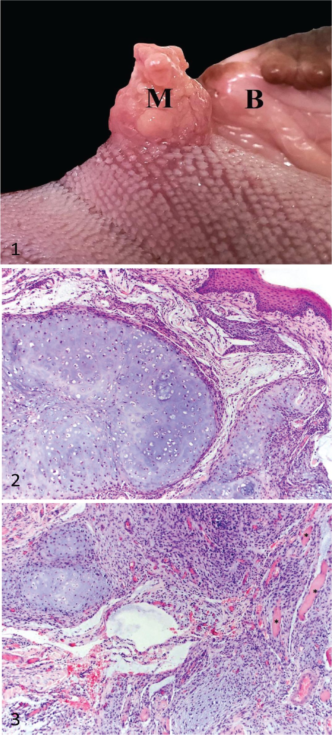
Extraskeletal chondrosarcoma of the tongue in a Labrador Retriever.
Histologic examination revealed an invasive neoplasm effacing large areas of glossal skeletal muscle. The mass consisted of multiple lobules of oval-to-spindle–shaped cells within lacunae separated by variable amounts of a basophilic chondroid matrix (Fig. 2). Frequently, adjacent to collagenous islands were moderately dense populations of poorly differentiated spindle-to-stellate–shaped cells often arranged in streams (Fig. 3). There was minimal anisokaryosis or anisocytosis in both the chondrocyte and spindle cell population, with no mitoses observed. Overlying the mass, there was focal ulceration of the mucosa with infiltration of the underlying submucosa by low numbers of neutrophils. Adjacent to the mass, small-caliber vessels were cuffed by small aggregates of lymphocytes. Distinctive features of the mass, coupled with the absence of identified primary masses in this dog, were compatible with a diagnosis of extraskeletal chondrosarcoma.
Chondrosarcomas are tumors of malignant mesenchymal cells that produce a chondroid matrix. Such tumors are one of the most common neoplasms of the canine appendicular and axial skeleton and can also originate as primary tumors in tissues such as the heart, liver, and spleen, where they are diagnosed as extraskeletal chondrosarcomas.2,6,8,13–15,19 Extraskeletal chondrosarcomas are very rare tumors and are estimated to account for ~ 1% of all chondrosarcomas in the dog. 11 Given that only 2–4% of all oropharyngeal tumors are reported to occur in the tongue,5,7 the presence of an extraskeletal chondrosarcoma in the tongue is unique. Additionally, of the 646 malignant tongue neoplasms reported in one study, none was classified as chondrosarcoma. 18 Extraskeletal chondrosarcoma of the tongue has been described in the horse and humans.1,20 In both cases, the tumor was excised with appropriate surgical margins, and the tumor had not recurred or metastasized at the time of publication, 10 mo post-removal for the horse and 20 mo post-removal for the human case.
The process by which chondrosarcoma could arise de novo from mesenchymal cells in the tongue, which do not normally generate or support cartilaginous structures, is unclear. The lyssa, a firm fibrous structure lying along the median ventral aspect of the tongue, would be one conceivable site of generation for the extraskeletal chondrosarcoma; however, the lyssa contains no cartilage and its location is separate from the chondrosarcoma that we describe. 4 Discussions of chondrosarcoma in the human tongue hypothesize that remnants of fetal cartilage within the lingual frenulum of the tongue may be responsible for the development of chondromas, which can subsequently develop into malignant tumors.3,17 This proposed pathogenesis would fit with the benign chondrolipoma described in the lingual frenulum of the tongue of a dog. 9 However, the dorsolateral location of our described extraskeletal chondrosarcoma, and distance from the frenulum, may better align with an alternative hypothesis proposing that repeated trauma to an area of the tongue results in metaplasia and subsequent transformation into a malignant neoplasia. 17
Our retrospective analysis of 236 tongue masses submitted to the ISU veterinary pathology surgical biopsy service between January 1, 2011 and July 1, 2019 expands currently available information on the proportion of tongue masses that are non-neoplastic, benign, or malignant, and the frequency of specific diagnoses or tumor types (Table 1).
Age distribution and histologic diagnoses of lingual mass biopsies evaluated at the Iowa State University veterinary pathology surgical biopsy service between January 1, 2011 and July 1, 2019.
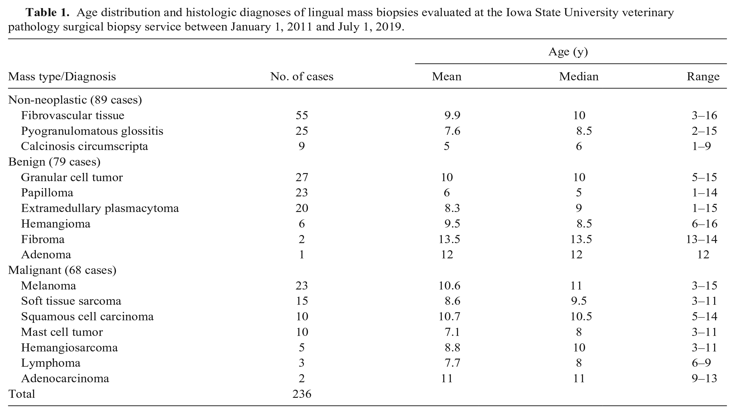
Nearly 40% (89 of 236 cases) of the evaluated glossal masses were classified as non-neoplastic, with granulomatous inflammation in response to foreign material and proliferative fibrovascular tissue as the most common findings. This was not surprising, given that dogs consistently use their tongues for the prehension of foreign objects or the removal of plant material trapped in fur, which may have the ability to puncture or lacerate the tongue. Together, these 2 diagnoses accounted for 90% of all non-neoplastic tongue masses. The remaining 10% of cases were diagnosed as calcinosis circumscripta (tumoral calcinosis), which is the mineralization of soft tissue, likely the result of previous, and perhaps repetitive, trauma.
Benign tumors accounted for 79 of the 236 evaluated lingual masses. Consistent with past retrospective analyses of dog tongue masses, granular cell tumors were the most prevalent neoplasm, accounting for over one-third of all benign tumors and nearly 20% of all neoplasms. 7 Granular cell tumors are thought to arise from neuroectodermal precursor cells, and, in contrast to their comparatively high frequency in the tongues of dogs, these are rare tumors in the rest of the body, but have been described in the heart and brain.10,16 Our analysis also identified a large number of papillomas and extramedullary plasmacytomas, which, together with granular cell tumors, comprised nearly 90% of all benign tumors. Interestingly, the age distribution of papilloma cases was relatively even, with 8 of 23 cases occurring in animals ≤ 2 y old, 7 cases in animals 3–7 y old, and 8 cases in animals ≥ 8 y old (Table 1). Although oral papillomatosis is thought to occur mainly in young dogs, it is also reported in immunosuppressed or immunodeficient animals. 12
Of the 236 tongue masses submitted to the ISU surgical biopsy service over the past 8 y, 68 (29%) were diagnosed as a malignant tumor. Melanoma was identified most commonly and accounted for approximately one-third of all diagnosed malignant tongue masses. Melanomas are the most common malignant oral tumor in dogs and are frequently seen in the tongue.7,18 Soft tissue sarcomas were the second most frequently diagnosed glossal malignant tumor. Tumors placed into this category included undifferentiated sarcomas, fibrosarcomas, and individual cases of liposarcoma and rhabdomyosarcoma. Squamous cell carcinoma and mast cell tumors each accounted for ~ 15% of all identified malignant neoplasms. The remaining 15% of malignant masses were divided among rare cases of hemangiosarcoma, lymphoma, and adenocarcinoma.
Footnotes
Declaration of conflicting interests
The authors declared no potential conflicts of interest with respect to the research, authorship, and/or publication of this article.
Funding
The authors received no financial support for the research, authorship, and/or publication of this article.