
Abstract
A 7.5-year-old raccoon dog (Nyctereutes procyonoides) from the Henry Doorly Zoo (Omaha, Nebraska) presented to the veterinary hospital for lethargy and weight loss. On physical examination, splenomegaly and hepatomegaly were noted on palpation and were confirmed by radiographic evaluation. Radiography also demonstrated a mass in the cranial mediastinum. A complete blood cell count revealed marked leukocytosis (115,200 cells/µl), with a predominance of lymphoid cells. The animal was euthanized due to a poor prognosis. Necropsy revealed splenomegaly, hepatomegaly, and a large multiloculated mass in the cranial mediastinum. The histologic and immunohistochemical diagnosis was multicentric T-cell lymphoma with a leukemic phase.
A 7.5-year-old male raccoon dog (Nyctereutes procyonoides) was reported by zookeepers as looking thin and lethargic. Upon visual inspection, mild weight loss and lethargy were confirmed, and the fur was short and rough. The animal lived with a conspecific in an indoor enclosure of the Henry Doorly Zoo (Omaha, Nebraska), and had no significant medical problems since its arrival at the zoo as a 2-year-old. The last routine physical examination performed 2.5 months earlier revealed no significant abnormalities other than mild symmetrical alopecia affecting the ventral neck and chest, caudolateral abdomen, and distal hind limbs; hematologic and biochemical analyses at that time were within normal limits for the species. 12
The following day, the raccoon dog was anesthetized using sevoflurane a delivered by facemask, and a complete physical examination was performed. Blood was collected from the cranial vena cava for hematologic and biochemical analyses. Abdominal palpation revealed splenomegaly, hepatomegaly, and an irregular mass in the cranial abdomen. Fur abnormalities were considered to be normal due to seasonal variation and molting. The body weight was 2.66 kg, a 30% reduction from 3.84 kg weight recorded 1.5 years previously. The rest of the physical examination was unremarkable. Lateral and ventrodorsal radiographs revealed splenomegaly, hepatomegaly, a semilunar radio-opaque structure in the cranial abdomen, a mass in the cranial thorax displacing the trachea dorsally, and congested lungs (Fig. 1). A peripheral blood count revealed marked leukocytosis (115,200 cells/µl, reference [ref.] interval: 12,400–17,400 cells/µl), and atypical lymphoid cells represented 82% of the total leukocyte count (94,500/µl, ref. interval: 3,000–4,600/µl; Fig. 2A). There was also a neutrophilia (segmented neutrophils 16,100/µl, ref. interval: 7,200–10,100/µl) with a presumed left shift (band neutrophils 2,300/µl, no ref. interval available), a monocytosis (2,300/µl, ref. interval: 500–1,000/µl), a mild polycythemia (red blood cells 7.49 × 10⁶/µl, ref. interval: 5.6–7 × 10⁶/µl), and a thrombocytopenia (platelets 152,000/µl, ref. interval: 300,000–478,000/µl). The platelet count was also decreased when compared to previous results for the same individual performed 2.5 months before clinical presentation (330,000/µl). Serum chemistry abnormalities included increased activity of aspartate aminotransferase (330 U/l, ref. interval: 20–40 U/l) and alanine aminotransferase (ALT; 270 U/l, ref. interval: 16–54 U/l). 12 In addition, alkaline phosphatase activity (1190 U/l), total bilirubin concentration (1.4 mg/dl), and urea nitrogen concentration (84 mg/dl) were increased when compared to previous results from this and other raccoon dogs kept at the Henry Doorly Zoo; however, reference intervals for these parameters could not be found in the literature. A presumptive diagnosis of lymphoid neoplasia was made, and the animal was humanely euthanized due to poor prognosis.
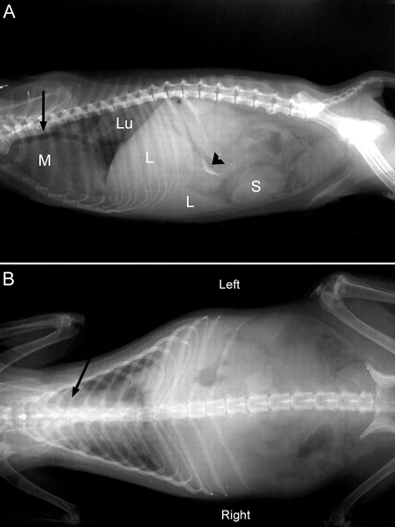
A, laterolateral radiograph. Note the enlarged spleen (S), enlarged liver (L), semilunar radio-opaque structure in the cranial abdomen (arrowhead), congested lungs (Lu), and a mass in the cranial mediastinum (M) displacing the trachea dorsally (arrow).
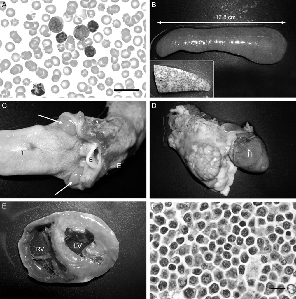
Necropsy revealed a thin animal, although a significant subcutaneous fat layer was present. Abnormal findings included congested lungs, splenomegaly (Fig. 2B), hepatomegaly with a marked reticulated pattern and coalescent pale areas (3–4 mm diameter) throughout the hepatic parenchyma (Fig. 2B), pale kidneys, enlarged mesenteric lymph nodes and pharyngeal tonsils (Fig. 2C), a large multiloculated mass (5 cm × 4 cm) in the cranial thorax attached to the base of the heart, large vessels and trachea (Fig. 2D), and a hard plastic foreign body in the stomach (corresponding to the semilunar structure detected by radiography and palpation). Transverse sections of the heart revealed dilatation of both ventricles (Fig. 2E). Submandibular and prescapular lymph nodes were slightly enlarged, but other peripheral lymph nodes were of normal size. Representative samples from multiple organs were collected, fixed in 10% neutral buffered formalin, and routinely processed for histology. Sections were cut at 4 µm and stained with hematoxylin and eosin. Histologically, the normal architecture of mediastinal lymph nodes (mass), liver, spleen, and pharyngeal tonsils was severely disrupted by a homogeneous population of neoplastic lymphocytes, characterized by scant cytoplasm, irregular nucleus, and clumped chromatin (Fig. 2F). A similar population of abnormal lymphocytes was focally present in bone marrow and peripheral lymph nodes.
Additional sections of the mediastinal mass, other lymph nodes, liver, and spleen were immunolabeled to determine the cell of origin of the neoplastic lymphocytes. Antigen retrieval was conducted by pressure-cooking in citrate buffer at pH 6.0. b For T-cell labeling, sections were tested with polyclonal rabbit anti-human cluster of differentiation (CD)3 as the primary antibody, c and biotinylated goat anti-rabbit immunoglobulin (Ig)G as the secondary antibody. d For B-cell labeling, monoclonal mouse anti-human CD79 was used as the primary antibody, e and biotinylated horse anti-mouse IgG as secondary antibody. f The secondary antibodies were labeled using streptavidin–horseradish peroxidase, g and 3,3-diaminobenzidine tetrahydrochloride (DAB) was used as the substrate for staining. h Sections were counterstained with hematoxylin. Neoplastic lymphocytes were positive for CD3 and negative for CD79, which characterized the neoplasia as a T-cell phenotype.
In addition, another section of affected tissues was submitted to heat-induced antigen retrieval in the presence of a citrate buffer solution. i An immunohistochemical stain for CD34 j was performed using an automated immunostainer. k Detection was performed with a DAB kit. l The lymphoid cells were negative for CD34. Endothelial cells from a human being and the raccoon dog were used as controls for CD34; the human control was positive, but the raccoon dog control was negative.
The term “lymphoma” encompasses a diverse group of malignancies arising in lymphoid tissue outside the bone marrow. 3 Lymphoma is the most common hematopoietic malignancy in animals, and has been reported in all domestic and many nondomestic species. 1,3,5,6,9,10,13,15 Lymphomas are classified depending on anatomic location (multicentric, alimentary, mediastinal, other), immunophenotype (B lymphocyte vs. T lymphocyte vs. non-B/non-T), cellular morphology (size, nuclear features, mitotic rate), histologic pattern (diffuse or follicular), and biologic behavior (indolent or aggressive). 3 In the case of the raccoon dog presented herein, the neoplasm was classified as T-cell multicentric lymphoma with a leukemic phase; this neoplasm was well differentiated and aggressive.
It is interesting to note the quick progression of clinicopathologic changes, as a health examination performed on the animal 83 days previous to clinical presentation did not reveal any abnormality other than increased ALT activity (238 U/l, ref. interval: 16–54 U/l). Upon clinical presentation, marked leukocytosis (with a predominant lymphocytosis), thrombocytopenia, increased liver enzyme activity, and an increased urea concentration were noted in this individual. The increased liver enzyme activity may have been as result of the destruction of the normal hepatic architecture by the invading neoplastic lymphocytes, which could have decreased functional hepatic mass leading to hyperbilirubinemia. Increased urea nitrogen concentration was probably the result of protein catabolism, as this animal was losing weight.
Among lymphomas in domestic dogs (members of the family Canidae, as raccoon dogs are), those of T-cell origin behave more aggressively and progress more rapidly than those of B-cell origin. 3,4 In addition, craniomediastinal lymphomas have a poor prognosis (less than 3 months of life span even with treatment) when compared to lymphomas in other locations. 4
Bone marrow and blood involvement, as seen in the present case, may be present in advanced cases of lymphoma. 3 The distinction between lymphoma with a leukemic phase and lymphoid leukemia may be difficult and arbitrary when extensive lymph node, bone marrow, and peripheral blood involvement is present. 3 As an example, chronic lymphocytic leukemia (CLL) in dogs share some characteristics with the case presented herein: CLL occurs in older animals, it produces high blood lymphocyte counts, most cases are of T-cell origin, and thrombocytopenia is a common findings. 14 However, CLL in dogs is usually an indolent, slowly progressive disease, typically originating from the spleen, and produces uniform splenomegaly, hepatomegaly, and lymphodenopathy. 3,14 Since some characteristics of CLL were not observed in the present case, lymphoma with a leukemic phase was considered most likely. Unfortunately, CD34 labeling could have helped differentiate between lymphoma and acute leukemia (the latter typically being CD34 positive), but it was concluded that the technique did not work in the raccoon dog tissue. In the domestic dog, lymphoma is typically a disease of middle-aged to older animals, and B-cell multicentric lymphoma is the most common form. The raccoon dog from the present case was considered aged (7.5-year-old), as maximum life span in wild populations has been determined to be 7–8 years of age. 7
Raccoon dogs are frequent subjects of wildlife medicine literature, either as native, introduced, or research species. 2,8,11,12 However, this species is not common in zoos, and there is a lack of references of medical conditions that can affect this species in captivity. The present case report describes clinical and pathological characteristics of lymphoma with a leukemic phase in raccoon dogs.
Footnotes
Acknowledgements
The authors thank Ms. Abbie Butler for conducting the immunohistochemistry, and the zookeepers and veterinary staff of the Henry Doorly Zoo for their help managing this case.
a.
SevoFlo, Abbott Animal Health, Abbot Park, IL.
b.
Antigen Retrieval Citra Solution, HK086-9K; BioGenex Laboratories Inc., San Ramon, CA.
c.
A0452, 1:800; Dako Denmark A/S, Glostrup, Denmark.
d.
BA-1000, 1:100; Vector Laboratories Inc., Burlingame, CA.
e.
M7051, clone HM57, 1:20; Dako Denmark A/S, Glostrup, Denmark.
f.
BA-2001, 1:100; Vector Laboratories Inc., Burlingame, CA.
g.
LSAB®2 Streptavidin-HRP, K1016; Dako Denmark A/S, Glostrup, Denmark.
h.
K3468, Dako Denmark A/S, Glostrup, Denmark.
i.
CC1 mild, Ventana Medical Systems Inc., Tucson, AZ.
j.
Catalog number 790-2927, clone QBEnd/10, mouse IgG1, no dilution; Ventana Medical Systems Inc., Tucson, AZ.
k.
Ventana Medical Systems Inc., Tucson, AZ.
l.
ultraView™ Universal DAB detection kit, Ventana Medical Systems Inc., Tucson, AZ.
The author(s) declared no potential conflicts of interest with respect to the research, authorship, and/or publication of this article.
The author(s) received no financial support for the research, authorship, and/or publication of this article.