
Abstract
Malignant neoplasms occur commonly in cattle, with lymphosarcoma being the most common. Chondrosarcoma rarely has been described and only in mature cattle. The present report describes a chondrosarcoma of the left scapula of an 8-month-old Holstein steer. Histologic examination of the mass revealed an unencapsulated, multilobular neoplasm composed of neoplastic spindle cells embedded in irregular islands of chondroid matrix, consistent with a diagnosis of chondrosarcoma.
A wide variety of malignant neoplasms have been reported in cattle, with lymphosarcoma having been cited as the most prevalent. 2 Chondrosarcoma is rarely encountered in the bovine; in several abattoir surveys, the frequency of benign or malignant cartilaginous neoplasms in cattle was estimated at less than 0.1–1% of all identified neoplasms.1,3,4,11,17 Of all domestic animals, chondrosarcoma is reported most frequently in the dog, at approximately 10% of all canine primary bone tumors and second only to osteosarcoma in incidence.5-8,10,16,17,19
An approximately 8-month-old Holstein steer (228 kg), from a feedlot in north central Kansas, was examined for a swelling on the craniolateral aspect of the left scapula (Fig. 1). On initial evaluation, the mass was firm and apparently nonpainful, and the steer was not lame. A fine-needle aspirate was performed, and sanguineous fluid, consistent with peripheral blood, was obtained. Presumptive diagnosis was a traumatic hematoma, and the steer was administered flunixin meglumine a (1.1 mg/kg, intravenously). Over the next 3 months, the steer performed consistent with his peers and showed no lameness, but the mass significantly enlarged. Radiographs revealed destruction of the proximal and cranial portions of the spine and body of the scapula. Based on the evidence of bony lysis, primary bone or soft tissue neoplasm was suspected, and the steer was euthanized.
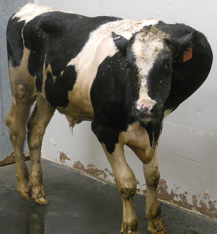
Holstein steer with a large mass on the craniolateral aspect of the left scapula.
Gross postmortem examination revealed an approximately 30 cm × 45 cm × 45 cm multilobular mass extending from the lateral aspect of the left scapula and infiltrating the adjacent soft tissues and skeletal muscle. The mass was firm, mottled gray to pale tan to light red, gritty, and difficult to cut with a knife. Cut surface revealed numerous multifocal to coalescing nodules (up to 4 cm in diameter) that were pale tan to white, with central foci of mineralization (Fig. 2).
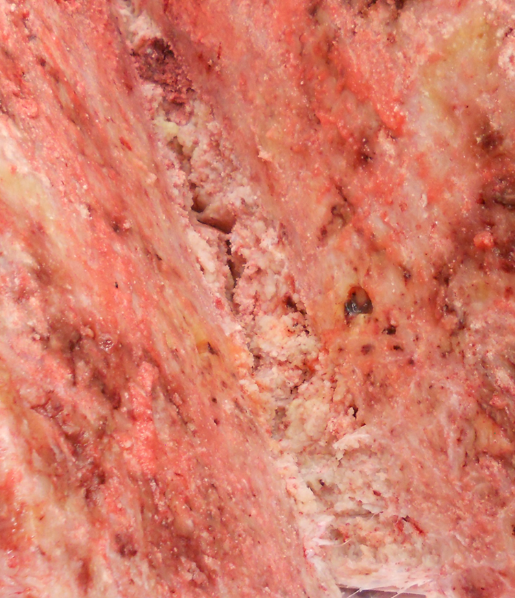
Holstein steer. Cut surface of the mass associated with the craniolateral aspect of the left scapula.
Tissue samples from the mass were fixed in 10% buffered formalin, processed routinely for sectioning, and then stained with hematoxylin and eosin. Histologic examination of the mass revealed an unencapsulated, poorly demarcated, moderately cellular, multilobular neoplasm composed of haphazardly arranged plump neoplastic spindle cells embedded in irregular islands of abundant pale amphophilic to basophilic chondroid matrix (Fig. 3). The lobules were separated by moderately thick bands of fibrovascular connective tissue, and within the lobules, neoplastic cells were arranged both individually and in small clusters within the matrix (Fig. 4). Neoplastic cells had indistinct cell borders and a small to moderate amount of vacuolated pale eosinophilic to amphophilic cytoplasm. Nuclei were oval to elongate, with finely stippled chromatin and generally inconspicuous nucleoli. Anisocytosis and anisokaryosis were mild, and the mitotic rate averaged less than 1 per 10 high powered fields. There was multifocal mineralization of the chondroid matrix and fibrovascular connective tissue, and large multifocal to coalescing regions of necrosis, admixed with abundant hemorrhage and fibrin.
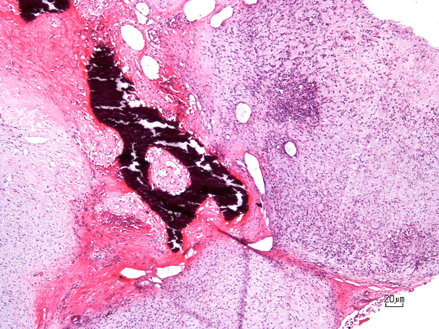
Holstein steer. The mass associated with the left scapula is multilobular and composed of neoplastic spindle cells embedded in irregular islands of pale basophilic chondroid matrix. Lobules are separated by bands of fibrovascular connective tissue with multifocal mineralization. Hematoxylin and eosin. Bar = 20 μm.
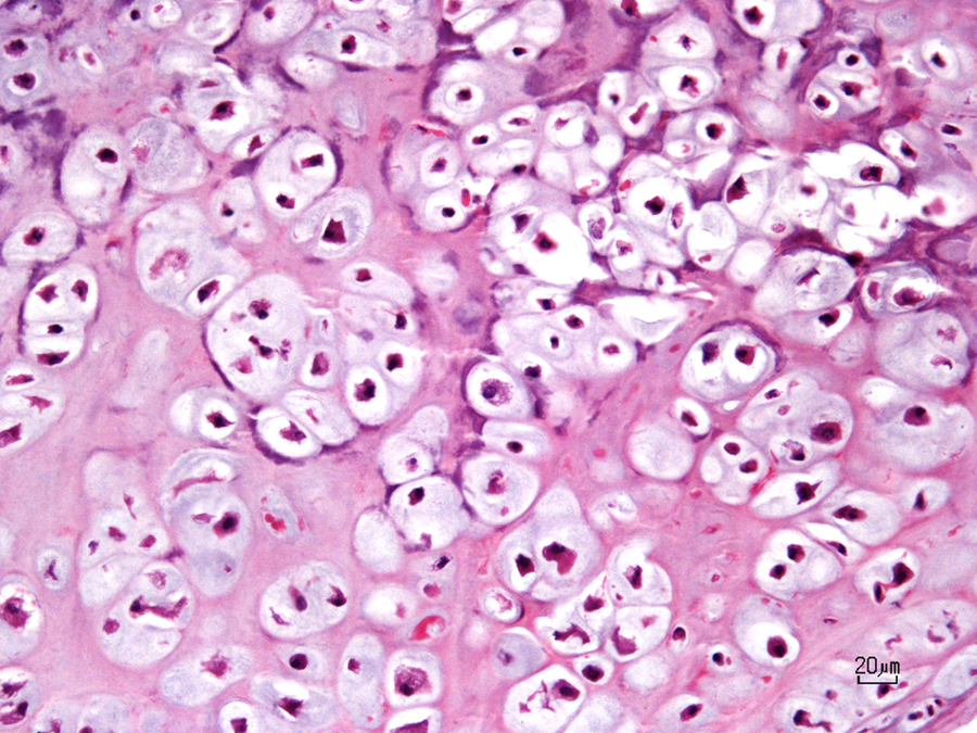
Holstein steer. Within neoplastic cartilaginous lobules, neoplastic cells are arranged both individually and in small clusters within the chondroid matrix. Hematoxylin and eosin. Bar = 20 μm.
Chondrosarcoma is a malignant mesenchymal neoplasm in that tumor cells produce varying amounts of neoplastic chondroid matrix, but not osteoid.3,6,8,16,19 Although bone may be present within the tumor, it is formed by endochondral ossification of tumor cartilage, rather than being produced by malignant osteoblasts as in osteosarcoma.18,19 Chondrosarcomas are classified as primary or secondary based on the origin of the neoplasm.7,8,18 Primary tumors arise from a previously normal bone, either from within the bone (central or medullary chondrosarcoma) or from the periosteum (peripheral or periosteal chondrosarcoma).7,8,18,19 Secondary chondrosarcomas arise by progression of a preexisting benign lesion of bone.7,8,18,19 In addition, there are reports of extraskeletal chondrosarcomas in animals, a rare form in which the neoplastic cells exhibit chondroid differentiation, but are present within soft tissue and lack association with the skeletal system.3,7,9,12,13,15 Most chondrosarcomas in animals are considered primary bone tumors of medullary origin.8,18,19
Chondrosarcomas tend to arise from any site where normal cartilage exists, and in all species, flat bones are more commonly involved than long bones.3,5,8,10,11,17,18,19,21 The most commonly reported locations in the dog include costochondral junction of the ribs, nasal turbinates, and pelvis, while cartilage of the sternocostal complex (followed by scapula and tuber coxae) is the most commonly identified location in sheep.1,6,7,12,17,19 Chondrosarcomas in cattle and horses are most often reported in flat bones, but occasionally occur in long bones or extraskeletal locations.1,3,7,14,19-22 In a retrospective study of 67 feline chondrosarcomas, 46 out of 67 cases were associated with bone, with the scapula being the most commonly affected flat bone (15% of all chondrosarcomas arising from the skeleton). 5 However, to the authors’ knowledge, there has been only one detailed report of chondrosarcoma arising within the scapular cartilage of a cow. 11
Compared to osteosarcoma, chondrosarcomas tend to grow more slowly and develop metastatic lesions much later and at a much lower frequency.18,19,21 The metastatic rate in dogs is reported to be approximately 20–25%, and spread is hematogenous, most often involving the lungs, although other organs (kidney, liver, heart, skeleton) can be affected.1,5,7,19 Local invasion is common, and recurrence is frequent, especially in areas where adequate surgical excision is difficult.5,19
Grossly, these tumors can resemble hyaline cartilage, with a pale gray, tan, or bluish-white appearance and often have variably sized regions of mineralization or ossification within. 3 Distinguishing a chondrosarcoma from a chondroma can be difficult histologically, but invasive behavior, large foci of necrosis, neoplastic chondrocytes with large or multiple nuclei, cellular anaplasia, and the presence of even a single mitotic figure suggest malignancy.1,3,7,8
Footnotes
Acknowledgements
The authors wish to thank Ryan Engel for radiologic technical assistance and Dr. Elizabeth DesChene Bochtrup for radiographic interpretation.
a.
Banamine®, Schering-Plough Animal Health, Union, NJ.
Declaration of conflicting interests
The author(s) declared no potential conflicts of interest with respect to the research, authorship, and/or publication of this article.
Funding
The author(s) received no financial support for the research, authorship, and/or publication of this article.