
Abstract
The naked mole-rat (NMR; Heterocephalus glaber)—a small, eusocial, subterranean rodent native to East Africa—is distinguished by its capability to live long and resist changes associated with the aging process. Notably, a growing amount of research has been dedicated to NMRs’ multifactorial capacity to resist cancer. Since 2016, however, zoos have begun to document various neoplasms in a handful of individuals. We present herein radiographic, gross anatomic, and histopathologic features of a case of a sacral chordoma in a geriatric female. Chordomas originate in notochordal remnants. These spinal tumors are most commonly seen in ferrets; chordomas are rare in humans, can be difficult to treat, and need wide surgical margins.
Keywords
The naked mole-rat (NMR; Heterocephalus glaber) holds the record for the longest-lived rodent. Some individuals reach > 30 y in age, or 5-times longer than expected based on body size. 5 NMRs have a longevity quotient (the ratio of actual maximum species lifespan to that predicted by body mass) similar to humans. 1 It has been thought that this unique species can give us new insights into various evolutionary theories of aging as well as the mechanisms that happen during the aging process. 2
Although the risk of malignant neoplasia generally heightens with age, the extraordinarily long-lived NMR displays a number of efficient DNA-repair and anti-cancer mechanisms. 12 NMR skin fibroblasts were found to resist carcinogenesis after being transduced with SV40 and RasG12V, which are oncogenes that cause cells of other mammalian species to form malignant tumors. 9 One component of NMR cancer defense appears to be hypersensitivity to contact inhibition, a process involving activity of pRb (cell cycle arrest) and p53 (apoptotic response) when cells reach a high density (Seluanov A, et al. Hypersensitivity to contact inhibition provides a clue to cancer resistance of naked mole-rat. Proc National Acad Sci 2009:19352–19357). Furthermore, NMR fibroblasts secrete and accumulate extremely high molecular weight hyaluronan that has been shown to prevent malignant transformation in this species. 15 Compared to mice, NMRs have more robust genome maintenance, with higher copy numbers of CEBPG (a regulator of DNA repair) and TINF2 (a protector of telomere integrity). 10 Also, NMR cells entering senescence can prevent potential pathogenesis by inhibiting transcription and mitochondrial translation (Zhao Y, et al. Naked mole rats can undergo developmental, oncogene-induced and DNA damage-induced cellular senescence. Proc National Acad Sci 2018:1801–1806). Given that the NMR genome was sequenced in 2011, discoveries such as expression of alpha2-macroglobulin (A2M) molecules and long noncoding RNAs (lncRNAs) will continue to shed light on mechanisms of cancer resistance in this remarkable species.6–8
Despite research reports starting in 1967, including a retrospective study of 138 autopsies over a 15-y period, the occurrence of cancer in NMRs was not officially chronicled until 2016.3–5 Since then, 2 reports have described a combined 7 instances (1 presumptive) of NMR neoplasms. The first discoveries, from the Brookfield Zoo and the National Zoological Park (NZP), were of an axillary adenocarcinoma, possibly of mammary or salivary origin, and a gastric neuroendocrine carcinoma, respectively. 4 The second article reported one case each of metastatic hepatocellular carcinoma, nephroblastoma (Wilms tumor), multicentric lymphosarcoma, cutaneous hemangioma, and presumptive esophageal adenocarcinoma (Barrett esophagus-like). 14 With our recent finding of sacral chordoma in a NMR, we offer another example of a neoplasm in this species previously thought to be cancer-resistant.
A geriatric (15+ y old) female naked mole-rat was evaluated because of a history of acute-onset pelvic limb ataxia, difficulty righting in the hind-end, and uncharacteristic aggression when handled. This individual had previously been treated for facial excoriations with topical chlorhexidine 2% solution (Vetoquinol, Fort Worth, TX) and silver sulfadiazine 1% cream (Ascend Laboratories, Montvale, NJ). The NMR was a member of a 1-male, 2-female colony that inhabited a 1.1 × 4.5 × 2.6 m enclosure along with another NMR colony of 7 individuals and a Damaraland mole-rat colony of 2 individuals. Average ambient temperature was 31.6°C and average humidity was 37% in the exhibit. This individual’s daily diet contained 10 g each of leafy greens, carrots, radishes, sweet potatoes, turnips, corn, green beans, and apples, along with one cinnamon-flavored primate biscuit.
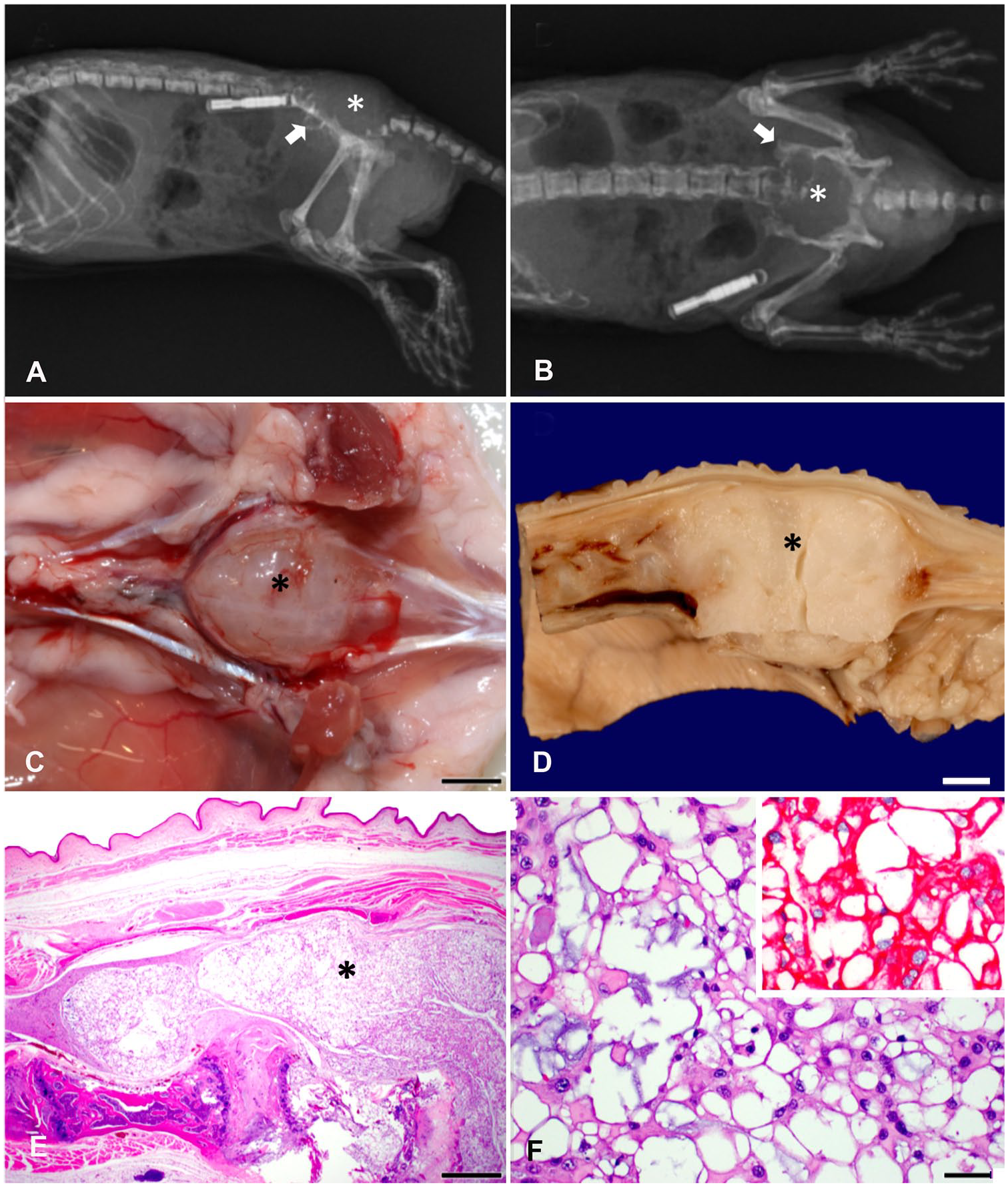
A physical examination under anesthesia revealed a diffuse, purple, soft, gelatinous, subcutaneous swelling along the pelvis and caudal lumbar area that extended past the coxofemoral joints bilaterally. Radiographs showed a 1.7 × 1.2 cm soft tissue opacity within the region of the pelvis, complete obliteration of the sacrum, and severe osteolytic lesions on vertebrae L6 and L7, dorsal wings of both ilia, left dorsal acetabular ridge, and first coccygeal vertebra (Fig. 1A, 1B), and there was a metallic foreign body consistent with a microchip along the right distolateral, dorsal body wall. Lumbar ultrasound revealed a hypoechoic mass with a ventral rim of hyperechoic proliferation (suspected bony proliferation). Given quality of life concerns, euthanasia was performed via intracardiac injection of barbiturate.
Sacral chordoma in a naked mole-rat.
A postmortem examination was performed and revealed an ~1 cm diameter, soft, gelatinous, pale gray-white sacral mass that bulged ~ 3 mm into the pelvic canal (Fig. 1C, 1D). The microchip noted on whole body radiographs did not have any communication with the sacral mass. On cut section, the mass was translucent white and oozed clear watery fluid. Following fixation of tissues in 10% neutral-buffered formalin, routine histology was performed. The sacral lesion was morphologically diagnosed as chordoma with vertebral and spinal cord effacement, but no evidence of distant metastasis. Histologically, it was characterized by lobules of proliferating physaliferous cells (highly vacuolated clear cytoplasm and a central nucleus) dissected by thin trabeculae of fibro-osseous to myxomatous stroma (Fig. 1E). The tumor resulted in severe effacement of the surrounding tissues, including vertebral bone resorption, infiltration of the spinal canal, and compression of the spinal cord and nerve roots. In addition to causing pelvic limb paresis, the lesion likely impaired the innervation to the urinary bladder resulting in inadequate voiding and ascending bacterial infection, as evidenced by moderate-to-severe, diffuse, chronic, lymphoplasmacytic cystitis and vaginitis with intraluminal bacteria. AE1/AE3 pancytokeratin immunohistochemistry was performed on the tumor to aid in characterization of the cell of origin. Tumor cells exhibited strong cytoplasmic cytokeratin reactivity, ruling out the possibility of chondrocyte (chondrosarcoma) or adipocyte (liposarcoma) origin and strengthening the diagnosis of chordoma (notochord origin; Fig. 1F).
Chordomas originate from notochordal remnants in the nucleus pulposus of the intervertebral disk. 17 Histologically, these tumors feature intracellular, bubble-like vacuoles that are uniquely described as physaliferous. 16 Although rare in humans, with an incidence of 0.08 in 100,000, chordomas are locally invasive, highly recurrent, and normally carry a poor prognosis. 16 In an assessment of 400 human cases, median survival time was 6.29 y. 11 Human chordomas have a roughly equal distribution between the skull base, mobile spine, and sacrum, and constitute over half of primary sacral tumors. Treatment consists of excision with wide margins and radiation therapy, which can be challenging given large tumor size, poor margination, and location near important structures. 16
Chordomas have been documented in several domestic and exotic species as well. Although seen predominantly in ferrets, these tumors have also been reported in dogs, cats, rats, mink, and zebrafish. Ferret chordomas are usually benign, highly differentiated, and most commonly located at the tip of the tail. 17 Furthermore, in 2016, researchers found chordomas in 43% of autopsied Perdido Key beach mice, a critically endangered subspecies of oldfield mouse. The high prevalence of this tumor in Perdido Key beach mice is suggestive of a germline mutation in this highly inbred species. 13 It remains to be seen whether our report of chordoma in a NMR, another highly inbred species given its eusociality, is an anomaly.
Our report demonstrates a new example of neoplasia in the naked mole-rat; chordomas have not been described previously in this species, to our knowledge. Although our report does not change the current status of the NMR as remarkably long-lived and cancer-resistant, it sheds more light on the occurrence of neoplasia in this species.
Footnotes
Acknowledgements
We thank the National Zoological Park’s Small Mammal House staff and volunteers for their care and support of the naked mole rat colony at NZP.
Declaration of conflicting interests
The authors declared no potential conflicts of interest with respect to the research, authorship, and/or publication of this article.
Funding
The authors received no financial support for the research, authorship, and/or publications of this article.