
Abstract
In August 2007, a 4-year-old, intact, female Domestic Shorthair cat was examined for a mass on the tip of the tail. Histological examination performed after apical caudectomy revealed a neoplasm affecting the distal part of the last coccygeal vertebra. The neoplasm consisted of lobules of physaliferous cells surrounding cartilaginous tissue and a central core of trabecular bone. A diagnosis of chondroid chordoma was made based on histomorphological features and immunohistochemical results. Chondroid chordoma has been previously reported in humans, rats, ferrets, and mink. To the authors' knowledge, chondroid chordoma has not been reported in cats. Neither recurrence nor metastasis was reported 7 months after surgery.
In August 2007, a 4-year-old, intact, female Domestic Shorthair cat was examined by the referring veterinarian for a 2.5 × 2 cm mass affecting the distal part of the last coccygeal vertebra. The mass was noticed 10 months prior and recently showed rapid growth. A complete physical examination excluded other clinical abnormalities. Following the veterinarian's advice, the owner decided to have the mass removed. Apical caudectomy was performed with a surgical margin of 2 coccygeal vertebrae (Fig. 1). The tissue was fixed in 10% neutral buffered formalin and routinely processed and paraffin embedded for histopathological examination.
Histologically, a well-demarcated nodular mass involving the last coccygeal vertebra and the surrounding subcutaneous tissue was observed. The nodule was composed of 3 concentrically arranged components: lobules of vacuolated polygonal cells (physaliferous cells) at the periphery, an internal layer of cartilage, and a central core of trabecular bone that contained marrow and hematopoietic cells (Fig. 2). The physaliferous cells were focally surrounded by a mucinous extracellular matrix that gradually blended into the cartilaginous zones (Fig. 3). Periodic acid-Schiff staining highlighted the cartilage component of the tumor and a small quantity of intensely pink granules in the cytoplasm of the vacuolated cells. Oil red O staining performed on frozen sections of the tumor was negative for lipid.
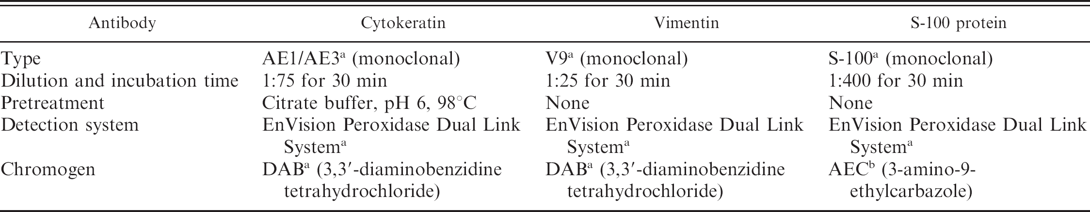
Immunohistochemistry was performed on 3.5-μm tissue sections to evaluate the presence of cytokeratin, vimentin intermediate filaments, and S-100 protein, as indicated in Table 1. All sections were counterstained with Mayer's hematoxylin.
Neoplastic cells showed dual expression of cytokeratin and vimentin intermediate filaments. Specifically, the lobules of physaliferous cells were intensely and diffusely positive for cytokeratin (Fig. 4) and vimentin (Fig. 5). S-100 protein was sporadically and intensely positive in physaliferous and chondroid cells, respectively (Fig. 6). A diagnosis of chondroid chordoma was made. No recurrence of the tumor was present 7 months after surgery.
Chordoma is an uncommon neoplasm arising from remnants of the notochord. The notochord defines the cranial-caudal axis of the embryo, induces the formation of the head and central nervous system, and serves as a development center organizing vertebral bodies and the basal portions of the sphenoid and occipital bones. 10 The nucleus pulposus is believed to be the only derivative of notochordal tissue. 13 Three distinct types of chordoma are recognized in humans: 1) the classic chordoma, 2) chondroid chordoma, and 3) chordoma with a malignant spindle cell component. The classic chordoma is a slow-growing, locally aggressive neoplasm with a high rate of recurrence, particularly in those of sacrococcygeal or vertebral origin; chondroid chordoma arises primarily in the spheno-occipital region and is characterized by chondromatous and chordomatous features.
Chondroid chordoma has a better prognosis than a classic chordoma. 14 In animals, only a few chordomas have been described in dogs, 7 cats, mice, 4 rats, and mink. Larger numbers have been reported in ferrets. 9 Chondroid chordoma has been reported in rats, mink, and ferrets 3 and is typically located on the tail. Only 1 case of intramuscular cervical noncartilaginous conventional chordoma with metastasis to prescapular lymph nodes has been reported in a cat. 2 In that case, neoplastic cells were negative for vimentin and cytokeratin but were positive for S-100 protein and neuron-specific enolase. 2 The feline case reported in the present study showed morphological and immunohistochemical features similar to those of chondroid chordomas described in ferrets. 3
Several immunohistochemical studies in various species have demonstrated both epithelial and mesenchymal characteristics of a chordoma, indicated by the dual expression of cytokeratin and vimentin intermediate filaments. This feature, also possessed by the notochord, offers a clear differentiation from a chondrosarcoma, which does not express cytokeratin. 1,5 Furthermore, the latter shows cellular features of malignancy, such as prominent nucleoli, irregularly shaped nuclei, and frequent mitotic figures. In a previous study, the positivity of S-100 in chordomas was found to be related to the presence of stromal glycosaminoglycans of the chondroitin sulfate type. 8 In addition, S-100 has been reported in an ever-increasing number of neoplasms of unrelated histogenesis. Neuron-specific enolase was found in cells with features of high metabolic activity. Consequently, S-100 protein and neuron-specific enolase were not considered to have any specific histogenetic implications but were considered as possible markers indicating cell-stromal interactions. 8 The distinction between conventional chordoma and the chondroid subtype seems to be of prognostic value. In fact, human patients with chondroid chordomas have survival rates 3 times higher than those with conventional chordomas. 6 Chondroid chordomas described in ferrets and mink have not been reported to metastasize, whereas the conventional type observed in cats 2 and rats have a high rate of metastasis. 2,12 Conversely, conventional canine chordomas appear to have a low metastatic potential, 11 which is a reason why they should be differentiated from tumors that are likely to metastasize, such as poorly differentiated chondrosarcomas, liposarcomas, and mucinous adenocarcinomas.
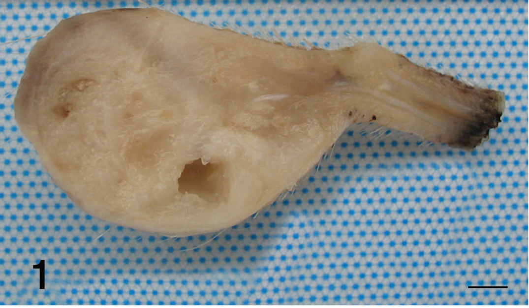
Cut section of the mass affecting the tip of the tail. Bar = 1 cm.
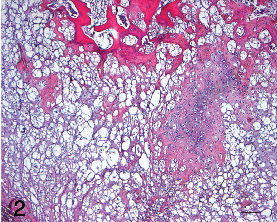
Physaliferous cells surrounding cartilage and trabecular bone, which contains bone marrow and hematopoietic cells. Hematoxylin and eosin stain. Bar = 200 μm.
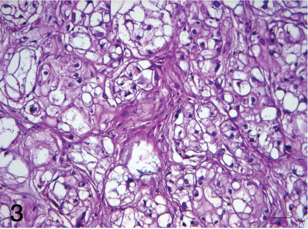
Lobules of large vacuolated cells intermingled with fibrous tissue. Marked cellular pleomorphism and presence of nests of cells with mucinous cytoplasm. Hematoxylin and eosin stain. Bar = 25 μm.
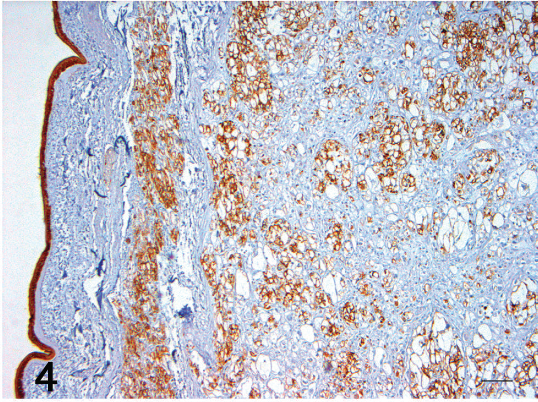
Cells at the periphery of the lesion showing strong immunoreaction for cytokeratin comparable to that of the epidermis. AE1/AE3, monoclonal. a Bar = 100 μm.
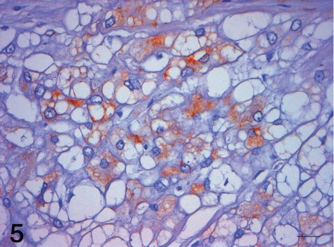
Immunohistochemical labeling of vimentin in cytoplasm of physaliferous cells. V9, monoclonal. a Bar = 25 μm.
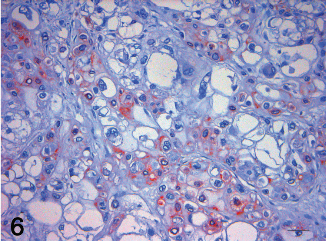
S-100 protein immunolabeling. Note a marked expression of the protein in cartilaginous tissue and weak reaction of the surrounding vacuolated cells. S-100, monoclonal. a Bar = 25 μm.
Antibodies, dilutions, pretreatments, detection system, and chromogen used for immunohistochemical study.
Furthermore, chordomas arising from the tail seem to have a better prognosis in all reported cases. Finally, only an accurate histological and immunohistochemical diagnostic approach will allow proper distinction of chondroid chordoma from other tumors of the tail with cartilaginous differentiation. To the authors' knowledge, the current report is the first case of chondroid chordoma described in a cat.
Acknowledgements. Alberto Masiero is kindly acknowledged for his assistance with the photographs.
Footnotes
a.
Dako North America Inc., Carpinteria, CA.
b.
Sigma-Aldrich, St. Louis, MO.