
Abstract
Apocrine cystomatosis is a rare condition characterized by clusters of cystically dilated sweat glands or other specialized apocrine glands. Cystic dilation of cutaneous sweat glands has been described in humans, dogs, and cats, but not in pigs, to our knowledge. We describe herein linear, brown, colloid-filled cavities < 1 cm diameter within the subcutaneous fat of the entire dorsal region of carcasses of three 6-mo-old pigs. These incidental findings were detected during meat inspection in 3 different slaughterhouses in Catalonia, Spain. Histopathology revealed multiple cystic cavities lined by flattened glandular epithelium, filled with proteinaceous material, and corresponding to cystic hyperplasia of sweat glands.
Keywords
Skin disorders in pigs, which are relatively common, may manifest only in the skin or may be signs of a generalized infection.2,12 Skin diseases in a swine herd can result in economic losses through decreased feed efficiency and growth rate, and increased mortality. Additionally, because the skin of pig carcasses has economic value, lesions in this organ can be a cause of partial carcass condemnation. 5 However, this economic impact is far less important than complete carcass condemnation that occurs because of respiratory, digestive, or reproductive diseases.
The origin of skin diseases can be divided into congenital, infectious, nutritional, environmental, neoplastic, and miscellaneous disorders. 2 Additionally, there are skin diseases with unknown causes.2,12
Hyperplastic lesions are unusual in pig skin, and grossly they may appear to be neoplasms. However, neoplasms are infrequent in swine given that most pigs are slaughtered before reaching maturity, when tumors typically develop. 4 The most commonly reported swine tumors affect young pigs, and include lymphosarcoma, nephroblastoma, and melanoma.3,4 Porcine neoplasms and hyperplastic lesions do not frequently cause clinical signs and are typically diagnosed as incidental lesions in diagnostic specimens or in carcasses at slaughter.6,8
Apocrine cystomatosis, also known as cystic hyperplasia of apocrine sweat glands, is a rare non-neoplastic condition characterized by clusters of cystically dilated sweat glands. 7 This condition has been described in humans, dogs, and less frequently, in cats, but not in pigs or any other livestock species, to our knowledge.
We describe herein generalized apocrine cystomatosis in three 6-mo-old pigs, which were unrelated (from different farms), slaughtered during the period 2018–2019 in 3 different inspected abattoirs in Catalonia, Spain. Veterinary inspectors reported no antemortem alterations in any of the 3 cases. During postmortem inspection, linear, brown, colloid-filled punctate and non-prominent nodules or vesicles < 1 cm diameter were noticed within the subcutaneous fat of the entire dorsal region in all 3 animals (Figs. 1, 2). The carcasses were rejected, and no internal organs were inspected. A sample of the skin of each animal was collected, refrigerated, and submitted to the Slaughterhouse Support Network (Servei de Suport a Escorxadors [SESC]) 10 for diagnosis. After fixation in 10% neutral-buffered formalin, tissues were processed routinely and stained with hematoxylin and eosin. Immunohistochemical staining with antibodies against Ki67, a marker for cell proliferation, was performed (Fig. 5).
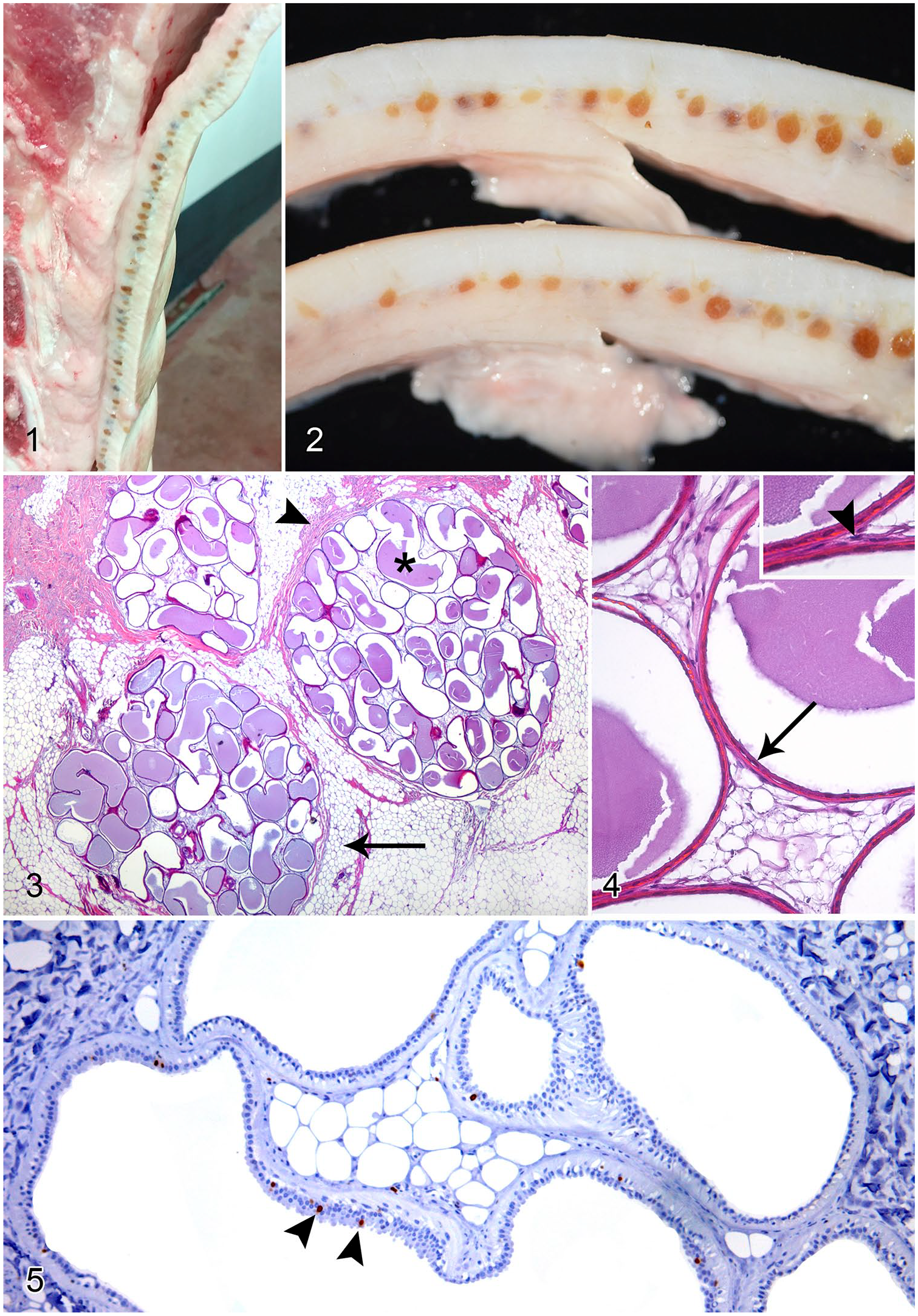
Apocrine cystomatosis in a 6-mo-old domestic pig.
The skin samples evaluated from the 3 animals had the same histologic alteration, namely clusters of distended apocrine glands forming tortuous cystic cavities and, occasionally, tubular structures of variable size within the subcutaneous fat. Adipocytes of the adjacent parenchyma were atrophic and compressed, and the dilated glands were surrounded by a thin rim of fibrous connective tissue (Fig. 3). Some cysts lost their fluid content during the processing of samples, but most contained dense eosinophilic colloid within their lumina (Fig. 3) admixed with occasional sloughed epithelial cells. Generally, the cysts were lined by a single layer of flattened epithelial cells without evidence of mitosis, multiple layers of epithelium, or tubulopapillary projections. A layer of flat myoepithelial cells surrounded the epithelium (Fig. 4). The adjacent dermis was free of inflammatory infiltrates. The epidermis could not be evaluated as a result of the coagulation artifacts caused by the scalding of the carcasses. Ki67 immunohistochemistry revealed a few glands with occasional cells of the apocrine epithelium labeling positive (1–6 per dilated gland; Fig. 5).
To our knowledge, apocrine cystomatosis has not been reported previously in pigs or any other livestock species. The location of these hyperplastic glands is different in dogs and cats. In dogs, the cysts can be solitary (referred to as apocrine cysts) or numerous and are mainly distributed over the head and neck.7,11 In cats, cystomatosis of the ceruminous glands is the most common presentation; cysts develop from the ceruminous glands of the lining of the concave pinna and can involve the external auditory canal opening, and occasionally the auditory canal. 7
There is a lack of agreement about the etiology and nature of apocrine cystomatosis in veterinary medicine. 11 Many hypotheses have been suggested. It was initially proposed that cyst formation might be attributed to the occlusion of excretory ducts. Later, it was suggested that apocrine cysts are the result of episodes of glandular hyperplasia followed by an involution phase and the inability to reabsorb the content of dilated glands. 12 More recently, single or multiple apocrine cysts have been described either as benign sweat gland tumors, or simply as an idiopathic, non-neoplastic, senile degenerative change or, in some cases, a congenital condition. 11 In our cases, an age-related cause is not considered likely given that the pigs were slaughtered before reaching maturity. A congenital origin cannot be ruled out, but cannot be confirmed, given that the condition was not observable clinically. However, the absence of normal apocrine glands in the sections evaluated, and the lack of evident dilation or obstruction of glandular ducts and/or inflammation or other alterations in the surrounding subcutaneous tissue, may be elements pointing toward a congenital origin.
The differential diagnosis of apocrine gland hyperplasia, of both sweat and special apocrine glands (mammary or ceruminous glands), must include adenomas. A diagnosis of adenoma requires histologic identification of cellular proliferation characterized by multiple layers of lining cells and acinar or papillary aggregates of cells projecting into cyst lumens.9,11 The proliferative activity of the epithelial cells can be further characterized by assessing the expression of Ki67 protein. 1 In our cases, Ki67 immunohistochemistry indicated a lack of cell proliferation, given that very few cells were labeled within affected glands. The expression of epidermal growth factor receptor (EGFr) in the apocrine epithelium, as well as the intracystic cation content (Na+/K+ ratio) and pH, have been shown to be helpful in determining malignancy in studies regarding human apocrine breast cysts. 11
Footnotes
Acknowledgements
We thank Blanca Pérez and Ghizlane El Korchi (Servei de Diagnòstic de Patologia Veterinaria, Veterinary School of Universitat Autònoma de Barcelona, Spain) for their technical support.
Authors’ note
Declaration of conflicting interests
The authors declared no potential conflicts of interest with respect to the research, authorship, and/or publication of this article.
Funding
The authors received no financial support for the research, authorship, and/or publication of this article.