
Abstract
The objective of the study was to develop an immunohistochemical (IHC) assay for rapid detection of Erysipelothrix rhusiopathiae. Serotypes 1a, 1b, and 2 are most frequently associated with clinical disease in pigs. Antiserum against serotypes 1a, 1b, and 2 was produced in rabbits, pooled, and applied to formalin-fixed, paraffin-embedded tissue sections of pigs (lungs, heart, spleen, and skin). The results obtained with the IHC assay were compared with direct culture on tissue samples from experimentally inoculated pigs either treated (n = 6) with antibiotics or untreated (n = 8) as well as on samples from field cases (n = 170) submitted to the Veterinary Diagnostic Laboratory at Iowa State University. The agreement between direct culture and IHC staining was found to be substantial. The results of the present study indicate that the IHC assay is highly sensitive and specific in detecting E. rhusiopathiae antigen in formalin-fixed, paraffin-embedded tissues. Results indicated that the IHC is particularly useful in cases in which pigs had been treated with antibiotics prior to submission and in which direct cultures of organs were negative. In addition, the IHC was found to be useful for detection of E. rhusiopathiae antigen in skin lesions, which are often culture negative.
Keywords
Erysipelothrix rhusiopathiae is a small, facultative anaerobic, Gram-positive rod that is the cause of swine erysipelas. In addition to pigs, E. rhusiopathiae has also been isolated from many species of both domestic and wild mammals, including humans, in which the disease is referred to as erysipeloid. 4,10 Swine erysipelas is distributed worldwide and is of economic significance. Thirty percent to 50% of pigs are known to harbor E. rhusiopathiae; however, these pigs are frequently healthy despite the presence of the organism in tonsils and lymphatic tissues. The reservoir for acute erysipelas is thought to be subclinically infected swine shedding E. rhusiopathiae in feces, urine, saliva, and nasal secretions. 10 Three clinical manifestations of swine erysipelas have been described: 1) the acute form, which may induce substantial morbidity and mortality within days, is characterized by sudden illness and/or death often associated with rhomboid skin lesions; 2) the subacute form, which is similar to the acute form but is typically less severe and commonly remains undetected; and 3) the chronic form, which may result in the development of arthritis and endocarditis. 10
The genus Erysipelothrix spp. contains 4 species and their associated serotypes: E. rhusiopathiae (serotypes 1a, 1b, 2, 4–6, 8, 9, 11, 12, 15–17, 19, 21, N), E. tonsillarum (serotypes 3, 7, 10, 14, 20, 22, 23), Erysipelothrix sp. strain 1 (serotype 13), and Erysipelothrix sp. strain 2 (serotype 18). 9 The pig is known to be susceptible to at least 17 of the described serotypes, but infections are most often attributed to E. rhusiopathiae serotypes 1a, 1b, and 2. Diagnostic investigations of U.S. field cases have indicated that E. rhusiopathiae serotype 1a is typically the cause of acute septicemic erysipelas, and serotype 2 is most common in subacute and chronic cases of swine erysipelas. 8 The other described serotypes of Erysipelothrix spp. are thought to have low levels of virulence for swine. 7
In the United States, diagnosis of swine erysipelas is most often confirmed by direct culture; however, direct culture can be both time-consuming (≥48 hr) and complicated. Difficulties can arise especially when compounded by specimen contamination, as Erysipelothrix spp. are characterized by small colony size and a slow rate of growth. In addition, the sensitivity of direct culture isolation may be adversely affected by tissue conditions and antimicrobial treatment of the pig. 1 The aim of the current study was to develop and validate an immunohistochemical (IHC) assay for detection of Erysipelothrix rhusiopathiae in formalin-fixed, paraffin-embedded tissue sections.
For the IHC assay, polyclonal antiserum against serotypes 1a, 1b, and 2 was produced in rabbits. For antiserum production, 2 rabbits were used for each serotype and immunized using a previously described protocol. 11 In brief, the rabbits were immunized by 5 successive intravenous injections of formalin-killed whole cells of the reference strains HC-585 (serotype 1a), 422–1 (serotype 1b), or NF-4 (serotype 2). Ten milliliters of a beef brain-heart infusion broth a culture from each of the 3 reference strains was incubated for 24 hr and placed into 1 liter of preincubated beef brain-heart infusion broth. a After incubation for 24 hr at 37°C, subcultures were made on agar plates to test for culture purity and colony smoothness. The broth cultures were killed by adding 1% formalin b (v/v) and incubated for 12 hr at room temperature. Cells were harvested by centrifugation, washed twice with chilled physiologic saline solution b containing 0.5% formalin, b and suspended to an optical density of 0.5 at 600 nm in a colorimeter. Sterile glass beads with a diameter of 4 mm were added to keep the cells in suspension, and the cell suspension was stored at 4°C until usage. Hyperimmune serum samples were confirmed to be specific by testing the sera against all available reference strains (serotypes 1–26, N) as previously described. 11
Comparison of direct culture and immunohistochemical (IHC) staining in pigs experimentally inoculated with different Erysipelothrix rhusiopathiae serotypes. *
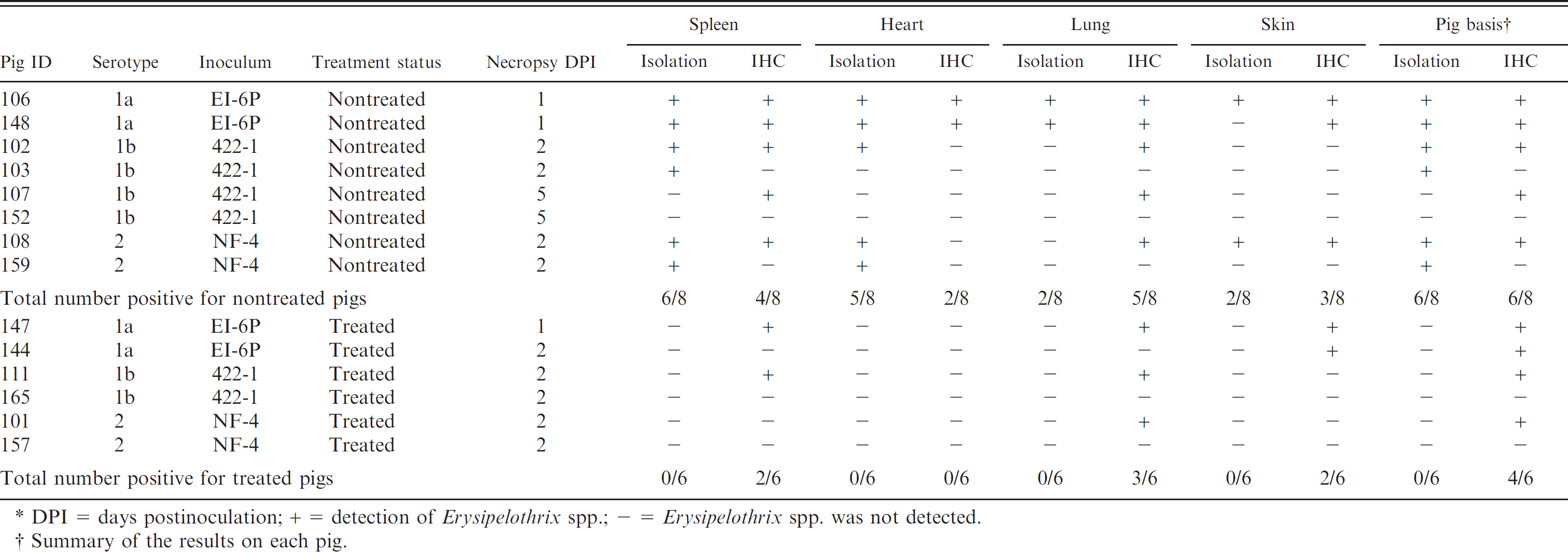
DPI = days postinoculation; + = detection of Erysipelothrix spp.; — = Erysipelothrix spp. was not detected.
Summary of the results on each pig.
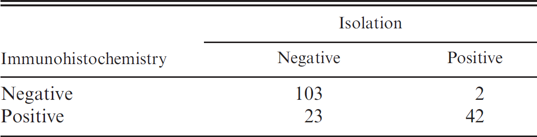
Comparison of direct culture and immunohisto-chemical staining on field samples.
For IHC detection of E. rhusiopathiae–specific antigen, formalin-fixed, paraffin-embedded tissue sections including liver, skin, lung, heart, synovium, tonsil, lymph nodes, intestines, spleen, and kidney were dewaxed and rehydrated with distilled water followed by quenching with 3.0%hydrogen peroxide b for 5 min. The slides were rinsed 3 times in deionized water, and digestion was completed with 0.05% protease type XIV (bacterial, from Streptomyces griseus) b for 2 min. After rinsing the slides 3 times with deionized water, the E. rhusiopathiae polyclonal antiserum cocktail containing equal volumes of the 3 antisera was applied in a 1:1,000 dilution for 1 hr at 37°C. Further processing was done by using a labeled streptavidin–biotin detection kit. c Known E. rhusiopathiae–positive tissue sections as well as known E. rhusiopathiae–negative tissue sections were used as controls for each IHC run. Figure 1A shows a skin section positive for E. rhusiopathiae. Negative control staining by omitting the E. rhusiopathiae antiserum on the same tissue section is shown in Figure 1B. Erysipelothrix rhusiopathiae antigen was commonly identified in bacteria-like structures in the lung interstitium (Fig. 2), in lumina of small vessels in lung tissues (Fig. 3), between hepatocytes (Fig. 4), or in the dermis most commonly in and around superficial blood vessels (Figs. 1A, 5).
Specificity of the E. rhusiopathiae IHC procedure was evaluated by testing sections from formalin-fixed, paraffin-embedded blocks from pigs known to be positive for other pathogens (i.e., Swine influenza virus, Porcine reproductive and respiratory syndrome virus, Porcine circovirus-2, E. tonsillarum, Erysipelothrix sp. strain 1, Erysipelothrix sp. strain 2, Mycoplasma hyopneumoniae, Streptococcus suis, Salmonella spp., Lawsonia intracellularis, Actinobacillus suis, Actinobacillus pleuropneumoniae, and Haemophilus parasuis). There was no evidence of cross-reaction with any of the pathogens tested, and the specificity was determined to be 100%.
Sensitivity was evaluated by comparing the IHC results to those obtained with E. rhusiopathiae direct culture. In brief, the external surface of the tissue specimens were seared with a heated spatula to remove contaminants, the specimen was incised using a sterile scalpel blade, and a sterile swab was inserted for bacterial collection for culture. 2 The swabs were inoculated on trypticase soy agar plates containing 5% sheep blood. d Plates were aerobically incubated at 35°C and examined at 24 and 48 hr postinoculation. Suspect colonies with a characteristic appearance similar to Erysipelothrix spp. were subcultured to trypticase soy agar plates containing 5% sheep blood, d incubated for 24 hr, and then biochemically confirmed using standard laboratory methods. 11
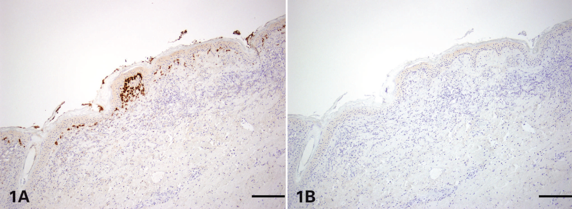
Skin, porcine. Immunohistochemical staining with (

Lung, porcine. Immunohistochemical staining using a polyclonal antiserum against Erysipelothrix rhusiopathiae revealing dark brown staining of bacteria-like organisms in alveolar septa and capillaries. Streptavidin–biotin–peroxidase complex method counterstained with hematoxylin. Bar = 20 μm.

Lung, porcine. Immunohistochemical staining using a polyclonal antiserum against Erysipelothrix rhusiopathiae revealing dark brown staining in bacteria-like organisms in the lumen of small vessels. Streptavidin–biotin–peroxidase complex method counterstained with hematoxylin. Bar = 20 μm.
For further evaluation of the assay, tissue samples were used from 14 pigs experimentally inoculated with E. rhusiopathiae serotypes 1a, 1b, or 2 and collected between 1 and 5 days postinoculation. The details of the experiment have been previously described. 1 A portion of the pigs (6/14) were treated with antibiotics 24 hr postinoculation. The results obtained by direct culture and IHC assay are summarized in Table 1 individually by tissue (lungs, spleen, heart, and skin) and on a per pig basis. The tissues for the comparison were selected based on availability for isolation and IHC assay. Erysipelas was confirmed in 6 of 8 untreated, experimentally inoculated animals with both direct culture and IHC. Using direct culture, E. rhusiopathiae most often was isolated from the spleen and heart tissues, whereas E. rhusiopathiae antigen was demonstrated by IHC staining more commonly in lung and skin sections. In contrast, E. rhusiopathiae was not isolated using direct culture from any of the tissues (lungs, spleen, heart, and skin) in animals treated with antibiotics. However, E. rhusiopathiae antigen was demonstrated in 4 of 6 treated animals (Table 1), indicating that treatment did not interfere with IHC detection.
In addition to tissues from experimentally inoculated pigs, diagnostic submissions were also included in the current investigation. The IHC procedure was tested on 170 field cases with clinical history and lesions suggestive of bacterial septicemia. Forty-four of these cases (25.9%) were confirmed to be positive by direct culture, and 126 of 170 cases (74.1%) were negative by direct culture. A comparison of results between direct culture and IHC are summarized in Table 2. The 2 tests agreed on 145 of the cases. Kappa statistics were calculated to measure statistical agreement between the 2 tests using a statistical software package. e Values for kappa range from −1 to 1, where −1 indicates agreement worse than expected by chance, 0 equals agreement no better than expected by chance, and 1 equals perfect agreement. 6 The following arbitrary standards for the strength of agreement as described by Landis and Koch were used: ≤0 = poor, 0.01–0.2 = slight, 0.21–0.4 = fair, 0.41–0.60 = moderate, 0.61–0.80 = substantial, and 0.81–1 = almost perfect. 5 Under the experimental conditions described in the present report, the kappa statistic was 0.67 ± 0.06, indicating substantial agreement between direct culture and IHC assay.
Disagreement between direct culture isolation and the IHC assay was found in 25 of 170 (14.7%) cases. Two of the 25 cases were direct culture positive but IHC negative. In both cases, tissues were microscopically unremarkable, and low numbers of E. rhusiopathiae were isolated from lung tissues. Twenty-three of the 25 cases with conflicting results were direct culture negative but IHC positive. Interestingly, all 23 IHC-positive and direct culture–negative cases had microscopic lesions consistent with bacterial septicemia (fibrin thrombi in small vessels, suppurative and necrotizing vasculitis, and necrosuppurative dermatitis). Erysipelothrix rhusiopathiae antigen was demonstrated in sections of skin in all cases that showed dermal lesions and where skin was submitted (21/23; Fig. 4), implying that affected sections of skin are ideal samples for IHC detection.
Immunohistochemistry approaches for detection of E. rhusiopathiae antigen in arthritic joints of pigs experimentally inoculated with E. rhusiopathiae serotype 2 have been described earlier. 3 However, to the authors' knowledge, the IHC assay described in the current study for detection of E. rhusiopathiae antigen is the first used for routine diagnostics. In summary, the IHC test was found to be highly sensitive and specific in detecting E. rhusiopathiae antigen in formalin-fixed, paraffin-embedded tissues. The authors concluded that IHC is particularly useful in cases in which the animals had been treated with antibiotics prior to submission and in which the cultures of organs were negative. In addition, the E. rhusiopathiae IHC was found to be useful for detection of erysipelas antigen in skin lesions, which are often culture negative.
Acknowledgements. The authors wish to thank Dr. Brian VanderLey, Matt Boogerd, and the bacteriology staff in the Veterinary Diagnostic Laboratory at Iowa State University for assistance with animal work, culturing, and typing of isolates. The authors also thank Joseph Brodie for assistance with development of the IHC assay; Dr. Richard Wood, Joann Kinyon, Nels Nord, and Dr. Paul Hauer for assistance in production of the rabbit antiserum; and Dr. Darin Madson for assistance with photography. Swine erysipelas research at Iowa State University is currently funded by Intervet-Schering-Plough Animal Health Inc. and the Iowa Pork Producers Association.

Liver, porcine. Immunohistochemical staining using a polyclonal antiserum against Erysipelothrix rhusiopathiae revealing dark brown staining of bacteria-like organisms accumulating between hepatocytes. Streptavidin–biotin–peroxidase complex method counterstained with hematoxylin. Bar = 20 μm.

Skin, porcine. Immunohistochemical staining using a polyclonal antiserum against Erysipelothrix rhusiopathiae revealing abundant bacteria-like organisms (dark staining) in the dermis. Streptavidin–biotin–peroxidase complex method counterstained with hematoxylin. Bar = 50 μm.
Footnotes
a.
BD Diagnostic Systems, Sparks, MD.
b.
Sigma-Aldrich, St. Louis, MO.
c.
Dako North America Inc., Carpinteria, CA.
d.
Thermo Fischer Scientific Inc., Waltham, MA.
e.
JMP® Version 7, SAS Institute Inc., Cary, NC.