
Abstract
A 19-mo-old Holstein heifer was inactive and dyspneic. Physical examination revealed wheezing, exophthalmos, a cervical mass, and lymphadenopathy. Cytology of the cervical mass and lymph nodes showed predominantly large atypical lymphocytes. Lactate dehydrogenase and thymidine kinase activities were elevated. Although nested PCR for bovine leukemia virus (BLV) using blood was positive, quantitative PCR showed a low number of provirus copies. Autopsy revealed enlargement of most lymph nodes examined, as well as white masses of various sizes in muscles of the left hindlimb and thoracic and abdominal organs. Histopathology revealed severe infiltration with neoplastic lymphocytes in these organs. The cervical mass was immune-positive for B-cell markers. The final diagnosis was thymic B-cell lymphoma with BLV infection.
There are 2 forms of bovine leukosis: sporadic bovine leukosis (SBL) of unknown cause, and enzootic bovine leukosis (EBL) caused by bovine leukemia virus (BLV) infection. SBL is typically classified into 3 forms: calf or juvenile form, thymic form, and cutaneous form.2,10,29 SBL usually affects cattle <3 y old; EBL usually affects those >3 y old.2,29 In the calf or juvenile form, sudden onset of lymphoid hyperplasia, weight loss, fever, tachycardia, dyspnea, bloat, and paraparesis are common clinical features in cattle up to 24 mo of age.2,23,29 In the thymic form (seen in animals 6–24 mo old), the cervical and/or intrathoracic thymus is usually affected, and clinical signs depend on the location and size of the tumor.2,21,27,29 Cervical swelling, dyspnea, bloat, jugular distention, tachycardia, forequarters edema, and fever are typical in the thymic form.2,21,27,29 In the cutaneous form (seen in animals up to 30 mo old), cutaneous plaques of 1–5 cm diameter can be found on the neck, back, rump, and thigh, and regional lymph nodes are also enlarged.2,29 In cases of EBL, central or peripheral lymph node enlargement is commonly observed.2,29 Although the calf or juvenile form and EBL mainly involve B-cells, thymic and cutaneous forms mainly involve T-cells. 29
We describe herein a rare case of atypical thymic B-cell lymphoma. A free-grazing 19-mo-old Holstein heifer had respiratory difficulty and inactivity (day 1). The heifer was first diagnosed with bacterial pneumonia and was treated by a local veterinarian with antibiotics for 3 d, after which signs resolved. However, 23 d after the last treatment, signs of bilateral conjunctivitis appeared and the heifer’s condition worsened each day, resulting in bilateral exophthalmos that did not resolve with treatment. The heifer developed inappetence, anorexia, inactivity, and wheezing, as well as enlargement of various lymph nodes. The ventral aspect of the neck was also hard and slightly enlarged. Masses ventral to the rectum were noted by palpation. Blood chemical analysis revealed increased total protein and lactate dehydrogenase (LDH), and decreased blood urea. To confirm the diagnosis by further examination, ownership of the heifer was transferred to the Veterinary Teaching Hospital at Obihiro University of Agriculture and Veterinary Medicine (Japan) on day 32.
Physical examination revealed emaciation, with a body score of 2.25 out of 5, and binocular protrusion, conjunctival swelling, white nasal discharge, and enlargement of multiple lymph nodes. Wheezing was noted on auscultation, and the ventral neck was enlarged and hard upon palpation (Fig.1). There were small periorbital masses and no pupillary light reflex from the right eye; response from the left eye was delayed. Ultrasound examination of the neck revealed a lobulated hyperechoic structure. Cytology results from fine-needle aspiration (FNA) of the ventral neck region (cervical mass) and enlarged iliac lymph nodes showed predominantly large atypical lymphoid cells with mitoses. These cells had a high nuclear-to-cytoplasmic ratio with multiple distinct nucleoli and finely stippled chromatin. Hematology revealed lymphocytosis (13.0 × 10⁹/L; reference interval [RI]: 1.6–5.6 × 10⁹/L), 9 with 79% atypical lymphocytes. Serum LDH activity was increased (1,813 U/L; RI: 697–1450 U/L).12,14 LDH isoenzyme analysis revealed increases in LDH-2 (739 U/L; RI: 137–503 U/L) and LDH-3 (387 U/L; RI: 82–262 U/L).12,14 Thymidine kinase activity was also increased (10.4 U/L; RI: <5.4 U/L).24,28 A serum ELISA to detect antibodies against BLV was positive.
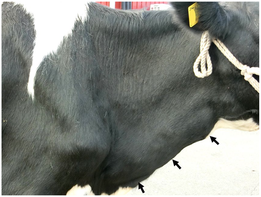
Disseminated thymic B-cell lymphoma in a Holstein heifer. The ventral neck is enlarged by firm nodules (arrows).
DNA was extracted from blood and FNA samples of the cervical mass and iliac lymph nodes (QIAamp DNA mini kit; Qiagen, Hilden, Germany), followed by nested PCR for BLV. 18 Distilled water and genomic DNA from the peripheral blood of a typical EBL cow were used as negative and positive controls, respectively. Nested PCR results were positive for BLV in all samples. The samples were then subjected to quantitative PCR (CoCoMo-BLV-qPCR; CoCoMo-BLV detection kit; Rikengenesis, Tokyo, Japan; TaqMan gene expression master mix; Thermo Fisher Scientific, Foster City, CA). CoCoMo-BLV-qPCR for samples from blood, the cervical mass, and an iliac lymph node yielded 17.9 copies/10 ng DNA, 1.2 copies/10 ng DNA, and <1 copy/10 ng DNA, respectively. In addition to CoCoMo-BLV-qPCR, we also performed inverse PCR to examine monoclonality and the proviral integration site. 19 Briefly, DNA samples were digested (PstI; Takara Bio, Shiga, Japan) and were then self-ligated (Mighty mix; Takara Bio). The resulting products were used as templates for PCR using inverse primers reported previously. 18 The negative result of inverse PCR suggested that BLV was not present in the tumor cells and was therefore not likely to be associated with tumorigenesis in our case.
The heifer was euthanized, and an autopsy was performed on day 34 after initial diagnosis according to ethical and animal welfare requirements under the guidelines of the Care and Use of Agriculture Animals of Obihiro University (Approval 29-38). The largest mass was found along the trachea from the lower neck to the chest, corresponding to the area of the thymus, which consisted of 3 parts of 22 × 8 × 6, 30 × 16 × 12, and 20 × 10 × 60 cm (Fig. 2A); however, normal thymus was not identified grossly. All lymph nodes including the abdominal, thoracic, and peripheral lymph nodes were markedly enlarged. Additionally, numerous white masses (0.5–10 cm diameter) were found throughout the body, infiltrating organs and tissues such as the liver, kidney, heart, lung, adrenal gland, gall bladder, rumen, abomasum, uterus, peritoneum, biceps femoris, semitendinosus muscles of the left hindlimb, ocular conjunctiva, periorbital fat, and subcutaneous fat of the left thoracic region. Cross-sections of all masses were similar in appearance with solid, milky white surfaces and some multinodular structuring in the masses along the trachea (Fig. 2B). Other gross findings included multiple erosions and ulcers in the mucosa of the reticulum and omasum.
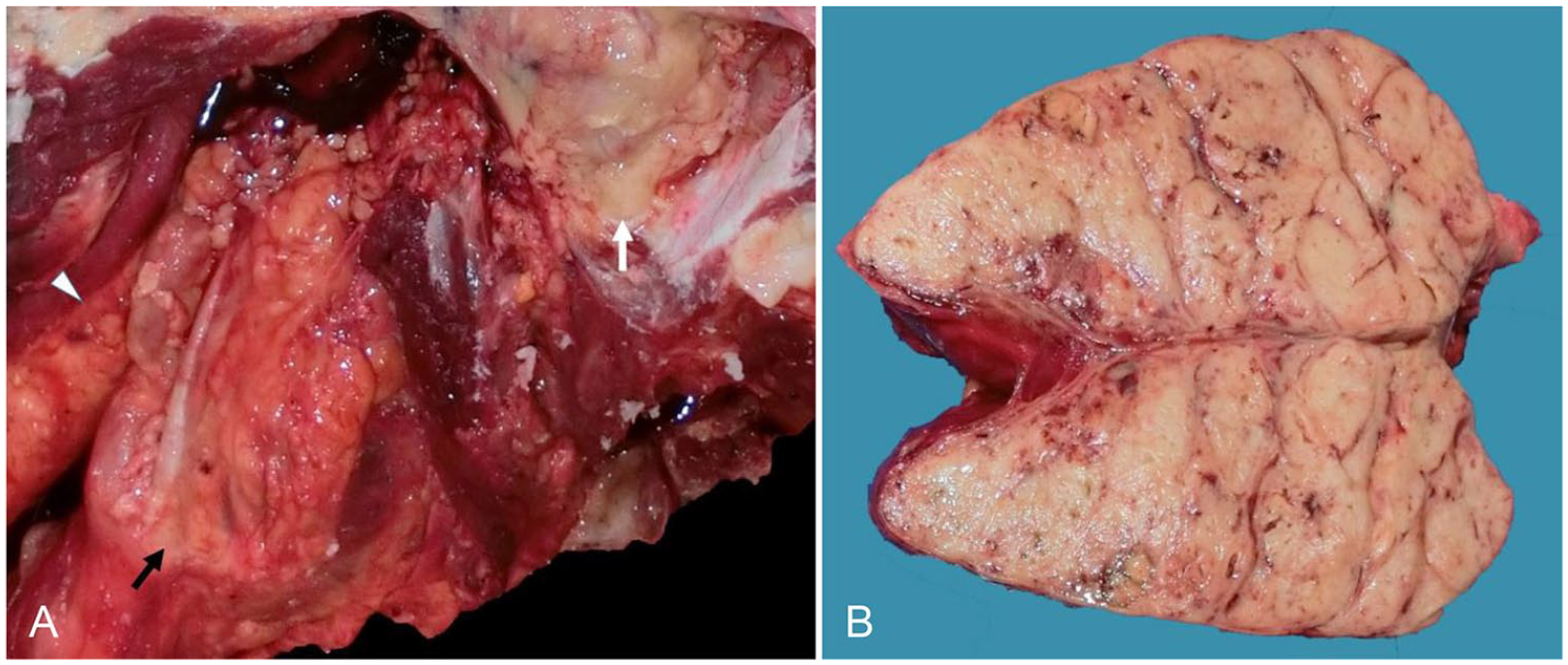
Disseminated thymic B-cell lymphoma in a Holstein heifer.
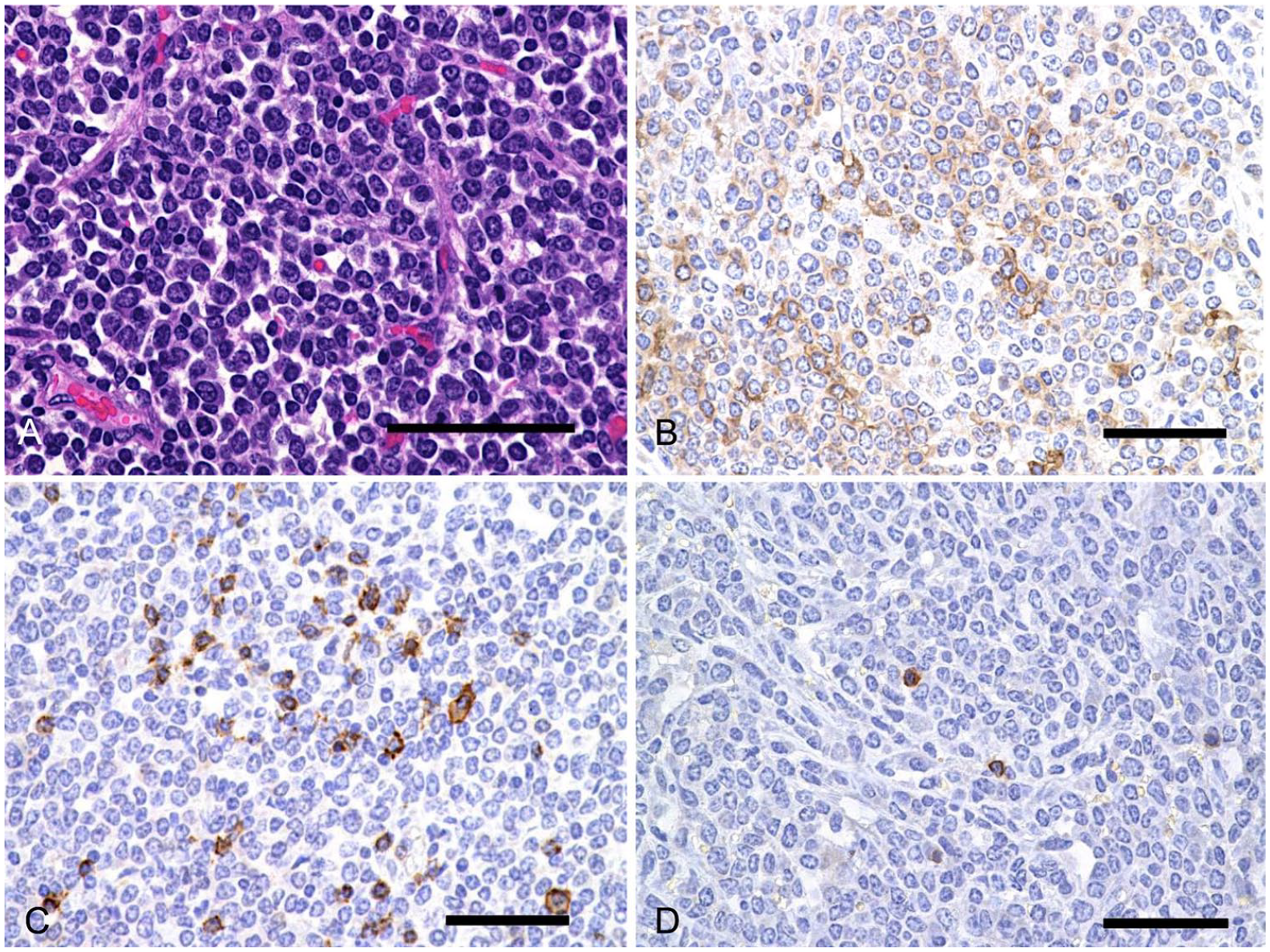
Microscopic examination revealed that the enlarged lymph nodes and all tissues with white masses were markedly infiltrated by neoplastic lymphoid cells. The neoplastic cells varied in size but were generally large (1.7–2.8 times the size of erythrocytes), with scant cytoplasm and polymorphic nuclei (Fig. 3A). There were up to 6 mitotic figures per high-power field (400×), and histologic appearance did not differ significantly between the neoplastic masses. In organs with neoplastic infiltrates, the parenchymal structure was extensively destroyed or replaced by neoplastic cells. Immunohistochemistry was conducted on the left iliac lymph node, periorbital mass, and cervical masses using antibodies for BLA36 (marker for B-cell lymphocytes; BioGenex Laboratories, Fremont, CA), CD20 (marker for B-cells; Thermo Fisher Scientific, Fremont, CA), CD3 (marker for T-cells; Dako, Tokyo, Japan), and cytokeratin AE1/AE3 (marker for epithelial cells; Dako). The majority of the neoplastic cells were immunopositive for BLA36 (Fig. 3B); some also stained positive for CD20 (Fig. 3C). On the other hand, neoplastic cells were negative for CD3 (Fig. 3D). In the neoplastic masses along the trachea, cytokeratin AE1/AE3 revealed within neoplastic foci a framework of epithelial cells, which were identified to be remnant epithelial cells of the thymus. Based on these results, the neoplasm was diagnosed as a B-cell lymphoma with primary involvement of the thymus.
Disseminated thymic B-cell lymphoma in a Holstein heifer.
Based on the autopsy and histopathologic findings, a preliminary diagnosis of bovine leukosis was offered. Given the location of the largest mass in the thymic area, T-cell lymphoma was suspected; however, immunohistochemistry revealed that the neoplastic mass originated from B-cells.
The heifer was positive for BLV antibodies and antigen in serology and molecular tests. The number of DNA copies was <18 copies/10 ng DNA, which was much lower than in a previous report in which EBL cattle harbored >1,000 copies/10 ng DNA. 26 The negative result of our inverse PCR suggested that BLV was not integrated into the tumor cells. This finding eliminated the possibility of EBL in our case. BLV infection is not limited to EBL, given that SBL cattle can also be infected by BLV. 7 Indeed, several clinical cases that were animal-positive but tumor-negative for BLV have been reported.1,4,11,15,16,20,22,25,32
Autopsy revealed that neoplastic masses were widely distributed in most organs, with the largest mass located in the ventral neck near the chest. Other than the cervical mass, macroscopic lesions can develop in the lymph nodes, bone marrow, liver, spleen, kidney, and lungs in thymic lymphoma.21,23,27 We found neoplastic lymphoid cells in all lesions examined. The cervical mass was considered to be of thymic origin and confirmed by cytokeratin staining, in which remnants of epithelial cells were found in neoplastic foci and neoplastic cells were positive for a B-cell marker. This suggests that the neoplasm was a B-cell lymphoma that originated from the thymus, given that the largest mass found was the cervical mass. To our knowledge, this is only the second report of thymic B-cell lymphoma in cattle, including a case series of familial B-cell lymphoma with thymic localization 6 ; the occurrence of this type of lymphoma is apparently uncommon. Thymic B-cell lymphoma is also a rare condition in other species, 30 including humans,4,5,13,31 dogs, 17 cats, 8 and horses. 3 Our case provides additional evidence that thymic B-cell lymphoma can occur in cattle.
Footnotes
Declaration of conflicting interests
The authors declared no potential conflicts of interest with respect in the research, authorship, and/or publication of this article.
Funding
This work was supported in part by the Japan Society for Promotion of Science KAKENHI grant 16K15044.