
Abstract
A 21-mo-old Japanese Black beef cow had swollen mandibular and superficial cervical lymph nodes. Fine-needle aspiration cytology of the superficial cervical lymph node revealed large lymphoblasts with mitoses present. The bovine leukemia virus (BLV) proviral load was relatively high, and phylogenetic analysis of the whole BLV genome classified the BLV strain as one with high viral replication activity. Genotyping of bovine leukocyte antigen genes indicated that the cow was susceptible to enzootic bovine leukosis (EBL). The bone morphogenetic protein 6 (BMP6) gene promoter region was hypermethylated. Monoclonal proliferation of B cells and monoclonal integration of the BLV provirus in the bovine genome were detected by a clonality test of B cells and an inverse PCR assay, respectively. At autopsy, generalized swelling of lymph nodes and spinal canal invasion by tumor tissue at vertebrae L5-6 were observed. Histologic analysis revealed diffuse proliferation of large round neoplastic cells that were positive for BLA36 and negative for CD3. The cow was definitively diagnosed with EBL based on these findings. Infection with a highly pathogenic strain of BLV, susceptibility of the BoLA-DRB3 alleles, and hypermethylation of the BMP6 gene may have contributed to the development of EBL in our case.
Bovine leukosis is divided into 2 types: enzootic bovine leukosis (EBL) caused by bovine leukemia virus (BLV; genus Deltaretrovirus) in cattle >3-y-old, and sporadic bovine leukosis (SBL) in younger cattle.1,4,21 However, early onset of EBL in cattle <3-y-old has also been reported, 16 and the incidence of EBL in young beef cattle has increased in Japan. 19 Therefore, it is difficult to distinguish EBL from SBL with BLV infection in young cattle, and detailed mechanisms for EBL onset in young cattle remain unclear.
Both viral and host factors must be considered when elucidating viral infectious diseases. Differences in pathogenicity of BLV strains, 14 sensitivity to EBL onset based on bovine leukocyte antigen (BoLA) DRB3 polymorphism, 8 and methylation status of bone morphogenetic protein 6 (BMP6) gene 10 have been reported as viral and host factors related to EBL onset. However, there are few reports of individual cases involving the combination of these factors. We describe here a clinical case of EBL caused by a highly pathogenic strain of BLV in a young cow with susceptible BoLA-DRB3 alleles and hypermethylation of the BMP6 gene.
A 21-mo-old Japanese Black beef cow was presented with emaciation and mandibular swelling. Initial examination by a local veterinarian (day 1) revealed a body temperature of 40.6°C (RI: 38.0–39.2°C) and heart rate of 100 beats/min (bpm; RI: 60–84 bpm). 3 Emaciation, difficulty standing, and mandibular swelling were noted. The cow was treated for mandibular inflammation with 10 mg/kg oxytetracycline and 2.5 mg/kg NSAID medication. By day 6, superficial cervical and subiliac lymph nodes were enlarged. Bovine leukosis was suspected by a local veterinarian, and, on day 12, the cow was transferred to the Animal Teaching Hospital at the Obihiro University of Agriculture and Veterinary Medicine (Obihiro, Hokkaido, Japan) to confirm the diagnosis.
On initial physical examination at the hospital, high rectal temperature (40.2°C), normal heart rate (66 bpm), polypnea (42 breaths/min; RI: 18–28 breaths/min), 3 emaciation, and difficulty standing were noted. Swelling in lymph nodes, including parotid, mandibular, superficial cervical, and subiliac lymph nodes, was observed (Fig. 1). Rectal palpation revealed masses (1–4 cm) in the pelvic cavity. Fine-needle aspiration (FNA) cytology of superficial lymph nodes revealed medium-to-large lymphoblasts with a small amount of cytoplasm and occasional mitoses (Suppl. Fig. 1).
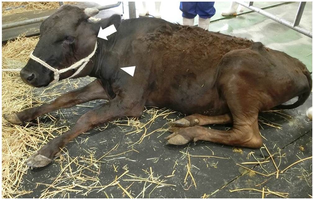
Swelling of parotid (arrow) and prescapular (arrowhead) lymph nodes on day 12 in a Japanese Black cow with enzootic bovine leukosis.
Hematologic examination showed mild anemia (RBC, 6.08 × 10¹²/L, RI: 5.10–7.60 × 10¹²; Hb, 70 g/L, RI: 85–122 g/L; PCV, 0.20 L/L, RI: 0.22–0.33 L/L). 3 WBC (11.2 × 10⁹/L; RI: 4.9–12.0 × 10⁹/L) and platelet count (606 × 109/L; RI: 193–637 × 109/L ) were within RIs. 3 In peripheral blood smears, 40% (4.48 × 10⁹/L ) of WBCs were medium-to-large atypical lymphoid cells (Suppl. Fig. 2). Serum biochemical analysis revealed increased activities of lactate dehydrogenase (3,660 U/L; RI: 697–1,450 U/L) and thymidine kinase (818 U/L; RI: <5.4 U/L).6,18
The BLV proviral load of DNA extracted from the peripheral blood sample was quantified (CoCoMo-BLV primer/probe, Riken Genesis; TaqMan gene expression master mix, Thermo Fisher) according to the manufacturer’s instructions. The BLV copy number in peripheral blood was 2,720 copies/50 ng DNA. The DNA from peripheral blood was used as a template for the PCR assay, which was performed using PrimeSTAR GXL DNA polymerase (Takara Bio) and 2 primer pairs (BLV 1-17 F: 5′-TGTATGAAAGATCATGC-3′, BLV 4565-4586 R: 5′-AATCTGATTGTGAGTCCAGAGG-3′; BLV 4416-4436 F: 5′-CAGTTCGGAGTTTCCCTTTCT-3′, BLV 8703-8720 R: 5′-TGTTTGCCGGTCTCTCCT-3′). 14 The PCR reaction was as follows: amplification with 30 cycles of denaturation at 98°C for 10 s, annealing at 55°C for 15 s, extension at 68°C for 5 min, and final extension at 68°C for 2 min. PCR products were treated with ExoSAP-IT express (Thermo Fisher). DNA libraries were prepared (QIAseq FX DNA library kit; Qiagen) following the manufacturer’s protocol, and sequenced (iSeq system; Illumina) using 2 × 150 bp paired-end reads. Quality control procedures were performed (Trim Reads tool, Genomics Workbench v.20.0; CLC bio). Unless otherwise stated, all software was used with default values applied. Mapping of quality-filtered reads against a reference BLV genome (EF600696.1) was performed using the CLC mapping tool, and 568 BLV genome sequences reconstructed by the sequencing reads (568 coverage). The whole genome sequence of BLV in our case was identified. A phylogenetic tree was constructed by neighbor-joining methods (1,000 bootstrap replications) using the whole genome sequence of BLV in our case (LC659972) and complete genome sequences of BLV obtained from GenBank (LC164086, AP018006–AP018032). BLV in our case was classified as a strain with high viral replication activity (group A; Fig. 2).
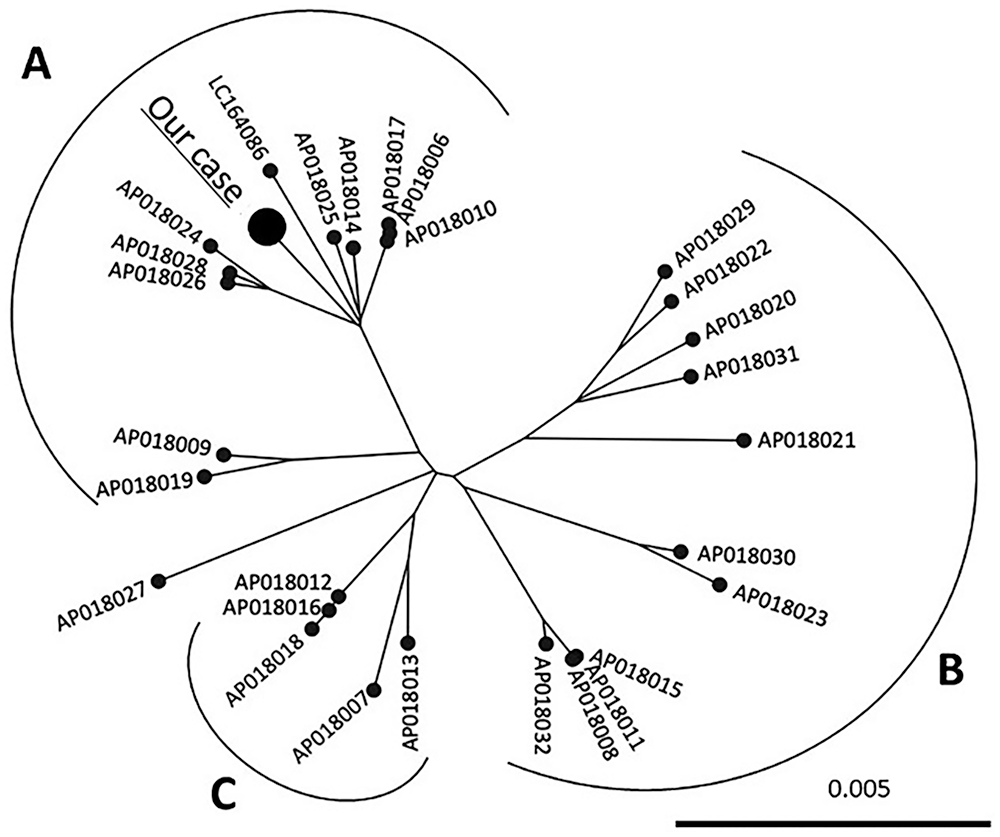
Phylogenetic analysis of the whole bovine leukemia virus (BLV) genome sequence in our case. A maximum-likelihood phylogenetic tree was constructed from whole BLV genome sequences of our case and 28 reference sequences. The BLV strains were divided into A–C based on a previous study. The bar denotes distance.
BoLA-DRB3 genotyping of our case was performed. The DNA from peripheral blood was used as a template for tailed PCR, which was performed using rTaq DNA polymerase (Toyobo) and primer pairs (NGS-DRB3FRW: 5′-TCGTCGGCAGCGTCAGATGTGTATAAGAGACAGCGCTCCTGTGAYCAGATCTATCC-3′, NGS-DRB3REV: 5′-GTCTCGTGGGCTCGGAGATGTGTATAAGAGACAG CACCCCCGCGCTCACC-3′). 13 The amplification program was carried out as follows: initial incubation at 94°C for 2 min, followed by 35 cycles of denaturation at 94°C for 30 s, annealing at 57°C for 30 s, extension at 72°C for 1 min, and final extension at 72°C for 2 min. PCR products were treated with ExoSAP-IT express (Thermo Fisher). DNA libraries were prepared (Nextera XT DNA library prep kit; Illumina) following the manufacturer’s protocol, and sequenced (MiSeq system; Illumina) using 2 × 300 bp paired-end reads. Quality control procedures were performed (Trim Reads tool; CLC). BoLA-DRB3 sequences were downloaded from the Immuno Polymorphism Database for sequences of the major histocompatibility complex (IPD-MHC; https://www.ebi.ac.uk/ipd/mhc/group/BoLA/), and mapping of quality-filtered reads against BoLA-DRB3 sequences was performed using the CLC mapping tool, resulting in 70.7% (6,878 of 9,735) reads mapped to the BoLA-DRB3*016:01 allele (Suppl. Table 1). Mapping rates to BoLA-DRB3 alleles other than BoLA-DRB3*016:01 were <4.4% (Suppl. Table 1). Hence, the BoLA-DRB3 genotype of our case was BoLA-DRB3*016:01/016:01.
Methylation status of the BMP6 gene promoter region was analyzed by the bisulfite sequencing method, as described previously. 10 Briefly, the sodium bisulfite modification of gDNA from peripheral blood was performed (MethylEasy Xceed rapid DNA bisulphite modification kit; Takara Bio) according to the manufacturer’s instructions. Semi-nested PCR on bisulfite-treated DNA samples for CpG islands in the BMP6 gene promoter region was performed. PCR products were cloned into the pCRTM4-TOPO vector (Invitrogen) and transformed into Escherichia coli (One Shot TOP10 chemically competent E. coli; Thermo Fisher). Plasmid DNA samples from 10 independent clones were sequenced with the M13R primer. The DNA sequence was determined (Prism BigDye terminator v.3.1 cycle sequencing kit, Prism 3500 genetic analyzer; Applied Biosystems) following the manufacturer’s instructions. Methylation rate of the BMP6 gene promoter region was 45.7% (256 of 560) in peripheral blood (Suppl. Fig. 3).
B-cell clonality of peripheral blood and swollen superficial lymph node tissue obtained by FNA was examined using PCR for immunoglobulin heavy-chain gene V-D-J junctional variability. 9 The DNA from peripheral blood and superficial lymph nodes aspirate was used as a template for PCR (HotStarTaq DNA polymerase, Qiagen; primer pair BoVHF1, BoVHR1). Genomic DNA from a typical EBL cow (7-y-old, BLV-positive, B-cell lymphoma) was used as positive control; cattle with endocarditis, persistent lymphocytosis, clinically health + BLV infection, and clinically health and no BLV infection were used as negative controls. The PCR products were electrophoresed in 3% agarose gel. A single band was detected in both peripheral blood and lymph node tissue (Suppl. Fig. 4A). In contrast, a smear or no visible bands were noted in negative controls.
BLV proviral integration clonality was analyzed using inverse PCR (iPCR). The procedure for iPCR was performed as reported previously. 11 DNA samples used for the B-cell clonality analysis were digested with PstI and were then self-ligated (Mighty Mix; Takara Bio). The products were used as templates for PCR using inverse primers (Pst-F, Pst-R). PCR products were electrophoresed in 2% agarose gel. A single band was detected in both peripheral blood and lymph node tissue (Suppl. Fig. 4B). In contrast, no visible bands were noted in negative controls.
The cow was euthanized on day 14 for autopsy according to the ethical and animal welfare requirements under the guidelines of the Care and Use of Agriculture Animals of Obihiro University (approval 29-38). Gross examination revealed swelling of systemic lymph nodes. Spinal canal invasion of tumor tissue at vertebrae L5-6 was observed. Histologic examination revealed infiltration of neoplastic lymphoid cells in enlarged lymph nodes, liver, spleen, heart, lung, adrenal, uterus, and the adipose tissue around the spinal cord (Fig. 3A). Immunohistochemically, tumor cells within enlarged lymph nodes stained positive for BLA36 (BioGenex; Fig. 3B) and negative for CD3 (BioGenex; Fig. 3C).
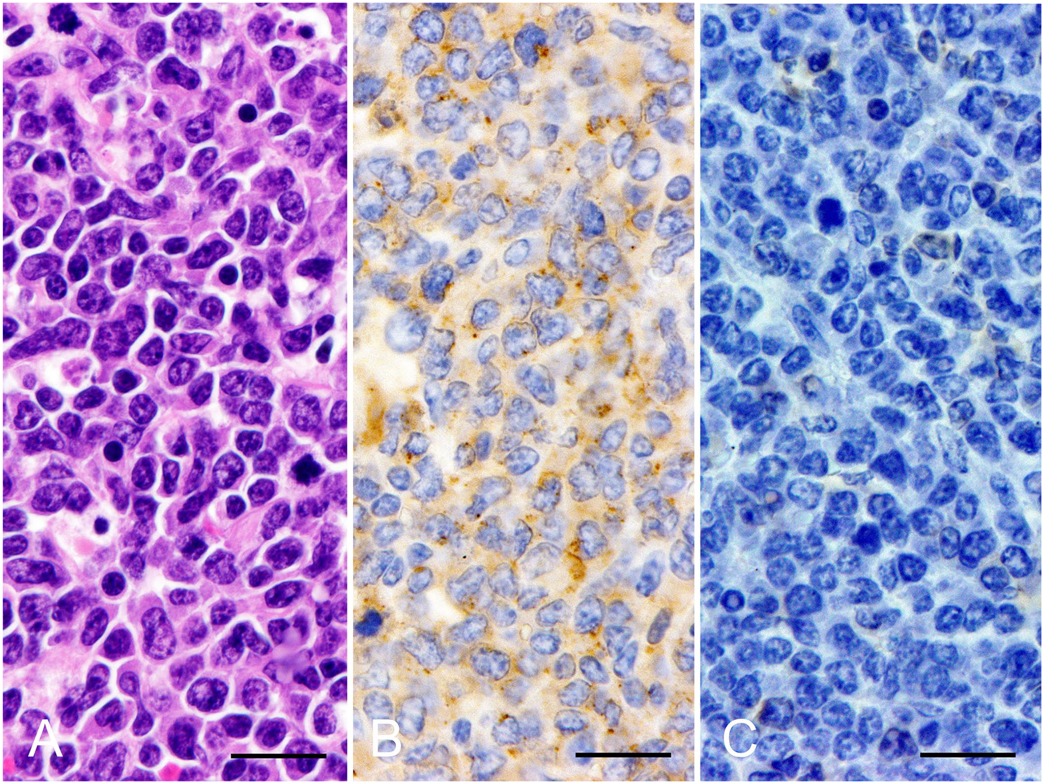
Histopathology and immunohistochemistry of superficial lymph node. Neoplastic cells had a round nucleus with nuclear atypia and granular patterned chromatin. These cells stained positive for BLA36 and negative for CD3.
EBL is typically observed in cattle >3-y-old,1,4,21 but cases of EBL in cattle <3-y-old have also been reported. 16 Moreover, onset of SBL in BLV-infected cattle has been reported. 5 Therefore, diagnosing EBL in young cattle is difficult. In our case, leukemia or lymphoma was suspected based on physical examination, FNA cytology, and hematologic examination. The BLV proviral load in our case was higher than the criterion for BLV infection associated with tumor development in a previous study (2,000 copies/50 ng DNA). 15 Monoclonal B-cell proliferation and monoclonal integration of the BLV provirus were detected by PCR for the Ig heavy-chain gene and iPCR, respectively. Histology demonstrated neoplastic proliferation of B cells. Based on these findings, we diagnosed EBL, despite the young age of the cow.
We also analyzed the BLV strain, BoLA-DRB3 polymorphisms, and methylation status in the BMP6 gene promoter region. Although genetic variation in BLV is limited, 12 BLV has been divided into 3 groups (A–C) based on the whole genome sequence, with high, moderate, and low viral replication activity in groups A, B, and C, respectively. 14 The same study found a high proviral load in cattle infected with group A strains and a low proviral load in those infected with group B or C strains. 14 Because proviral load was positively associated with disease progression, pathogenicity of group A strains might be higher than that of group B and C strains. 14 In our case, phylogenetic analysis of BLV demonstrated that the BLV classification was consistent with group A, suggesting high pathogenicity of the BLV strain in our case (Fig. 2; Suppl. Fig. 5). However, we did not evaluate the pathogenicity of the BLV in our case.
The MHC plays a crucial role in antigen presentation and immune responsiveness,17,20 and is also associated with infectious diseases including viral infection. The MHC system associated with disease susceptibility in cattle is known as BoLA. 13 The DNA sequence polymorphisms of BoLA-DRB3 are correlated with proviral load and onset of EBL, and the BoLA-DRB3*016:01 allele is associated with susceptibility to EBL in Japanese Black cattle. 8 In our case, analysis of the BoLA-DRB3 gene demonstrated that the genotype was BoLA-DRB3*016:01/016:01. Accordingly, our case could have been susceptible to EBL development.
BMP6 reduces proliferation and induces apoptosis in B cells. 7 Epigenetic modifications, including DNA methylation, can alter the transcription activity in mammals. 2 A study found that the BMP6 gene promotor region methylation rate in healthy cattle was 0.0–14.8%, and, by using reverse-transcription PCR, expression of the BMP6 gene mRNA was not found in samples from EBL cattle with those with >33.4% methylation. 10 Therefore, >33.4% methylation of the BMP6 gene promotor region was considered as hypermethylation resulting in inhibition of BMP6 gene expression. In our case, methylation of the BMP6 gene promoter region in peripheral blood was 45.7%. Hypermethylation of the BMP6 gene promoter region might have contributed to the development of EBL in our case.
Supplemental Material
sj-pdf-1-vdi-10.1177_10406387221102123 – Supplemental material for Enzootic bovine leukosis in a 21-month-old Japanese Black cow with high susceptibility
Supplemental material, sj-pdf-1-vdi-10.1177_10406387221102123 for Enzootic bovine leukosis in a 21-month-old Japanese Black cow with high susceptibility by Masaki Maezawa, Kana Sakaguchi, Yuka Tagaino, Yuki Fujii, Masataka Akagami, Junko Kawakami, Ken-ichi Watanabe, Yoshiyasu Kobayashi, Haruko Ogawa and Hisashi Inokuma in Journal of Veterinary Diagnostic Investigation
Footnotes
Acknowledgements
We thank all staff members of the Department of Veterinary Medicine at Obihiro University, those at Ibaraki Prefecture Kenpoku Livestock Hygiene Service Center for their technical assistance, and the Tokachi Agricultural Mutual Aid Association for bringing this clinical case to our attention.
Authors’ note
The data that support the findings of this study are available from the corresponding author upon request.
Declaration of conflicting interests
Laboratory of OSG Veterinary Science for Global Disease Management is an endowment laboratory, supported with a grant from OSG Corporation.
Funding
This work was supported by JSPS KAKENHI grants 20H03142 and 20J10567.
Supplemental material
Supplemental material for this article are available online.
References
Supplementary Material
Please find the following supplemental material available below.
For Open Access articles published under a Creative Commons License, all supplemental material carries the same license as the article it is associated with.
For non-Open Access articles published, all supplemental material carries a non-exclusive license, and permission requests for re-use of supplemental material or any part of supplemental material shall be sent directly to the copyright owner as specified in the copyright notice associated with the article.