
Abstract
A 5-y-old backyard Araucana–Americana rooster was presented to the regional diagnostic laboratory with a history of progressive lethargy and respiratory signs. Autopsy revealed a single large mass of testicular origin in the coelomic cavity, causing compression of other organs. Histologically, the mass was 1 neoplasm with mixed components of 2 different germ cell tumors, namely a teratoma composed of elements of all 3 primordial germ cell lines (ectoderm, mesoderm, and endoderm), and a seminoma consisting of round or polygonal cells arranged in sheets supported by a scant fibrovascular stroma. Teratomas and seminomas are both considered to be uncommon neoplasms in poultry medicine. A testicular teratoma is composed of mature embryonic tissue derived from at least 2 of the 3 germinal layers. Seminomas and teratomas both arise from the germinal epithelium of seminiferous tubules and are classified as germ cell tumors. This neoplastic mass thus is a rare case of a mixed germ cell tumor.
A 5-y-old backyard Araucana–Americana rooster was presented under a federal cooperative agreement grant to the regional animal health laboratory of the Virginia Department of Agriculture and Consumer Services (Harrisonburg, VA) for euthanasia and autopsy because of a history of progressive lethargy and respiratory signs. The rooster was euthanized with carbon dioxide gas. Autopsy revealed a single large 15 × 10 × 10 cm mass of left testicular origin in the coelomic cavity causing compression of other organs. The firm, nodular mass had a white and tan mottled appearance with notable vasculature (Fig. 1). On cut surface, the mass was compartmentalized with fluid-filled cystic areas plus other areas of fatty tissue and hard, bone or cartilage-like tissue (Fig. 2). The right testis was small and appeared atrophied. Other findings included mild pulmonary consolidation and a heavy parasite load, with Ascaridia galli and Heterakis gallinarum in the midgut and distal ends of the ceca, respectively. Differential diagnoses for an enlarged testicle include orchitis, testicular neoplasia, and edema caused by sodium toxicity.
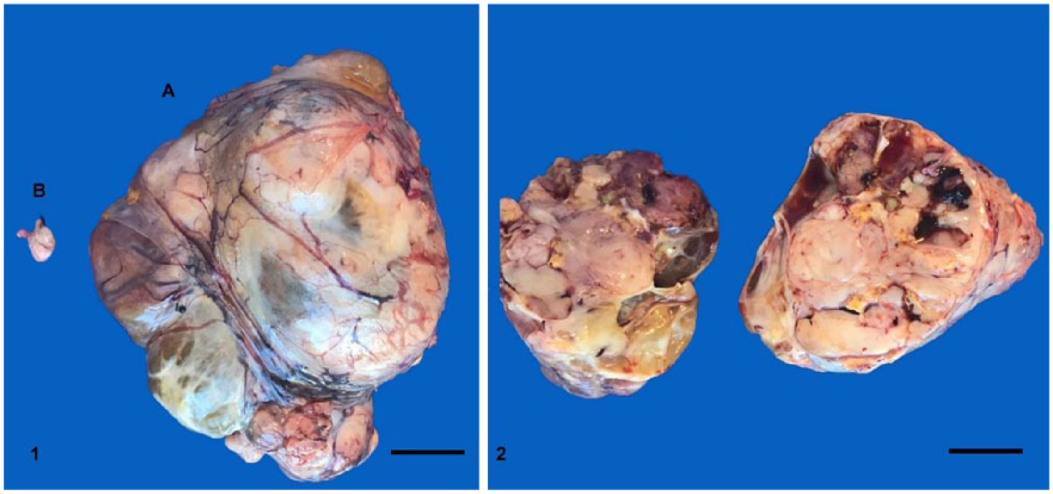
Testicular mass found in the coelomic cavity of a chicken.
Samples of ceca, small intestine, spleen, liver, kidney, heart, lung, bursa, right testis, left testicular mass, sciatic nerve, and ascending aorta were submitted for histologic analysis. Histologically, the cecal gut-associated lymphoid tissue was prominent, and there was mild diffuse lymphoplasmacytic enteritis. A single intraluminal nematode cross-section in the ceca was identified as Heterakis gallinarum and a single cestode embedded in the small intestinal mucosa was identified as Ascaridia galli. The spleen was reactive, with mild lymphoid hyperplasia, and the liver was characterized by minimal focal fibrosis with angiogenesis and hepatocyte loss. There was minimal, multifocal lymphoplasmacytic interstitial nephritis. Neither the liver nor the kidneys had recognizable lead inclusions. There was minimal focal lymphocytic and heterophilic myocarditis, and mild, multifocal histiocytic and granulocytic pneumonia. Multifocally, parabronchi were expanded by edema fluid, and anthracosilicosis was observed. There was age-related atrophy in the bursa, and multifocally, there was bursal glandular ectasia that may have been sectioning artifact.
The testicular mass consisted of components of a seminoma and a teratoma. Both neoplasms arise from the germinal epithelium of seminiferous tubules and are classified as germ cell tumors, thus a mixed germ cell tumor was diagnosed. The seminoma (Fig. 3) was a discrete, densely cellular, expansile mass that completely replaced seminiferous tubules, and consisted of round or polygonal cells arranged in sheets supported by a scant fibrovascular stroma. Individual cells had scant-to-abundant eosinophilic cytoplasm and well-defined cellular borders. Nuclei were round-to-oval, variably sized, and often centrally located, with single, prominent nucleoli. Mitotic figures were common (2–4 per 400× field), and were occasionally atypical. The teratoma was composed of tissues stemming from all 3 germ cell lines (Figs. 4–6). The ectodermal elements were neural tissue, similar to that of the central nervous system, and islands of squamous epithelium undergoing abrupt central keratinization with occasional associated adnexal glands. The endodermal element consisted of variably sized structures, lined by a single layer of cuboidal epithelium or ciliated epithelium with goblet cells (intestinal or respiratory epithelium). The mesodermal components were cartilage, bone, bone marrow, and adipose tissue. There was also granulocytic-to-mixed, multifocal, mild orchitis and multifocal necrosis in the residual left testis, and a right gonad resembling immature testis or ovary. Lesions were not evident in the sections of sciatic nerve or ascending aorta examined.
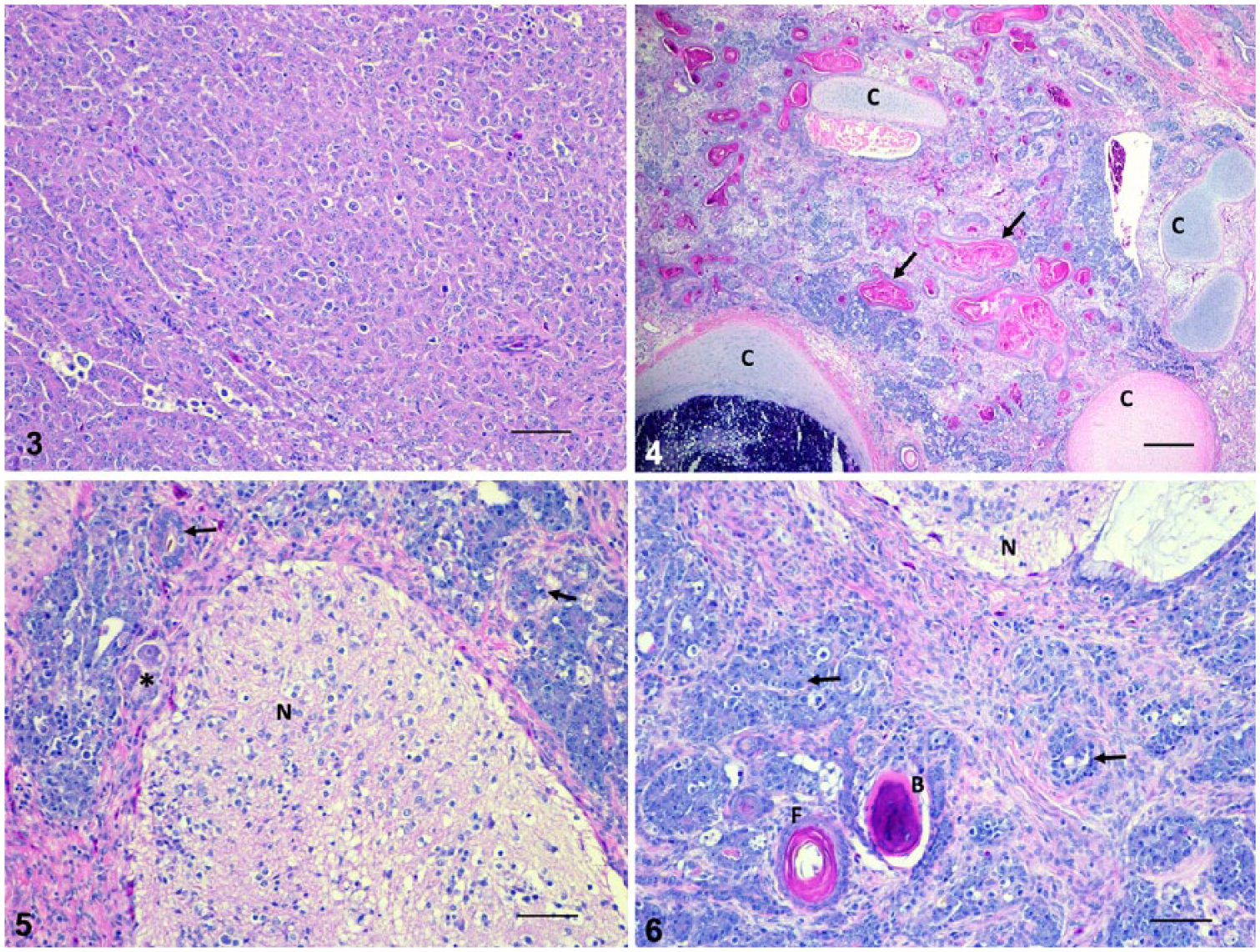
Mixed germ cell testicular tumor in a chicken.
Histologic diagnoses were mixed testicular germ cell tumor, anthracosilicosis, orchitis, and multiple organ inflammation. The testicular tumor, as a space-occupying mass, was the most likely cause of the clinical signs in this rooster. Teratomas and seminomas, the 2 neoplastic components identified in the mixed germ cell tumor, are both considered uncommon neoplasms, but have been reported in chickens. 13 Anthracosilicosis results from inhalation of carbon and material, such as sand or dust, containing silicon dioxide. It is seen in animals from a smoky, dirty, or dusty environment and is generally an insignificant finding at autopsy; the site of origin of this rooster was not provided. Orchitis, a possible cause of infertility in poultry, is commonly associated with bacteria including Salmonella and Escherichia coli. 3 The multiple organ inflammation is most likely the result of trauma from the compressive force of the testicular mass on the organs in the coelomic cavity.
Teratomas are tumors characterized by the histologic presence of mature embryonic tissue derived from at least 2 of the 3 germinal layers (endoderm, mesoderm, and ectoderm), hence the name tridermic teratoma. 8 The neoplasm in our case consisted of cell types from all 3 germinal layers. In poultry, teratomas have been described most commonly in the testis, but have also been documented in the adrenal gland, eye, kidney, ovary, pineal body, and spinal cord.4,5,13 Teratomas are generally benign, slow-growing tumors. Complications typically occur once the tumor becomes excessively large, causing compression of organs in the coelomic cavity and death. 9 These tumors have also been experimentally induced in poultry by injecting cadmium, copper, or zinc into active testes. 3 Advances in human medicine suggest that teratomas may be linked to factors such as inherited genes, fluctuating reproductive hormones, and low birth weights. 7 Furthermore, human medicine has reported that teratomas are present in >50% of mixed germ cell tumors in adults.2,15
Seminomas have been reported very rarely in chickens. 1 The rarity of these tumors may reflect the young age of birds at processing. Most seminomas in domestic animals are diagnosed in older individuals. 11 Seminomas tend to be large unilateral tumors that are typically benign; however, they can occasionally be bilateral and/or malignant with metastases reported. 13 Affected testes tend to be diffusely enlarged, have a thick capsule, and differ little from a normal testis in color and consistency. The contralateral testis is usually normal. 3 Seminomas tend to have a classical histologic appearance, as seen in our case, with sheet-like arrangements of clear cells with well-defined cytoplasmic borders and flattened nuclear membranes that are subdivided by fibrovascular septa into variably sized, smaller groups of cells. 14
In a retrospective study of 1,598 backyard chicken autopsy submissions to 2 veterinary diagnostic laboratories, reproductive neoplasms were diagnosed in 72 cases (4.5%). 6 Interestingly, all of the neoplasms identified in the review were ovarian adenocarcinomas. Adenocarcinomas are common malignant reproductive neoplasms diagnosed in chickens and are also referred to as carcinomatosis once direct extension metastasis has occurred. 13 This statistic is still important, however, because it helps to highlight the rarity of testicular neoplasms in the chicken. A retrospective study 12 from the University of Georgia assessed samples from 827 birds over a 5-y period. Of these samples, 76 had neoplastic processes, with only 4 (0.4%) associated with the reproductive tract. Of these, there was a seminoma in a northern cardinal, and no teratomas diagnosed. The authors noted that testicular neoplasms tend to be more common in budgerigars and cockatiels than other species of birds. 12 One of the major reasons for this rarity of diagnosed testicular neoplasms in poultry is likely the result of the over-representation of hens in backyard poultry operations in general, which leads to an over-representation of hens in laboratory submissions. Although several cases of testicular neoplasia have been described in wild birds, such as ducks, these may be under-reported as a result of death by unrelated causes and lack of diagnostic workups. 10
Testicular tumors are generally divided into germ cell tumors and sex cord-stromal tumors. 2 Germ cell tumors include seminomas, embryonal carcinomas, and teratomas; sex cord-stromal tumors include Sertoli-cell tumors and Leydig (interstitial) cell tumors. Mixed germ cell–sex cord-stromal tumors include neoplastic elements derived from both the germ cell and sex cord stromal elements of the testis. These are generally considered to be 2 independent tumors that are present concurrently. The most common example in domestic animals of a mixed tumor is a seminoma and Sertoli-cell tumor that are both present but are clearly separated and independent.2,11 A collision tumor within the testis is 1 mass with 2 distinct neoplastic processes of different cell origin. 11 Seminomas and Sertoli-cell tumors occasionally abut, thus creating one tumor with features of both a seminoma and Sertoli-cell tumor. These would be collision tumors because they have an interface between 2 distinct tumors. In true mixed tumors, both neoplastic elements are derived from the same cell population and are intimately mixed in variably sized tubular structures that are separated by a fibrous stroma of variable density.2,11 This presentation was seen in our case. Furthermore, some pathologists may view these as separate tumors because of the presence of a fibrovascular stroma; however, given the definitions above and the fact that both neoplastic components are derived from germ cells, we classified this as a mixed germ cell tumor.
To date, the etiology of germ cell tumors is largely unknown.14,15 In a 1950s survey of 2,000 poultry autopsy cases over 11 y, the author discussed links between various germ cell tumors involving seminomas that have appeared simultaneously in testicular masses. 4 The study proposed several mechanisms by which mixed germ cell tumors may arise. The most promising of these theories is the development of 2 individual cell types from a common ancestral stem cell, referred to as the germ cell theory in human literature. Advances in molecular biology and karyotyping in human medicine have shown that gonadal teratomas contain an identical pair of chromosomes, which suggests that they are the haploid descendants of embryonic germs cells that then undergo parthenogenesis.
Footnotes
Declaration of conflicting interests
The authors declared no potential conflicts of interest with respect to the research, authorship, and/or publication of this article.
Funding
The authors received no financial support for the research, authorship, and/or publication of this article.