
Abstract
Mixed germ cell tumors of the ovary have rarely been reported in veterinary species. A 3-year-old intact female Labrador Retriever dog was presented for lethargy, abdominal distention, and a midabdominal mass. An exploratory laparotomy revealed a large (23 cm in diameter) left ovarian tumor and multiple small (2–3 cm in diameter) pale tan masses on the peritoneum and abdominal surface of the diaphragm. Histological examination of the left ovary revealed a mixed germ cell tumor with a yolk sac component with rare Schiller–Duval bodies and a teratomatous component comprised primarily of neural differentiation. The abdominal metastases were solely comprised of the yolk sac component. The yolk sac component was diffusely immunopositive for cytokeratin with scattered cells reactive for α-fetoprotein and placental alkaline phosphatase. Within the teratomatous component, the neuropil was diffusely immunopositive for S100, neuron-specific enolase, and neurofilaments with a few glial fibrillary acidic protein immunopositive cells. Ovarian germ cell tumors may be pure and consist of only 1 germ cell element or may be mixed and include more than 1 germ cell element, such as teratoma and yolk sac tumor.
Tumors of the ovary have been described in many species and are considered to originate from 3 ovarian cell types with distinct embryologic origins: epithelial tumors of Mullerian origin (adenoma or carcinoma), sex cord stromal tumors (e.g., granulosa cell tumor and thecoma-fibroma), and germ cell tumors (dysgerminoma, teratoma, yolk sac tumors). 2 Germ cell tumors are a heterogeneous group of neoplasms and can occur in pure form or mixed, including more than 1 germ cell component. 16 Mixed germ cell tumors of the gonad are rare entities and, to the authors’ knowledge, in the veterinary literature have only been described in the ovary of 1 female cynomolgus macaque. 14 Extragonadal germ cell tumors are also rare and have been described almost exclusively in the suprasellar region of the brain of the dog 13 and in the eye of a dog. 6
The yolk sac is an embryonal structure whose function has evolved phylogenetically. In some species, it provides a supply of nutrients to the developing fetus. 5 The yolk sac also contains germ cells that migrate bilaterally to the urogenital crest during embryogenesis; this site will become the future gonad. Abnormal migration and persistence of germ cells at ectopic sites can result in extragonadal germ cell tumors in numerous locations along the midline (i.e., suprasellar, mediastinal, retroperitoneal). 13 While uncommon, ovarian teratomas have been described in veterinary species, particularly the dam; most ovarian teratomas are mature and behave as benign tumors. 2 Totipotent neoplastic germ cells can differentiate along embryonal or extra-embryonal lines. Embryonal somatic differentiation results in teratomatous elements; differentiation along a primitive embryonic cell line results in embryonal carcinoma. Extra-embryonal differentiation can occur along 1) the trophoblastic line, giving rise to choriocarcinoma, or 2) the yolk sac line, giving rise to a yolk sac tumor (“endodermal sinus tumor”). 8
Yolk sac tumors have been described sporadically in the veterinary literature1,3 but have been extensively described in the human medical literature.7,12,15 Mixed germ cell tumors differentiate along more than 1 cell line. To the authors’ knowledge, these tumors have only rarely been described in veterinary species. 14 The following is a detailed description of an ovarian mixed germ cell tumor with teratomatous and yolk sac components in a dog.
A 3-year-old, intact female Labrador Retriever dog was presented for lethargy, abdominal distention, and a midabdominal mass. The abdominal cavity contained approximately 3 liters of clear, watery, straw-colored fluid (transudate). An exploratory laparotomy revealed a large (23 cm in diameter) left ovarian tumor and multiple small (2–3 cm in diameter) pale tan masses on the peritoneum and abdominal surface of the diaphragm. An ovariohysterectomy was performed, and all visible peritoneal tumor implants were removed. The uterus, including both horns and attached ovaries, was submitted fresh along with formalin-fixed samples of peritoneum, diaphragm, and 1 hepatic hilar lymph node. The dog began chemotherapy and survived for 3 months postsurgery before being re-presented for thoracic metastases and substantial pleural effusion. No further diagnostic workup was performed, and the dog was euthanized; a necropsy was not performed.
The left ovary was replaced by a large (approximately 23 cm in diameter) multilobulated, soft to semi-firm neoplastic mass (Fig. 1). The capsular surface was reddish-brown, and the majority of the cut surface was beige (Fig. 2). The associated uterine tube (oviduct) and left uterine horn, the uterine body, right uterine horn, and right ovary were unremarkable. The cortex and medulla of the hepatic lymph node were effaced and replaced by a beige poorly demarcated mass.
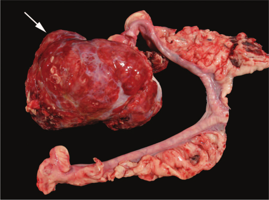
Three-year-old Labrador Retriever dog; ovaries and uterine horns. There is a 23-cm (at greatest diameter), dark red to brown, multilobulated mass arising from the left ovary (arrow). The right ovary and associated uterine tubes are unremarkable.
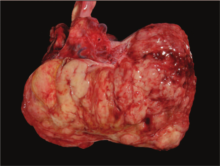
Macroscopic features of left ovarian mass from a 3-year-old Labrador Retriever dog. On cut section, the multilobulated fleshy mass has multifocal areas of hemorrhage.
Tissues were fixed in 10% neutral buffered formalin for 24 hr and processed routinely; sections were stained with hematoxylin and eosin. For immunohistochemistry, formalin-fixed tissue sections were routinely immunostained with the following antibodies at the University of Minnesota Medical Center, Fairview Immunohistochemistry Laboratory (Minneapolis, Minnesota): α-fetoprotein (AFP; polyclonal, dilution 1:1000), a placental alkaline phosphatase (PLAP; monoclonal, predilute), b cytokeratin AE1-AE3 (monoclonal, predilute), a and cluster of differentiation (CD)34 (monoclonal, predilute). c For the following antibodies, formalin-fixed, paraffin-embedded sections were routinely processed at the University of Minnesota Veterinary Diagnostic Laboratory (Saint Paul, Minnesota): glial fibrillary acidic protein (GFAP; polyclonal, dilution 1:600), a neurofilaments (NF; monoclonal, dilution 1:2000), a S100 (polyclonal, dilution 1:800), a neuron-specific enolase (NSE; monoclonal, dilution 1:200), a and synaptophysin (monoclonal, dilution 1:40). a
The ovarian tumor and metastases contained a number of different histologic patterns including yolk sac and teratomatous components. The left ovarian neoplasm contained 2 distinct components, that of the yolk sac tumor and that of the teratoma. The latter constituted the majority of the tumor bulk (approximately 90%) and differentiated almost exclusively toward neural tissue (Fig. 3A, 3B). Despite the yolk sac component comprising the minority of the tumor bulk in the left ovary, it was the sole representative of the metastases.
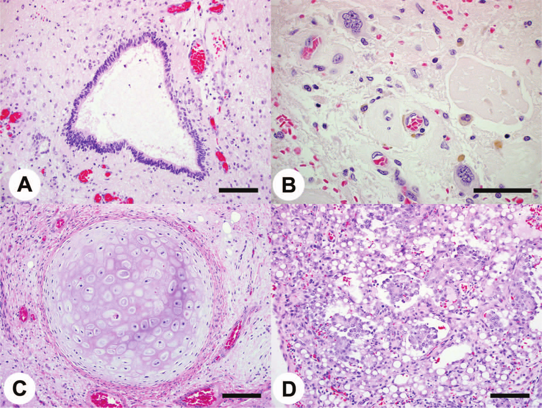
Three-year-old Labrador Retriever dog.
The nervous tissue of the teratoma consisted primarily of a poorly cellular, ill-defined, loose pale eosinophilic material interpreted as neuropil, which surrounded individual large cells (20–40 μm) with moderate amounts of pale eosinophilic to amphophilic cytoplasm and Nissl substance, and large central multilobulated nuclei with 1 distinct nucleolus (neurons; Fig. 3B). There were small cells (7–10 μm) with intensely dark round nuclei and little, if any, visible cytoplasm (glial cells) scattered throughout the neuropil and occasionally clustered (Fig. 3B). Scattered throughout the tumor were small- to medium-sized (100–200 μm in diameter) tubular structures lined by short columnar cells that had basally located nuclei and a moderate amount of lightly eosinophilic cytoplasm; these structures recapitulated respiratory epithelium (Fig. 3A). Smaller, round to ovoid tubules were lined by small stratified cells and were reminiscent of developing neural tubes. Mitotic figures were not observed. In some fields, medium-sized (1 mm in diameter) islands of well-differentiated hyaline cartilage (Fig. 3C), well-differentiated adipocytes (Fig. 3C), and small foci of immature hair follicles were identified. In addition, there were foci of cellular debris (necrosis/apoptosis), occasionally associated with neutrophils, mineral deposits, and golden brown pigment-laden cells. There were also multiple blood vessel profiles, many with intraluminal fibrin thrombi and surrounding hemorrhage.
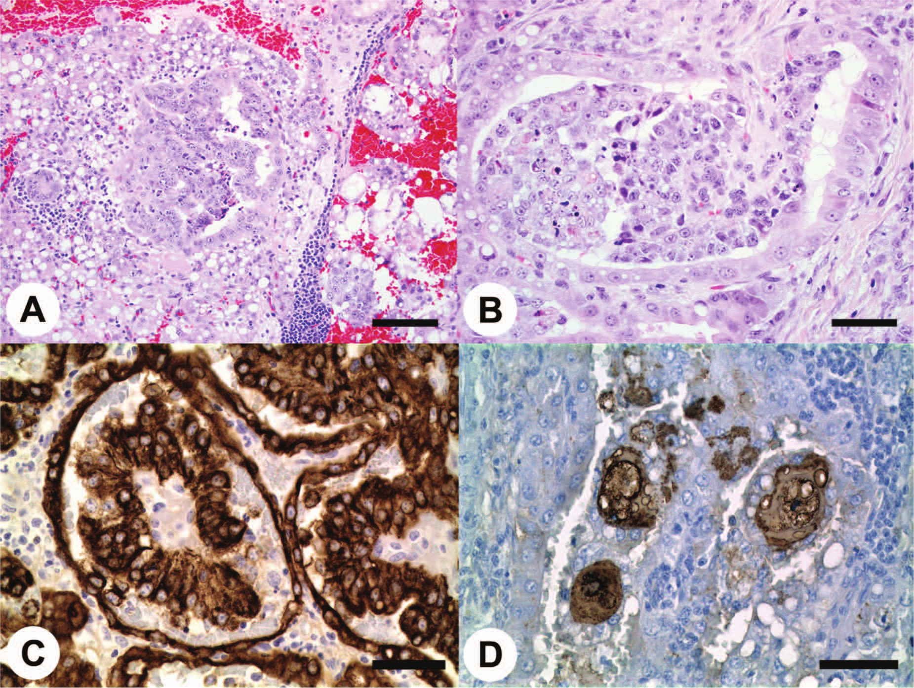
The yolk sac component in the left ovary and the metastases (hepatic lymph node, peritoneum, and diaphragm) consisted of a moderately to highly cellular, poorly demarcated mass arranged in tubulopapillary structures that occasionally condensed to form broad sheets (Figs. 3D, 4A, 4B). The tubulopapillary structures commonly formed festoons housed in cystic structures lined by cuboidal neoplastic cells (endodermal sinus pattern; Figs. 3D, 4A, 4B). Rare Schiller–Duval bodies were noted; the bodies consisted of small tufts of neoplastic cells surrounding a fibrovascular core, contained in a space lined by flattened to cuboidal cells (Fig. 4C). The neoplastic cells were large and polygonal, with a moderate amount of eosinophilic to amphophilic often-vacuolated cytoplasm; the cell nuclei were round to oval, vesicular, and contained a single prominent nucleolus. Mitotic figures within the yolk sac component were frequent, with an average of 4 per 40× high power field. Multifocal apoptotic bodies were observed.
Three-year-old Labrador Retriever dog.
The hepatic lymph node and biopsies of the diaphragm and peritoneum showed extensive metastasis that consisted exclusively of the yolk sac component of the primary tumor. The neoplastic cells were arranged primarily in a papillary pattern, with cytoplasmic and nuclear features similar to those observed in the left ovary.
The immunohistochemistry results for the ovarian tumor (teratomatous component) as well as the metastases (yolk sac component) are summarized in Table 1. The yolk sac component was diffusely immunopositive for cytokeratin AE1-AE3 (Fig. 4C); scattered cells were reactive for AFP (Fig. 4D) and PLAP. The yolk sac component was immunonegative for CD34. The neuropil of the teratomatous component was diffusely immunopositive for S100, NSE, and NF. Some large cells were immunopositive for GFAP. The teratomatous component was negative for synaptophysin.
Immunohistochemistry results of the ovarian tumor and metastases.*
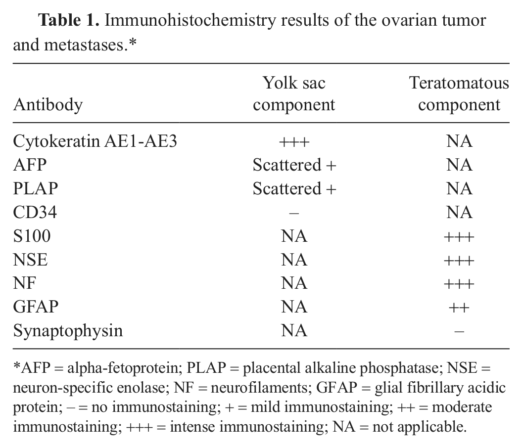
AFP = alpha-fetoprotein; PLAP = placental alkaline phosphatase; NSE = neuron-specific enolase; NF = neurofilaments; GFAP = glial fibrillary acidic protein; – = no immunostaining; + = mild immunostaining; ++ = moderate immunostaining; +++ = intense immunostaining; NA = not applicable.
Ovarian germ cell tumors may be pure and consist of only 1 germ cell element or may be mixed and include more than 1 germ cell element, such as teratoma and yolk sac tumor. The mixed germ cell tumor described in the current study contained yolk sac and teratomatous components. In the human medical literature, in 15% of cases, yolk sac elements coexist with another germ cell component, and yolk sac components are considered to be highly malignant. 15
The yolk sac component was a classical malignant tumor (poorly differentiated with high mitotic rate and metastases), which ultimately led to the euthanasia of this dog. All neoplastic germ elements in a mixed germ cell tumor, even if small, should be reported, as they will determine the behavior and treatment of the tumor. As a corollary, these tumors should be properly sampled and carefully analyzed. The route of metastasis of the yolk sac tumor was likely to have been via the lymphatic vessels due to the observed lymph node metastases and also transcoelomic because of the hydroabdomen (ascites) and serosal implants observed in biopsies of the diaphragm and peritoneum.
Yolk sac tumors of human beings have numerous histologic patterns, including endodermal sinus, reticular (or microcystic), polyvesicular vitelline, solid, parietal, papillary, glandular, hepatoid, and mesenchyme-like. 15 Schiller–Duval bodies are pathognomonic of this tumor. Combinations of these patterns are usually present, with the most common pattern being the microcystic or reticular pattern. 7 Of the variable histological patterns that may be observed in human yolk sac tumors, the patterns observed in the current case included papillary and reticular (microcystic) with Schiller–Duval bodies.
Immunohistochemically, most yolk sac tumors are at least focally positive for AFP, α1-antitrypsin, and cytokeratin, but not for epithelial membrane antigen (EMA). 15 The yolk sac tumor in the present case was focally immunopositive for AFP; immunostains for α1-antitrypsin or EMA were not performed. In addition, many yolk sac tumors have periodic acid–Schiff-positive, diastase-resistant hyaline globules; however, in the current case, hyaline globules were not observed. Most yolk sac tumors contain Schiller–Duval bodies, which are structures that are thought to arise from attempts at recapitulating the yolk sac epithelium. Such structures, initially interpreted by Schiller as recapitulating mesonephric glomeruli (“mesonephroma ovarii”), were reinterpreted by Teilum as recapitulating extraembryonic yolk sac allantoic structures of the rat’s placenta. 11 Teilum’s hypothesis has been supported by the observation that these tumors produce GATA-4, a transcription factor that regulates differentiation and function of murine yolk sac endoderm. 10 The current case had scattered Schiller–Duval bodies.
Ovarian teratomas have been described in most veterinary domestic species but are noted to be most common in dogs and cattle.2,4 Classically, elements of all 3 embryonal cell layers are present in teratomas; some tumors may include derivatives of 2 or only 1 layer (monodermal teratomas). With reference to the present case, the teratomatous component included adipocytic, respiratory, cutaneous adnexal, cartilaginous, and neural elements. In the human medical literature, teratomas with mature components are usually cystic (“dermoid cysts”) 15 and are usually benign as they are in veterinary species.2,9 The presence of immature teratomatous tissues, particularly if abundant, correlates with aggressive behavior; in the current case, mature and immature teratomatous elements were recognized. The present report presents a detailed gross, histologic, and immunohistochemical description of a canine mixed germ cell tumor.
Footnotes
Acknowledgements
The authors would like to acknowledge Drs. Judith Cooper and Michelle Ritt (University of Minnesota Veterinary Medical Center) for the submission of the specimen and for providing historical information. The authors would like to thank the Histology and Immunohistochemistry laboratories at the University of Minnesota Veterinary Diagnostic Laboratory and the University of Minnesota Medical Center.
a.
Alpha-fetoprotein, cytokeratin AE1-AE3, glial fibrillary acidic protein, neurofilaments, neuron-specific enolase, synaptophysin, S100; Dako North America Inc., Carpinteria, CA.
b.
Placental alkaline phosphatase, Leica Biosystems Richmond Inc., Richmond, IL.
c.
CD34, Ventana Medical Systems Inc., Tucson, AZ.
Declaration of conflicting interests
The author(s) declared no potential conflicts of interest with respect to the research, authorship, and/or publication of this article.
Funding
The author(s) received no financial support for the research, authorship, and/or publication of this article.