
Abstract
A 20-y-old female llama (Lama glama) was euthanized after a history of chronic dyspnea and osteoarthritis. At autopsy, the lungs were covered by clear gelatinous material and expanded by firm, variably discrete, tan-white nodules up to 8 cm diameter containing tan-white, viscous material. The tracheobronchial lymph nodes were firm and enlarged up to 6 × 4 × 3 cm; the thoracic aorta and carotid arteries were lined by hard, tan-white, mineralized intimal plaques. Histologic examination of lung revealed numerous 10–20 μm diameter yeasts with clear 1–2 μm thick double-contoured walls, central basophilic nuclei, and frequent broad-based budding, consistent with Blastomyces dermatitidis. DNA sequencing confirmed the diagnosis. B. dermatitidis should be considered in the differential diagnosis of pulmonary disease in llamas.
A 20-y-old, 155-kg, female llama (Lama glama) in good body condition with a history of chronic dyspnea and osteoarthritis was euthanized and presented to the University of Tennessee Veterinary Medical Center (Knoxville, TN) for autopsy. The thoracic cavity contained clear, gelatinous (fibrinous) material. Replacing 90% of the right lung and 40% of the left lung were firm, tan-white nodules of 2 mm to 8 cm diameter (Fig. 1).
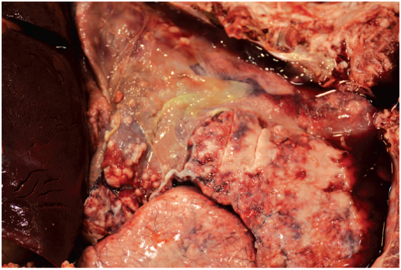
Lung from an adult female llama (Lama glama) with pulmonary blastomycosis. Gross photograph of pyogranulomatous pneumonia with thoracic fibrinous exudate.
On cut section, the larger nodules contained tan-white viscous-to-caseated material; the surrounding parenchyma was fibrotic. The tracheobronchial lymph nodes were firm and enlarged up to 6 × 4 × 3 cm; the largest had central mineralization. Lining the luminal aspect of the aorta and coronary arteries were multiple, variably sized, hard, tan-white plaques of intimal mineralization. Other gross findings included mild-to-moderate degenerative joint disease of the femorotibial and scapulohumeral joints. All other organ systems were grossly normal.
Sections of the lungs, lymph nodes, aorta, and carotid arteries were fixed in 10% buffered formalin and examined histologically. Distorting to effacing the pulmonary parenchyma were multifocal-to-coalescing, densely cellular infiltrates of macrophages, neutrophils, and lesser numbers of plasma cells with irregular random areas of necrosis. Within both the inflammatory foci and the necrotic tissue were numerous 10–20 µm diameter yeasts with a clear 1–2 µm thick double-contoured wall, a central basophilic nucleus, and frequent broad-based budding, consistent with Blastomyces dermatitidis (Fig. 2). Fungal yeasts were both extracellular and within multinucleate macrophages and stained positively with Grocott–Gomori methenamine silver (Fig. 3). Remaining alveolar septa were variably expanded by increased collagen and infiltrates of macrophages, neutrophils, and plasma cells. The tracheobronchial lymph nodes were expanded by fibrosis and central mineralization with loss of normal architecture; yeasts were not found in the lymph nodes. Within the aorta and carotid arteries, there was multifocal subintimal mineralization. Ziehl–Neelsen and Fite–Faraco staining for acid-fast bacilli in the lungs and lymph node were negative.
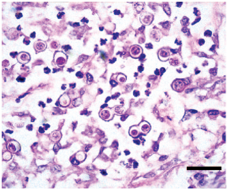
Blastomyces dermatitidis yeasts with frequent broad-based budding within an area of pyogranulomatous inflammation. H&E. Bar = 20 µm.
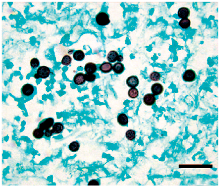
Numerous Blastomyces dermatitidis yeasts with frequent broad-based budding. Grocott–Gomori methenamine silver. Bar = 20 µm.
A 2014 report of disseminated blastomycosis in an alpaca raised the issue of genetic subgroups within the Blastomyces genus and called for active surveillance to expand the knowledge of the genetic and geographic diversity of this pathogen. 12 In addition, differentiation from Blastomyces gilchristii, which was identified in 2013 in a limited geographic range (Wisconsin, Minnesota, and Ontario) but associated with acute respiratory syndrome, was considered critical.5,7 In order to more completely define the organism in this case, we pursued DNA sequencing.
DNA was extracted (Qiagen, Valencia, CA) from formalin-fixed, paraffin-embedded tissue and used as template for polymerase chain reaction (PCR) using primers targeting the internal transcribed spacer (ITS)4 and ITS5 to amplify the entire ITS region, 5.8S ribosomal (r)RNA gene, and portions of the 18S and 26S rRNA genes, as described previously. 22 Direct Sanger sequencing of the PCR product was performed at the University of Tennessee Genomics Core Facility (Knoxville, TN). The sequence obtained was compared to those in GenBank using the BLASTn algorithm. 2 The sequence best matched those of Blastomyces dermatitidis (anamorph form) and Ajellomyces dermatitidis (teleomorph form) with 99% nucleotide similarities over 645 bp. Alignment of the locus within the ITS2 region, which is known to contain a single nucleotide polymorphism within the genus, confirmed that the sequence best matched that of B. dermatitidis rather than that of the newly described B. gilchristii. 5
B. dermatitidis is the anamorph (asexual) form of A. dermatitidis, which forms hyphae with fruiting bodies as a teleomorph (sexual) form under certain environmental conditions. 13 Infection is typically acquired via inhalation of the dimorphic Blastomyces anamorph form, which occurs as a yeast in tissue at ~37ºC; primary cutaneous inoculation also occurs rarely. 6 B. dermatitidis is common in multiple areas of the Americas and Africa, including the location of our report in northeastern Tennessee. 3
Blastomycosis occurs in a variety of domestic and wild mammals, most commonly humans and dogs. 6 Clinical disease is characterized by focal-to-disseminated sites of intense infiltrates of neutrophils, macrophages, and plasma cells, with necrosis to fibrosis in a variety of organs. In addition to the lungs, blastomycotic lesions have been reported in lymph nodes, the eye, testicles, brain, skin, and various internal organs in disseminated disease. 6
The presumptive clinical diagnosis for this llama prior to euthanasia was chronic obstructive pulmonary disease (COPD). Differential diagnoses for the gross appearance of the lungs at autopsy included infection caused by Mycobacterium spp., Rhodococcus equi, Coccidioides immitis, Cryptococcus neoformans, Cryptococcus gattii, Histoplasma capsulatum, Aspergillus spp., and B. dermatitidis. Llamas are susceptible to coccidioidomycosis, but this fungal agent was considered unlikely, given that endemic areas for C. immitis are in the southwestern United States and parts of Central and South America. 10 However, travel history for this animal was not provided. Bacteria within the Mycobacterium tuberculosis complex were considered as a possible etiology, given that tuberculosis has been described in llamas and camels.1,16 Of the differentials at autopsy (each of which have been recognized in other species at this locale), the paucity of reports regarding the occurrence of cryptococcosis, histoplasmosis, aspergillosis, rhodococcosis, and blastomycosis in camelids, specifically llamas, made these diseases less likely than tuberculosis.4,10–12,15,23 B. dermatitidis was identified microscopically and confirmed via DNA sequencing, and there was no evidence of mycobacterial infection.
Infection via inhalation of spores is probable in this llama, resulting in severe pyogranulomatous pneumonia. The enlargement and fibrosis of the tracheobronchial lymph nodes may be secondary to dissemination of blastomycosis. No fungal organisms or other etiologic agents were found despite a battery of stains (Gram, Fite–Faraco, Ziehl–Neelsen, periodic acid–Schiff, and Gomori methenamine silver) and diligent searching. However, given the chronicity of the lesion, other causes of chronic lymphadenitis resulting in fibrosis cannot be ruled out.
Mineralization of the aorta and other arteries is presumed secondary to blastomycosis in this animal. As a component of chronic disease, monocytes and macrophages can generate and release tumor necrosis factor–alpha (TNFα) that drives mineralization, and macrophages may accumulate basic calcium phosphate that can locally increase cytokine production (TNFα, interleukin [IL]-1, and IL-8) that further drives mineralization and stimulates endothelial cells to differentiate toward osteoblasts. 20 In dromedary camelids and cattle with Johne’s disease, which exhibits a predominantly granulomatous cellular response, proinflammatory cytokines (IL-1α, IL-1β, IL-6, IL-10, interferon-γ [IFNγ], and TNF-α), acute-phase and oxidative-stress proteins are significantly increased. 9 Hypercalcemia has been reported in dogs with granulomatous diseases, including blastomycosis, and is likely associated with the conversion of calcifediol (25-hydroxyvitamin D) to calcitriol (1,25-dihydroxyvitamin D) by activated macrophages.8,14 Elevated vitamin D levels cause hypercalcemia and hyperphosphatemia, which may result in the mineralization of soft tissues, including blood vessels, endocardium, kidneys, lungs, and gastric mucosa. 17 No other organs are known to be mineralized in our case; however, only limited tissues were examined histologically.
Other potential causes of arterial mineralization in livestock include ingestion of cholecalciferol rodenticide or calcinogenic plants, such as Cestrum diurnum, Solanum glaucophyllum (previously Solanum malacoxylon), and Trisetum flavescens, and chronic disease, including that caused by Mycobacterium avium ssp. paratuberculosis.18,19 This llama had no known exposure to calcinogenic plants or rodenticides. There were no gross lesions consistent with Johne’s disease; however, as no culture of the intestine or histology of the intestine and mesenteric lymph nodes were performed, subclinical Johne’s infection with increased macrophage production of TNFα and other cytokines cannot be ruled out completely. 21 Acid-fast staining of lung was negative for other mycobacterial species.
Blastomycosis is rarely reported in camelids. However, B. dermatitidis should be considered in the differential diagnosis of pulmonary disease in llamas, particularly in endemic areas.
Footnotes
Acknowledgements
We thank Dr. David Bemis and the UTCVM mycology and bacteriology laboratory for assistance with this case.
Declaration of conflicting interests
The authors declared no potential conflicts of interest with respect to the research, authorship, and/or publication of this article.
Funding
The authors received no financial support for the research, authorship, and/or publication of this article.